. 2020 Mar 16;2(1):vdaa038.
doi: 10.1093/noajnl/vdaa038. eCollection 2020 Jan-Dec.

Bevacizumab-based treatment as salvage therapy in patients with recurrent symptomatic brain metastases

Affiliations

Bevacizumab-based treatment as salvage therapy in patients with recurrent symptomatic brain metastases

Anna Sophie Berghoff et al. Neurooncol Adv. .

Abstract

Background: Salvage treatment for recurrent brain metastases (BM) of solid cancers is challenging due to the high symptomatic burden and the limited local treatment options.

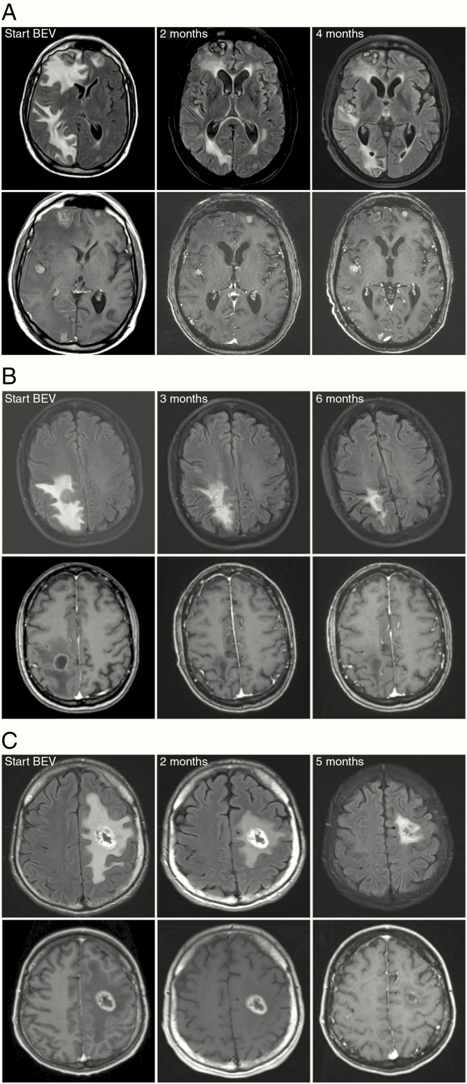
Methods: Patients with recurrent BM with no option for further local therapies were retrospectively identified from BM databases. Bevacizumab-based treatment was initiated as a salvage treatment. Radiological imaging before and after bevacizumab-based treatment was reevaluated for treatment response using the Response Assessment in Neuro-Oncology (RANO) BM criteria.

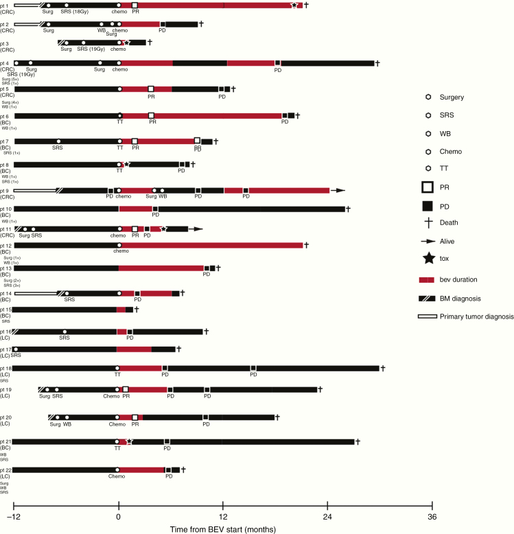
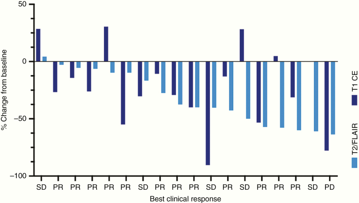
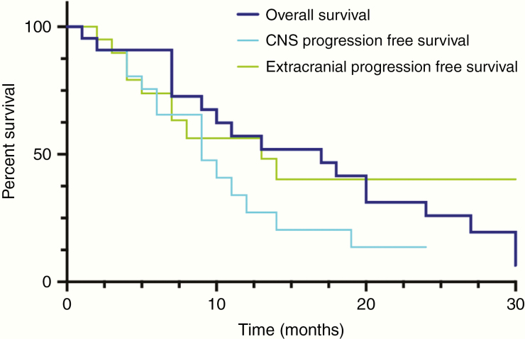
Results: Twenty-two patients (36.4% male) with recurrent BM from breast cancer (40.9%), colorectal cancer (31.8%), or lung cancer (27.3%) were identified. Previous BM-directed therapies were radiosurgery in 16/22 (72.7%) patients, whole-brain radiotherapy in 8/22 (36.4%), and neurosurgical resection in 11/22 (50.0%). Time since BM diagnosis to initiation of bevacizumab treatment was 16.5 months. Of 22 patients 14 (63.6%) received concurrent systemic therapies. Neurological symptom improvement could be achieved in 14/22 (63.6%) and stabilization in 6/22 (27.3%) patients, resulting in a clinical benefit in 20/22 (90.9%) patients. Steroids could be reduced or stopped in 15/22 (68.2%) patients. Rate of improvement on T1-weighted imaging was 15/19 (78.9%; median reduction: -26.0% ± 32.9) and 19/20 (95%; median reduction: -36.2% ± 22.2) on T2-weighted FLAIR imaging. According to RANO-BM best response was partial response in 7/19 (36.8%), stable disease in 9/19 (47.3%), and progressive disease in 3/19 (15.7%) patients. Median CNS-specific progression-free survival was 8 months and median overall survival after initiation of bevacizumab treatment was 17 months.

Conclusions: Bevacizumab-based treatment had clinically relevant intracranial activity in the vast majority of patients suffering from recurrent, symptomatic BM. The data supports a prospective clinical trial of bevacizumab as a salvage treatment in BM.

Keywords: bevacizumab; brain edema; brain metastases; recurrence; steroids.


Figures

Figure 1.
Figure 1.
Timelines demonstrating the clinical course of disease of all included patients. Each line represents an individual patient. Study inclusion with the start of bevacizumab treatment is set to timepoint 0. Patient history is shown up to 12 months prior to study inclusion. The primary tumor is indicated on the left, and prior therapies are listed below. LC, non-small cell lung cancer; BC, breast cancer; CRC, colorectal cancer; SRS, radiosurgery; WB, whole-brain radiotherapy; TT, targeted treatment; PR, brain-specific partial response; PD, brain-specific progressive disease; tox, treatment-associated toxicity; BM, brain metastases; bev, bevacizumab.
Figure 2.
Figure 2.
Waterfall plot demonstrating the intracranial response to bevacizumab-based treatment in patients with recurrent BM on T1 CE (after Gd-contrast enhancement) and T2/FLAIR MRI images. FLAIR sequence or if not available T2 sequence was used for response assessment. The best clinical response for each patient is given below. SD, stable disease; PR, partial response; PD, progressive disease.
Figure 3.
Figure 3.
(A) MR images of baseline (BL) and follow-up MRI of a lung cancer patient with a partial response after initiation of bevacizumab-based treatment. Partial response is seen both on FLAIR (upper row) and T1-weighted (lower row) images. (B and C) MR imaging examples of patients with breast cancer (B) and colorectal cancer (C) showing partial response on FLAIR and complete response on T1-weighted images after Gd-contrast administration (B) on follow-up MRI. Note that the tumor volume (central core) and perifocal edema can be differentiated on FLAIR images.
Figure 4.
Figure 4.
Kaplan–Meier plot showing overall survival, CNS-specific progression-free survival, and extracranial progression-free survival after initiation of bevacizumab-based treatment.

References

    1. Berghoff AS, Schur S, Füreder LM, et al. Descriptive statistical analysis of a real life cohort of 2419 patients with brain metastases of solid cancers. ESMO Open. 2016;1(2):e000024. - PMC - PubMed
    1. Soffietti R, Abacioglu U, Baumert B, et al. Diagnosis and treatment of brain metastases from solid tumors: guidelines from the European Association of Neuro-Oncology (EANO). Neuro Oncol. 2017;19(2):162–174. - PMC - PubMed
    1. Rana N, Pendyala P, Cleary RK, et al. Long-term outcomes after salvage stereotactic radiosurgery (SRS) following in-field failure of initial SRS for brain metastases. Front Oncol. 2017;7:279. - PMC - PubMed
    1. Specht HM, Combs SE. Stereotactic radiosurgery of brain metastases. J Neurosurg Sci. 2016;60(3):357–366. - PubMed
    1. Kellner CP, D’Ambrosio AL. Surgical management of brain metastases. Neurosurg Clin N Am. 2011;22(1):53–59, vi. - PubMed

LinkOut - more resources