. 2020 May 6;2(1):vdaa056.
doi: 10.1093/noajnl/vdaa056. eCollection 2020 Jan-Dec.

Molecular profiles and immunomodulatory activities of glioblastoma-derived exosomes

Affiliations

Molecular profiles and immunomodulatory activities of glioblastoma-derived exosomes

Juliana Hofstatter Azambuja et al. Neurooncol Adv. .

Abstract

Background: Glioblastoma is one of the most immunosuppressive human tumors. Emerging data suggest that glioblastoma-derived exosomes (GBex) reprogram the tumor microenvironment into a tumor-promoting milieu by mechanisms that not yet understood.

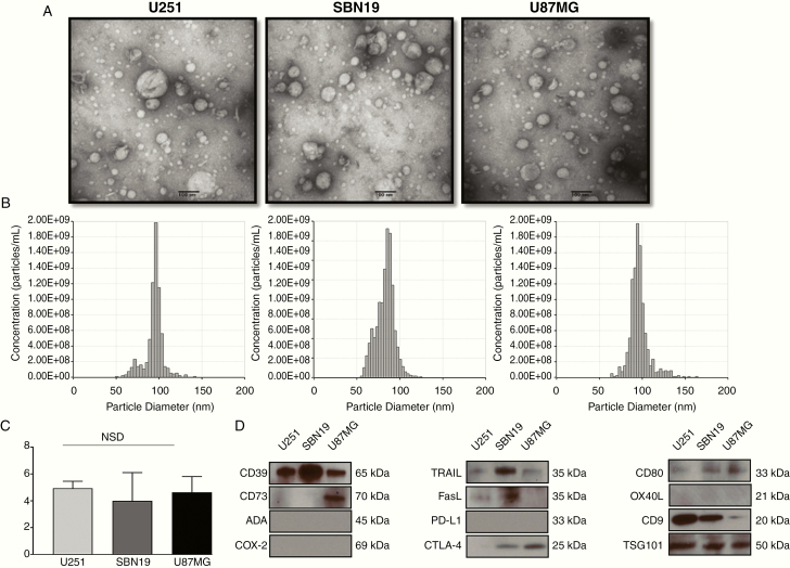
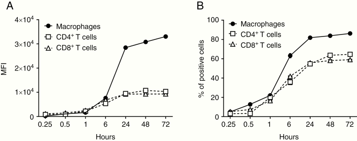
Methods: Exosomes were isolated from supernatants of glioblastoma cell lines by size exclusion chromatography. The GBex endosomal origin, size, protein cargos, and ex vivo effects on immune cell functions were determined. GBex were injected intravenously into mice to evaluate their ability to in vivo modulate normal immune cell subsets.

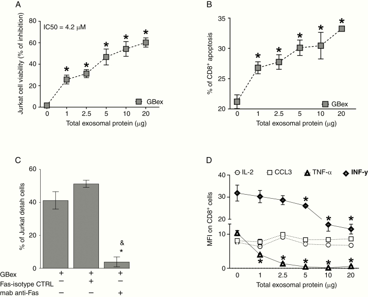
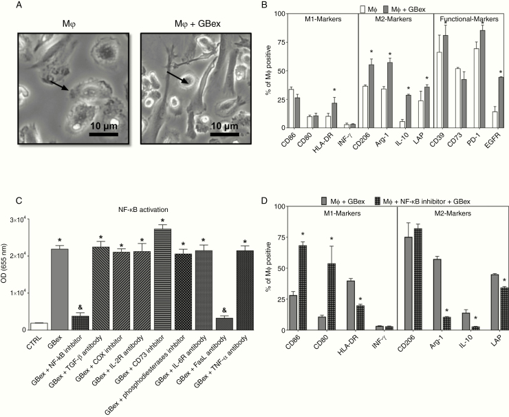
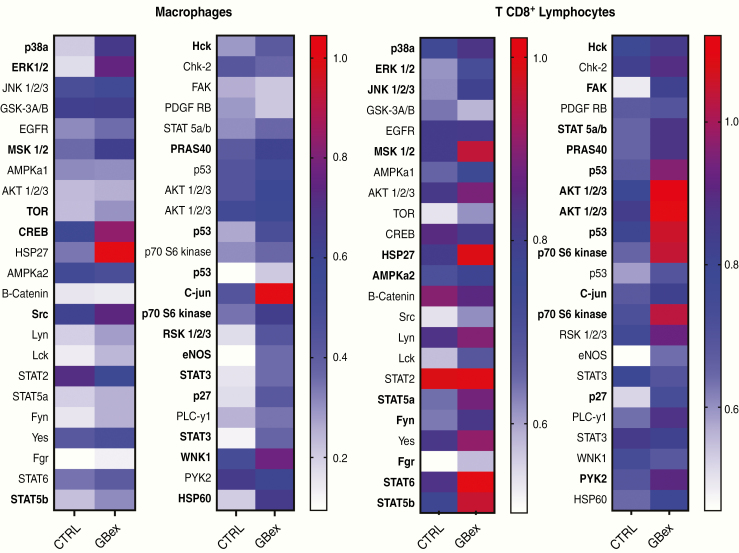
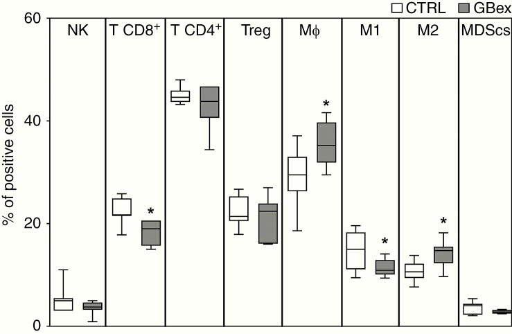
Results: GBex carried immunosuppressive proteins, including FasL, TRAIL, CTLA-4, CD39, and CD73, but contained few immunostimulatory proteins. GBex co-incubated with primary human immune cells induced simultaneous activation of multiple molecular pathways. In CD8+ T cells, GBex suppressed TNF-α and INF-γ release and mediated apoptosis. GBex suppressed natural killer (NK) and CD4+ T-cell activation. GBex activated the NF-κB pathway in macrophages and promoted their differentiation into M2 cells. Inhibition of the NF-κB pathway in macrophages reversed the GBex-mediated effects. GBex-driven reprogramming of macrophages involved the release of soluble factors that promoted tumor proliferation in vitro. In mice injected with GBex, the frequency of splenic CD8+ T cells, NK cells, and M1-like macrophages was reduced, while that of naïve and M2-like macrophages increased (P < .05).

Conclusions: GBex reprogrammed functions of all types of immune cells in vitro and altered their frequency in vivo. By creating and sustaining a highly immunosuppressive environment, GBex play a key role in promoting tumor progression.

Keywords: tumor microenvironment; exosomes; glioblastoma; immune system; macrophages.


Figures

Figure 1.
Figure 1.
Characteristics and the immunomodulatory profile of GBex. GB cells were plated and expanded for 72 h. Cell supernatants were harvested, pre-cleared, concentrated to 1 mL, and used for exosome isolation by mini-SEC. (A) Representative transmission electron microscopy images of GBex produced by the 3 GB cell lines (U87MG, SBN19, and U251). (B) qNano analyses of the isolated GBex. (C) Total protein levels isolated from supernatants of GB cells. The data are mean values ± SD from 3 experiments. Data were analyzed by ANOVA followed by Tukey post hoc. (D) Western blot profiles of GBex isolated from the supernatants of the GB cell lines. Each lane was loaded with 10 μg GBex protein. Note the presence of exosome markers (CD9 and TGS101) and the immunoinhibitory proteins (CD39, CD73, FasL, CTL-4, TRAIL).
Figure 2.
Figure 2.
Uptake of labeled GBex by cells. Human macrophages, CD4+ T and CD8+ T cells (1 × 10⁵ cells in wells of a 48-well plate) were co-incubated with GBex (10 µg) isolated from supernatants of U251 GB cell line and labeled with SYTO RNASelect Green Fluorescent cell stain dye as described in Methods. Flow cytometry was performed at the indicated time points, and the graphs show the mean fluorescence intensity (MFI) in (A) and % of positive cells in (B).
Figure 3.
Figure 3.
GBex induced apoptosis of primary CD8+ T cells and reduced cytokine expression. (A) Cell viability: CD8+ Jurkat cells were co-incubated for 24 h with increasing protein levels of GBex isolated from supernatants of the U251 GB cell line. (B) Annexin V-stained primary human-activated CD8+ T cells incubated with GBex or PBS as a control for 24 h. (C) Cell viability of CD8+ Jurkat cells preincubated with anti-FAS Mab and after 1 h with 4.2 µg of GBex protein isolated from supernatants of the U251 GB cell line. *Significantly different from control cells and &different from FAS-isotype control (CTRL). (D) Expression levels of IL-2, CCL3, TNF-α, and INF-γ measured by flow cytometry in CD8+ Jurkat cells co-incubated with GBex. Data were analyzed by ANOVA followed by post hoc comparisons (Tukey test). *Significantly different from control cells at P < .005 and &Significantly different from isotype CTRL.
Figure 4.
Figure 4.
GBex induced M2-like polarization via NF-κB activation in macrophages. (A) Representative phase-contrast microphotographs of macrophages after treatment with GBex for 72 h. Note alterations of macrophage morphology in Mφ + GBex (bar equals 10 µm). (B) Macrophage polarization after treatment with GBex for 72 h. The panel shows M1 markers (CD86, CD80, HLA-DR, and INF-γ), M2 markers (CD206, Arginase-1, IL-10, and LAP), and functional markers (CD39, CD73, PD-1, and EGFR). (C) Biological activity of GBex (20 µg) on the activity of the NF-κB pathway macrophages. *Significantly different from CTRL and &significantly different from GBex at P<0.05. (D) Macrophage polarization after treatment with the NF-κB inhibitor for 30 min followed by the addition of GBex. Values represent the mean ± SEM from 3 independent experiments. Data were analyzed by ANOVA followed by post hoc comparisons (Tukey–Kramer test). *Significantly different from macrophages+ GBex group at P < 0.05.
Figure 5.
Figure 5.
GBex-induced phosphorylation in multiple molecular pathways in macrophages and CD8+ T cells. Analysis of total proteins (300 μg) in lysates of macrophages or CD8+ T cells which were co-incubated with GBex for 15 or 12 min, respectively. Values are normalized to reference spots on the membranes. The arrays were quantified using ImageJ and the most important changes are highlighted in bold.
Figure 6.
Figure 6.
In vivo changes in immune cells in the spleen of normal mice injected intravenously with 1 mg/kg of GBex. Mice received intravenous injections of GBex in PBS (1 mg/kg or equivalent volume) every 3 days for 16 days (see the schedule in Supplementary Figure 1). Animals were sacrificed and spleens were harvested, dissociated to obtain a single-cell suspension. The frequency of immune cells was determined by immunostaining and flow cytometry. The percentages of NK (CD45+ NKp16+), CD8+ T cells (CD45+CD3+CD8+), CD4+ T cells (CD45+CD3+CD4+), T-regulatory cells (CD4+FOXP3+), macrophages (CD45+CD11b+F4/80+), M1-like macrophages (CD45+CD11b+CD80+CD86+), M2-like macrophages (CD45+CD11b+CD206+), and MDSCs (CD45+CD11b+Gr1+) were determined in the spleen. The values represent the mean values ± SEM from 7 animals. *Indicates a significant difference from the CTRL (P < .05) as determined by Student’s t-test.

References

    1. Molinaro AM, Taylor JW, Wiencke JK, Wrensch MR. Genetic and molecular epidemiology of adult diffuse glioma. Nat Rev Neurol. 2019;15(7):405–417. - PMC - PubMed
    1. Zhang Y, Chen J, Xue Q, et al. Prognostic significance of microRNAs in glioma: a systematic review and meta-analysis. Biomed Res Int. 2019;2019:4015969. - PMC - PubMed
    1. Wick W, Osswald M, Wick A, Winkler F. Treatment of glioblastoma in adults. Ther Adv Neurol Disord. 2018;11:1756286418790452. - PMC - PubMed
    1. D’Alessio A, Proietti G, Sica G, Scicchitano BM. Pathological and molecular features of glioblastoma and its peritumoral tissue. Cancers (Basel). 2019;11(4):469. - PMC - PubMed
    1. Stupp R, van den Bent MJ, Hegi ME. Optimal role of temozolomide in the treatment of malignant gliomas. Curr Neurol Neurosci Rep. 2005;5(3):198–206. - PubMed

LinkOut - more resources