Review
. 2020 Sep;11(9):630-640.
doi: 10.1007/s13238-020-00755-1. Epub 2020 Jul 8.

Cellular metabolism and homeostasis in pluripotency regulation

Affiliations
Review

Cellular metabolism and homeostasis in pluripotency regulation

Kun Liu et al. Protein Cell. 2020 Sep.

Abstract

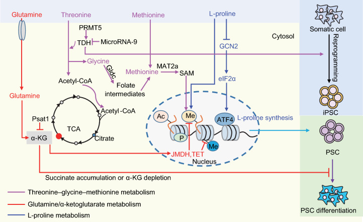
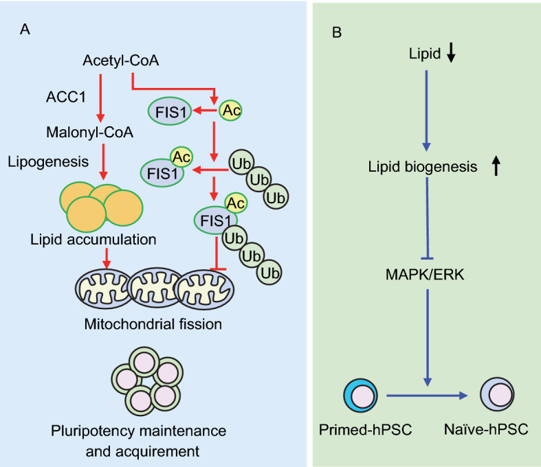
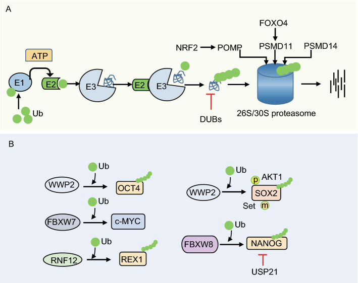
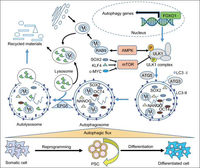
Pluripotent stem cells (PSCs) can immortally self-renew in culture with a high proliferation rate, and they possess unique metabolic characteristics that facilitate pluripotency regulation. Here, we review recent progress in understanding the mechanisms that link cellular metabolism and homeostasis to pluripotency regulation, with particular emphasis on pathways involving amino acid metabolism, lipid metabolism, the ubiquitin-proteasome system and autophagy. Metabolism of amino acids and lipids is tightly coupled to epigenetic modification, organelle remodeling and cell signaling pathways for pluripotency regulation. PSCs harness enhanced proteasome and autophagy activity to meet the material and energy requirements for cellular homeostasis. These regulatory events reflect a fine balance between the intrinsic cellular requirements and the extrinsic environment. A more complete understanding of this balance will pave new ways to manipulate PSC fate.

Keywords: amino acid metabolism; autophagy; lipid metabolism; pluripotent stem cell (PSC); ubiquitin-proteasome system (UPS).


Figures

Figure 1
Figure 1
Amino acid metabolism in pluripotency regulation. Threonine/methionine metabolism contribute to pluripotency regulation by providing SAM for DNA and histone methylation in PSCs. The threonine dehydrogenase TDH is highly expressed in mESCs, maintaining a high ratio of SAM/SAH that is correlated with high H3K4me3 levels. TDH expression is positively regulated by PRMT5 and negatively regulated by microRNA-9. Metabolism of glutamine and glucose regulates pluripotency through α-KG, which is a cofactor for Jumonji domain-containing histone demethylases (JMDH) and the ten-eleven translocation family of enzymes (TETs) that are involved in DNA demethylation. The cellular level of L-proline is fine-tuned by the amino acid starvation response (AAR) pathway Gcn2-Eif2α-Atf4. Excessive supplementation with L-proline leads to ESC differentiation. Appropriate intracellular synthesis of L-proline safeguards PSC pluripotency
Figure 2
Figure 2
Fatty acid metabolism in pluripotency regulation. Appropriate cellular levels of lipids safeguard pluripotency. (A) De novo synthesis of fatty acids, initiated by the enzyme acetyl-Coenzyme A carboxylase alpha (ACC1), promotes pluripotency maintenance and acquisition by enhancing mitochondrial fission. This conserved pathway is antagonized by ubiquitin-proteasome mediated degradation of the acetylated mitochondrial fission mediator FIS1. (B) In human PSCs, exogenous lipid deficiency induces intracellular lipogenesis which ultimately inhibits endogenous ERK and promotes pluripotency
Figure 3
Figure 3
Regulation of pluripotency by the ubiquitin-proteasome system (UPS). (A) PSCs exhibit high proteasome degradation activity, which is regulated by FOXO4-driven expression of the 19S proteasome subunit PSMD11 and corresponding enhanced assembly of 26S/30S proteasomes. (B) Levels of the pluripotency factors OCT4, c-MYC, REX1, SOX2 and NANOG are fine-tuned by UPS to maintain the precise quantity that facilitates pluripotency. p, phosphorylation; m, methylation
Figure 4
Figure 4
Regulation of pluripotency by autophagy. PSCs exhibit a high autophagic flux that is regulated by FOXO1, which coordinates the autophagy machinery gene program at the transcriptional level. High autophagic flux maintains appropriate levels of cellular pluripotency factors like OCT4, SOX2 and NANOG, and organelles like mitochondria (M). Inhibition of autophagy leads to accumulation of abnormal mitochondria and breakdown of pluripotency in spite of increased levels of pluripotency proteins. Activation of autophagy by AMPK is essential for both pluripotency maintenance and acquisition. Inactivation of mTOR by the pluripotency factors SOX2, KLK4 or c-MYC facilitates somatic cell reprogramming to pluripotency

References

    1. Alexander PB, Wang J, McKnight SL. Targeted killing of a mammalian cell based upon its specialized metabolic state. Proc Natl Acad Sci USA. 2011;108:15828–15833. - PMC - PubMed
    1. Ang YS, Tsai SY, Lee DF, Monk J, Su J, Ratnakumar K, Ding J, Ge Y, Darr H, Chang B, et al. Wdr5 mediates self-renewal and reprogramming via the embryonic stem cell core transcriptional network. Cell. 2011;145:183–197. - PMC - PubMed
    1. Bedford L, Paine S, Sheppard PW, Mayer RJ, Roelofs J. Assembly, structure, and function of the 26S proteasome. Trends Cell Biol. 2010;20:391–401. - PMC - PubMed
    1. Boyer LA, Lee TI, Cole MF, Johnstone SE, Levine SS, Zucker JP, Guenther MG, Kumar RM, Murray HL, Jenner RG, et al. Core transcriptional regulatory circuitry in human embryonic stem cells. Cell. 2005;122:947–956. - PMC - PubMed
    1. Buck MD, O’Sullivan D, Geltink RIK, Curtis JD, Chang CH, Sanin DE, Qiu J, Kretz O, Braas D, van der Windt GJW, et al. Mitochondrial dynamics controls T cell fate through metabolic programming. Cell. 2016;166:63–76. - PMC - PubMed

Publication types