. 2020 Aug 12;142(32):13718-13728.
doi: 10.1021/jacs.0c02789. Epub 2020 Jul 30.

Water-Gated Proton Transfer Dynamics in Respiratory Complex I

Affiliations

Water-Gated Proton Transfer Dynamics in Respiratory Complex I

Max E Mühlbauer et al. J Am Chem Soc. .

Abstract

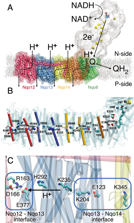
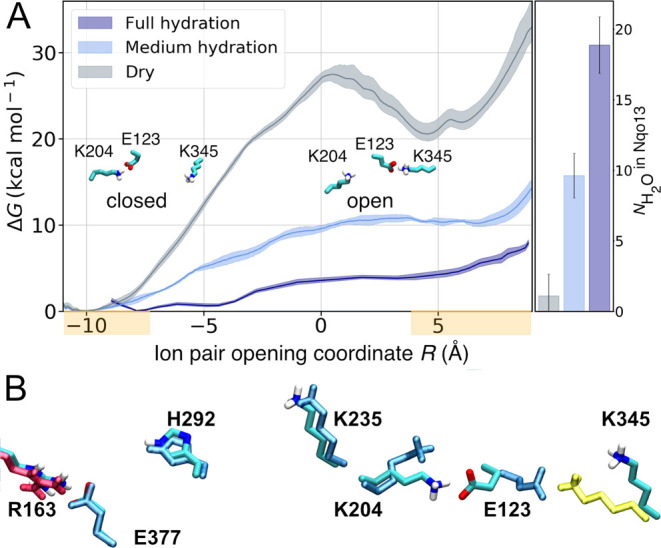
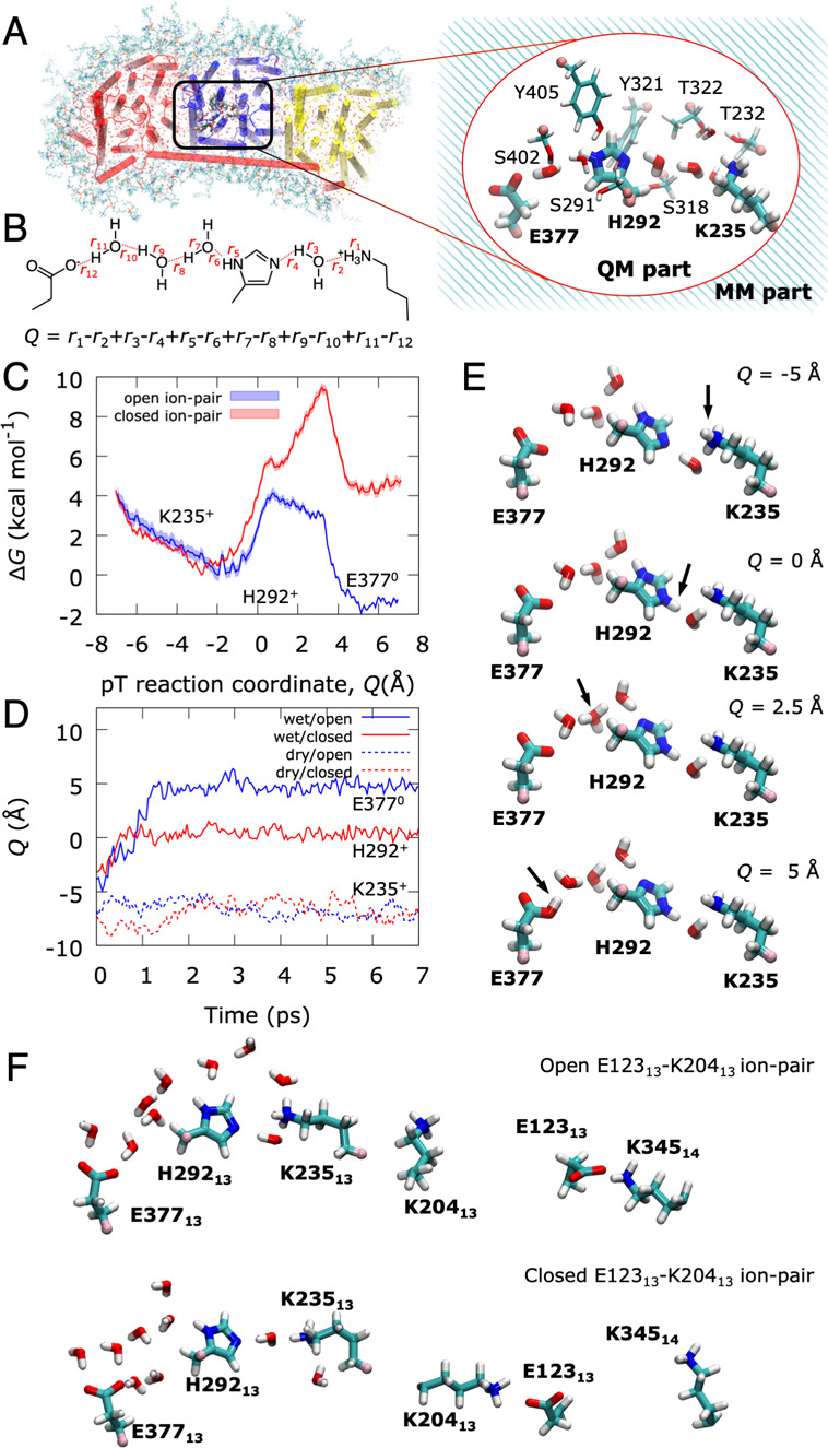
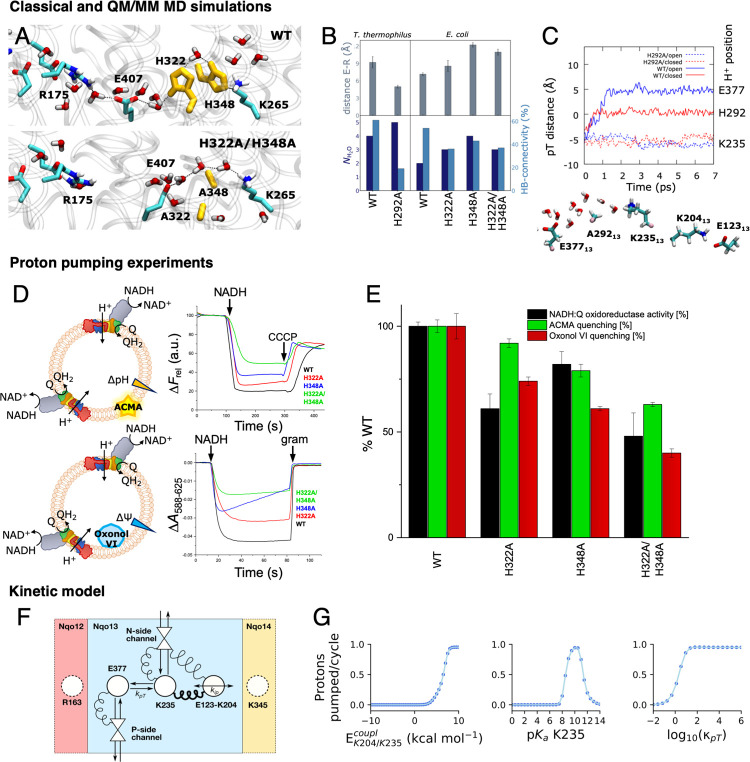
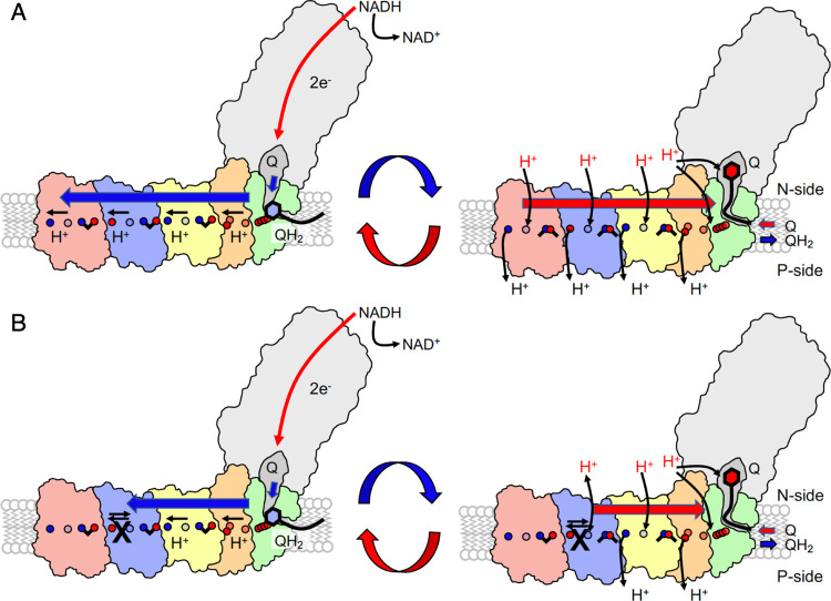
The respiratory complex I transduces redox energy into an electrochemical proton gradient in aerobic respiratory chains, powering energy-requiring processes in the cell. However, despite recently resolved molecular structures, the mechanism of this gigantic enzyme remains poorly understood. By combining large-scale quantum and classical simulations with site-directed mutagenesis and biophysical experiments, we show here how the conformational state of buried ion-pairs and water molecules control the protonation dynamics in the membrane domain of complex I and establish evolutionary conserved long-range coupling elements. We suggest that an electrostatic wave propagates in forward and reverse directions across the 200 Å long membrane domain during enzyme turnover, without significant dissipation of energy. Our findings demonstrate molecular principles that enable efficient long-range proton-electron coupling (PCET) and how perturbation of this PCET machinery may lead to development of mitochondrial disease.


Conflict of interest statement

The authors declare no competing financial interest.

Figures

Figure 1
Figure 1
(A) Complex I catalyzes electron transfer from NADH to Q in its hydrophilic domain (in white) and pumps protons across the Nqo12 (red), Nqo13 (blue), and Nqo14 (yellow), as well as in the Nqo8 (green) and Nqo7/10/11 (in orange) subunits of the membrane domain. (B) An axis of conserved charged and polar residues spans the membrane domain from the Q channel to the terminal Nqo12 subunit. (C) Nqo13 is connected to its neighboring subunits by charged interfaces (blue squares) that enable formation of ion-pairs between the subunits. The lateral pT from Lys23513 via His29213 to Glu37713 is marked with an arrow.
Figure 2
Figure 2
(A) Free energy profiles for opening the Lys20413/Glu12313 ion-pair at the Nqo13/Nqo14 interface in the fully hydrated state (dark blue), at medium hydration levels (light blue), and in the dry state (gray) of Nqo13. The Glu/Lys residues were modeled in their charged state (see Table S10). The ion-pair opening (shifting) reaction coordinate, R, is defined as d(E12313-K20413) – d(E12313-K34514). Closed ion-pair R ∼ (−9 ± 2) Å and open ion pair R∼ (+5 ± 2) Å are marked in orange on the R-axis. Ion-pair opening is favored by the increased hydration state of the proton channel. (B) In the closed conformation, the ion-pair interaction is established between Lys20413 and Glu12313, whereas in the open conformation, Glu12313 forms a contact with Lys34514 (in yellow).
Figure 3
Figure 3
Free energy and dynamics of lateral proton transfer in Nqo13. Prior to the pT reaction, all Glu/Lys residues were modeled in their charged states (see Table S10). (A) QM/MM models for the pT reaction in the Nqo13 subunit, showing the atoms in the QM region (inset). (B) The proton transfer (pT) reaction coordinate (Q) is represented by a linear combination of bond breaking and bond forming distances. (C) QM/MM free energy profiles for the lateral pT from Lys23513 via His29213 to Glu37713 in the fully hydrated state with closed (in red) and open (in blue) ion-pair conformations, respectively. (D) Unbiased QM/MM MD simulations for the pT process. In the medium-hydrated state (red and blue dashed lines), the pT stalls at Lys23513 in both open and closed ion-pair conformations. In the fully hydrated state with a closed ion-pair, the proton equilibrates between Lys23513 and His29213 within ∼1 ps (solid red line) but does not progress onward during the remaining simulation. In stark contrast, the ion-pair opening induces full pT to Glu37713 within ∼2 ps (solid blue line). (E) Snapshots of the QM/MM free energy profiles showing the transferred proton on Lys23513 (top), His29213 (second panel), at an intermediate Zundel ion (third panel), and Glu37713 (bottom). See Movie S1 for animation of the pT dynamics. (F) Snapshots of the open ion-pair conformation (top, with intact Glu12313/Lys34514 ion-pair) and the closed ion-pair conformation (bottom, intact Lys20413/Glu12313 ion-pair).
Figure 4
Figure 4
Perturbing the proton transfer dynamics in complex I. (A) Snapshots of pT wires in the WT (top) and H322A/H348A variant (bottom) of E. coli complex I after 100 ns MD simulations (see Figure S4). (B) Substitution of the bridging histidine residues leads to perturbation in the Glu37713/Arg16312 (Glu407M/Arg175L) and Lys23513/Glu37713 (Lys265M/Glu407M) distances in classical MD simulations of T. thermophilus (T) and E. coli (E) complex I. Additional water molecules close the gap between the lysine and glutamate residues but with a lowered occupancy relative to the WT. (C) pT dynamics from QM/MM MD simulations of the WT and H292A variant (top) and snapshot of the H292A variant, showing the position of the pT wire and the Lys20413-Glu12313 ion-pair in closed conformation (bottom). (D) ACMA (top) and oxonol-VI (bottom) assays from proton pumping experiments in E. coli complex I. The ACMA and oxonol-VI assays monitor the ΔpH and ΔΨ gradient across the proteoliposome membrane, respectively (see also Figure S6D). (E) Summary of NADH:Q oxidoreduction activity, ACMA, and oxonol quenching experiments of WT and alanine variants. All measurements were performed in triplicates, with errors given as standard deviations. All activities were measured from the maximum level of the optical changes without extrapolation to zero time point using the plateau levels as a base. The NADH:Q oxidoreductase activity was determined from monitoring NADH decrease at 340 nm. See Supporting Information Methods and Figure S6 for further details. (F) Schematic representation of the proton pumping kinetic model of complex I with N- and P-side channels, Glu37713, Lys23513, and the ion-pair Glu12313/Lys20413 (see Materials and Methods). (G) Effect of the parameters of the kinetic model on the proton pumping across the membrane (see Materials and Methods). Left: coupling energy between ion-pair and Lys23513. Center: the pKa of Lys23513. Right: the K235/E377 intrinsic pT rate. See Supporting Information Methods and Figures S7, S13 for a detailed description of the kinetic model.
Figure 5
Figure 5
Putative proton pumping mechanism in complex I. (A) Left: reduction of quinone (Q) to quinol (QH2) triggers stepwise proton transfer and ion-pair opening steps that propagate as a forward electrostatic pulse (blue arrow) to the terminal Nqo12 edge (in red) of the membrane domain. Right: The pumped protons are released by proton uptake from the N-side (H+ in red) and ion-pair closure during the backward signal (red arrow) to the membrane-bound Q binding site. Quinol/quinone exchange and reprotonation of the Q-site reset the pumping machinery for the next catalytic cycle. (B) Introduced mutations in Nqo13 (black cross) perturb the lateral pT from Nqo13 onward. The electrostatic backward pulse is reflected prematurely back from Nqo13 to the Q-channel, establishing pumping by Nqo14 (in yellow) and Nqo8/7/10/11 (green/orange) but at perturbed timing that affects the Q oxidoreduction activity.

References

    1. Kaila V. R. I. Long-Range Proton-Coupled Electron Transfer in Biological Energy Conversion: Towards Mechanistic Understanding of Respiratory Complex I. J. R. Soc., Interface 2018, 15, 20170916. 10.1098/rsif.2017.0916. - DOI - PMC - PubMed
    1. Hirst J. Mitochondrial Complex I. Annu. Rev. Biochem. 2013, 82, 551–575. 10.1146/annurev-biochem-070511-103700. - DOI - PubMed
    1. Brandt U. Energy Converting NADH: Quinone Oxidoreductase (Complex I). Annu. Rev. Biochem. 2006, 75, 69–92. 10.1146/annurev.biochem.75.103004.142539. - DOI - PubMed
    1. Sazanov L. A. A giant molecular proton pump: Structure and mechanism of respiratory complex I. Nat. Rev. Mol. Cell Biol. 2015, 16, 375–388. 10.1038/nrm3997. - DOI - PubMed
    1. Wikström M. Two protons are pumped from the mitochondrial matrix per electron transferred between NADH and ubiquinone. FEBS Lett. 1984, 169, 300–304. 10.1016/0014-5793(84)80338-5. - DOI - PubMed

Publication types