. 2020 Sep;9(17):6330-6343.
doi: 10.1002/cam4.3201. Epub 2020 Jul 9.

Switches of SOX17 and SOX2 expression in the development of squamous metaplasia and squamous intraepithelial lesions of the uterine cervix

Affiliations

Switches of SOX17 and SOX2 expression in the development of squamous metaplasia and squamous intraepithelial lesions of the uterine cervix

Jobran M Moshi et al. Cancer Med. 2020 Sep.

Abstract

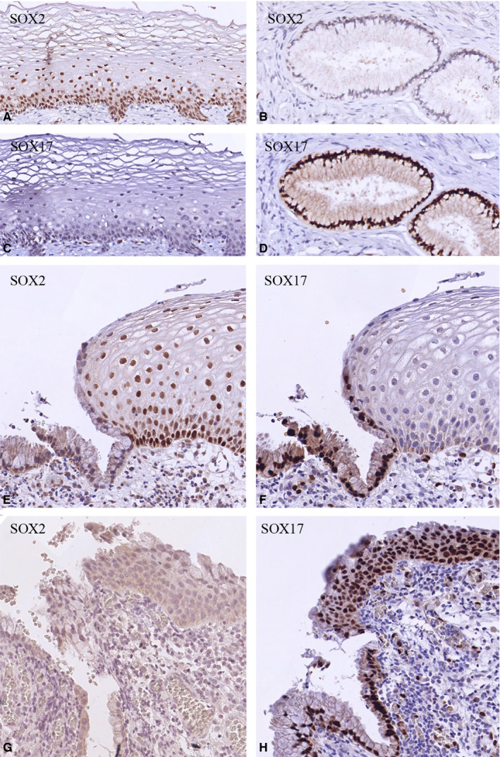
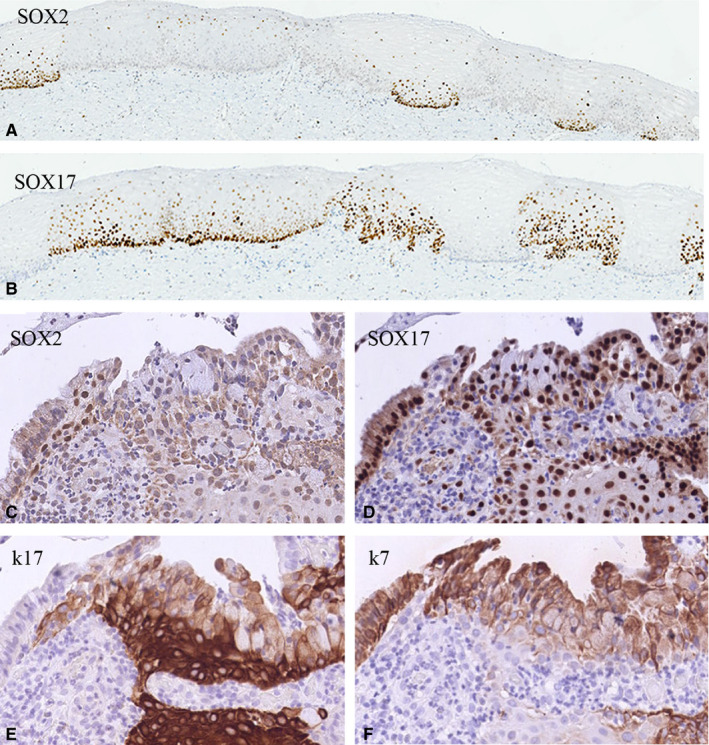
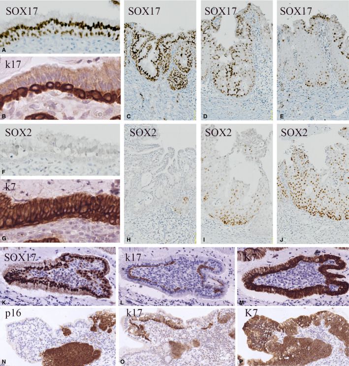
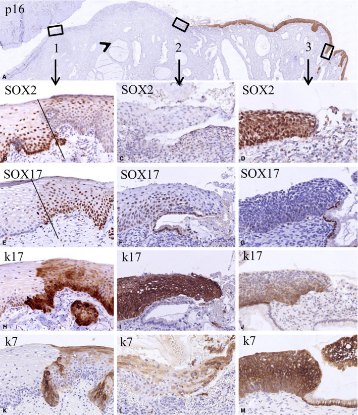
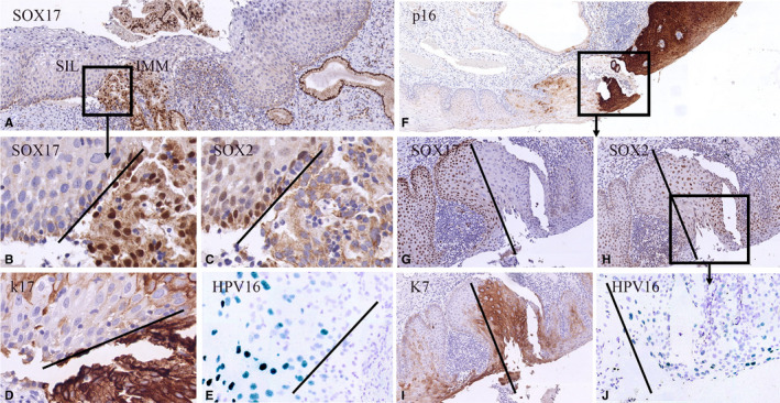
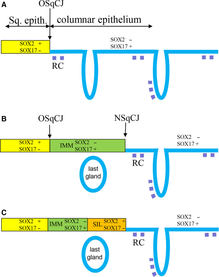
Aims: The dynamics and topographical distribution of SOX17 and SOX2 expression was studied in the transformation zone (TZ) of the uterine cervix. This TZ is a dynamic area where switches from glandular into squamous epithelium can be recognized, new squamocolumnar junctions are formed, and premalignant lesions originate. SOX17 and SOX2 show mutually exclusive expression patterns in the normal uterine cervix, with SOX2 being exclusively found in squamous epithelium, while SOX17 is detected in endocervical columnar cells and reserve cells.

Methods and results: Normal cervices and squamous intraepithelial lesions (SIL) were studied with immunohistochemistry, methylation of SOX17, human papilloma virus (HPV) genotyping, and in situ hybridization. In the TZ squamous metaplasia originating from these reserve cells can still show SOX17 expression, while also remnants of SOX17-positive immature metaplasia can be recognized in the normal squamous epithelium. SOX17 expression is gradually lost during maturation, resulting in the exclusive expression of SOX2 in the majority of (SIL). This loss of SOX17 expression is independent of methylation of the CpG island in its promotor region. HPV can be detected in SOX17-positive immature metaplastic regions in the immediate vicinity of SOX2-positive SIL, suggesting that switches in SOX17 and 2 expression can occur upon HPV infection.

Conclusions: This switch in expression, and the strong association between the distribution of reserve cells and squamous areas within the columnar epithelium in the TZ, suggests that reserve cell proliferations, next to basal cells in the squamous epithelium, are potential targets for the formation of squamous lesions upon viral infection.

Keywords: SOX17; SOX2; cervical preneoplasia; keratins; reserve cells; squamocolumnar junction; squamous intraepithelial lesions; transformation zone.


Conflict of interest statement

The authors are responsible for disclosing all financial and personal relationships between themselves and others that might bias their work. There are no potential conflicts.

Figures

FIGURE 1
FIGURE 1
SOX expression patterns in normal cervical epithelia. A‐D, Mutually exclusive SOX2 (A, B) and SOX17 (C, D) immunostaining patterns of normal ectocervical squamous epithelium (A, C) and endocervical glandular epithelium (B, D). This switch in SOX expression is clearly seen at the SqCJ (E, F). SOX2 is negative in immature metaplastic epithelium (G), while this epithelium is positive for SOX17 (H)
FIGURE 2
FIGURE 2
SOX expression patterns in metaplastic cervical epithelia. A and B, SOX2 (A) and SOX17 (B) immunostaining in corresponding areas morphologically classified as mature squamous epithelium, showing alternating and mutually exclusive expression of SOX2 and SOX17. C‐F, SOX2 is negative in immature metaplastic epithelium (C), while this epithelium is positive for SOX17 (D), with the strongest expression in the basal cell layers. Immature metaplastic squamous epithelium is characterized by a strong positivity for keratin 17 mainly in the basal cell layers (E) and a slightly weaker, more superficial positivity for keratin 7 (F)
FIGURE 3
FIGURE 3
SOX expression patterns in cervical reserve cells. SOX17 immunostaining of reserve cells in normal epithelium found in cases of microglandular hyperplasia (A‐J) and SIL (K‐P). A‐E, SOX17 immunoreactivity in endocervical glandular epithelium and reserve cells (A), microglandular hyperplasia (C), and in early immature metaplasia (D, E). F‐J, Increasing expression of SOX2 is seen upon development of immature metaplasia (F, H‐J). Keratin 17 (B) shows a specific immunostaining reaction in the reserve cell compartment, while keratin 7 (G) additionally also stains the columnar/glandular epithelial cells. K‐P, SOX 17 positivity (K) in keratin 17 (L) and keratin 7 (M)‐positive reserve cells in a case of high‐grade squamous intraepithelial lesions. These stretches of reserve cells may be positioned in close proximity to the HPV‐positive premalignant lesion as shown in (N‐P) after immunostaining for p16 (N), keratin 17 (O), and keratin 7 (P)
FIGURE 4
FIGURE 4
Switches in the expression of SOX2 and SOX17 in the transition of normal epithelium to high‐grade squamous intraepithelial lesions (HSIL). Low magnification of a section stained for p16 showing negative normal epithelia and a positive HSIL lesion (A). The boxes 1‐3 in this figure indicate epithelial transitions within the normal epithelium (box 1: a transition between normal and metaplastic squamous epithelium, possibly the original squamocolumnar junction), a new squamocolumnar junction (box 2), and the transition between HSIL and glandular epithelium (box 3). The fact that glands are seen beneath the squamous epithelium between boxes 1 and 2 (arrowhead) indicates that this region can be regarded as the transformation zone. The higher magnifications of these areas (B‐M) illustrate the mutually exclusive expression of SOX2 and SOX17 in these transitions. This is again evident in the keratin 17‐ and keratin 7‐negative normal ectocervical squamous epithelium in the p16‐negative area (left epithelial area in B, E, H, K). The keratin 17‐ and keratin 7‐positive metaplastic epithelium (right epithelial area in B, E, H, K) shows an extensive SOX17 positivity in the basal and intermediate compartment, and in this case SOX2 positivity in the superficial layers. In the strongly keratin 17‐positive squamous epithelium close to the new SqCJ only SOX17 is expressed, while SOX2 expression is absent (C, F, I, L). This SOX expression pattern is reversed in HSIL as identified by expression of keratins 7 and 17, where SOX2 expression is high and SOX17 expression is absent (D, G, J, M; p16 positive area)
FIGURE 5
FIGURE 5
Switches in expression of SOX2 and SOX17 in relation to high risk human papilloma virus infection in SIL. Low magnifications of high‐grade squamous intraepithelial lesions (HSIL) lesions immunostained for SOX17 (A) and p16 (F). These cases show switches in SOX expression in areas containing the keratin 7‐positive HSIL (I) lesions next to keratin 17‐positive (immature) metaplasia (D). The metaplastic areas show SOX17 positivity (B, G) and no expression of SOX2 (C, H). Only the SOX2‐positive regions show HPV 16 positivity as detected by in situ hybridization (CISH; E, J)
FIGURE 6
FIGURE 6
Schematic overview of switches in SOX expression during formation of metaplasia and SIL in the cervical transformation zone. IMM, immature metaplasia; LG, position of the last gland (sometimes seen as Nabothian cyst) beneath the squamous epithelium formed during the metaplastic process; NSqCJ, new squamocolumnar junction at the interface between metaplasia and columnar epithelium; OSqCJ, original squamocolumnar junction at the interface between squamous epithelium and columnar epithelium; RC, reserve cells

References

    1. Wright TC, Ferenczy A. Benign diseases of the cervix In: Kurman RJ, ed. Blaustein's Pathology of the Female Genital Tract. New York: Springer; 1994;203‐227.
    1. Mukonoweshuro P, Oriowolo A, Smith M. Audit of the histological definition of cervical transformation zone. J Clin Pathol. 2005;58:671. - PMC - PubMed
    1. Schiffman M, Castle PE, Jeronimo J, Rodriguez AC, Wacholder S. Human papillomavirus and cervical cancer. Lancet. 2007;370:890‐907. - PubMed
    1. Herfs M, Hubert P, Delvenne P. Epithelial metaplasia: adult stem cell reprogramming and (pre)neoplastic transformation mediated by inflammation? Trends Mol Med. 2009;15:245‐253. - PubMed
    1. Herfs M, Hubert P, Moutschen M, Delvenne P. Mucosal junctions: open doors to HPV and HIV infections? Trends Microbiol. 2011;19:114‐120. - PubMed

Publication types

MeSH terms