Review
. 2020 Aug 1;129(2):272-282.
doi: 10.1152/japplphysiol.00381.2020. Epub 2020 Jul 9.

Edward F. Adolph Distinguished Lecture. Skeletal muscle atrophy: Multiple pathways leading to a common outcome

Affiliations
Review

Edward F. Adolph Distinguished Lecture. Skeletal muscle atrophy: Multiple pathways leading to a common outcome

Sue C Bodine. J Appl Physiol (1985). .

Abstract

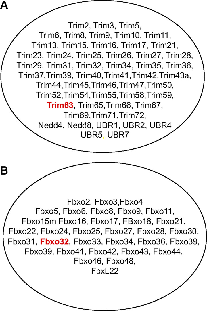
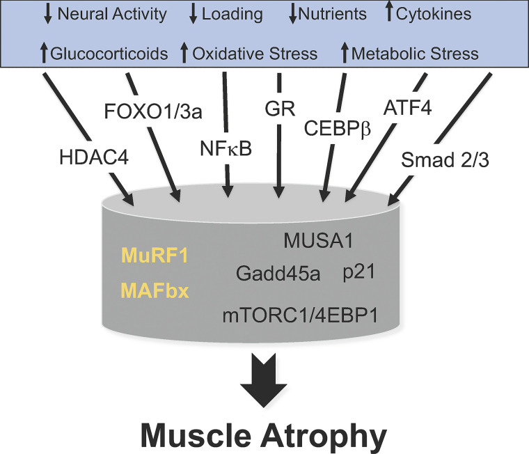
Skeletal muscle atrophy continues to be a serious consequence of many diseases and conditions for which there is no treatment. Our understanding of the mechanisms regulating skeletal muscle mass has improved considerably over the past two decades. For many years it was known that skeletal muscle atrophy resulted from an imbalance between protein synthesis and protein breakdown, with the net balance shifting toward protein breakdown. However, the molecular and cellular mechanisms underlying the increased breakdown of myofibrils was unknown. Over the past two decades, numerous reports have identified novel genes and signaling pathways that are upregulated and activated in response to stimuli such as disuse, inflammation, metabolic stress, starvation and others that induce muscle atrophy. This review summarizes the discovery efforts performed in the identification of several pathways involved in the regulation of skeletal muscle mass: the mammalian target of rapamycin (mTORC1) and the ubiquitin proteasome pathway and the E3 ligases, MuRF1 and MAFbx. While muscle atrophy is a common outcome of many diseases, it is doubtful that a single gene or pathway initiates or mediates the breakdown of myofibrils. Interestingly, however, is the observation that upregulation of the E3 ligases, MuRF1 and MAFbx, is a common feature of many divergent atrophy conditions. The challenge for the field of muscle biology is to understand how all of the various molecules, transcription factors, and signaling pathways interact to produce muscle atrophy and to identify the critical factors for intervention.

Keywords: MAFbx; MuRF1; mTORC1; protein synthesis; ubiquitin proteasome pathway.


Conflict of interest statement

Dr. Bodine holds equity in Emmyon, Inc., and serves on the Scientific Advisory Board.

Figures

Fig. 1.
Fig. 1.
Regulation of skeletal muscle mass throughout the life span. This diagram illustrates some of the critical factors that regulate skeletal muscle size at different life stages. At each life stage skeletal muscle undergoes specific changes that are regulated by different critical factors. During the embryonic stage growth factors, hormones and myocytes/satellite cells play a critical role in the formation and innervation of muscle fibers. During the postnatal stage, external loading and increased neural activation become important factors along with growth factors, hormones, and satellite cells in regulating increases in the length and cross-sectional area of muscle fibers. As adults, maintaining muscle mass and adapting to changes in external loading and neural activation become the dominant focus. With advancing age, decreases in external loading and activity, as well as, inflammation, increasing levels of cytokines, oxidative stress, and metabolic stress can lead to a decrease in muscle mass and strength.
Fig. 2.
Fig. 2.
Skeletal muscle atrophy is prevalent in many diseases. Skeletal muscle atrophy is a serious consequence of the diseases and conditions listed in this diagram.
Fig. 3.
Fig. 3.
Building a multidisciplinary muscle research program. A multidisciplinary research program was developed to discover novel mechanisms regulating skeletal muscle size. At the center of the program was the establishment of animal models of skeletal muscle atrophy (denervation, nerve crush-reinnervation, joint immobilization, hindlimb suspension, glucocorticoid excess) and hypertrophy (functional overload, reloading following disuse). Muscle tissues taken at different time points were used in the identification of novel gene and protein targets using differential gene expression and proteomics approaches. Muscle samples were also used to identify signaling pathways that were altered in response to stimuli that induced atrophy and hypertrophy. Furthermore, pharmacological agents, such as clenbuterol, IGF1, rapamycin, glucocorticoids, were utilized both in vivo and in vitro to manipulate selective pathways to induce changes in muscle size. Finally, technologies such as in vivo electroporation and mouse genetic engineering were utilized to modify gene expression in skeletal muscle.
Fig. 4.
Fig. 4.
Discovery of MuRF1 and MAFbx in skeletal muscle. A: animal models of disuse atrophy range in severity from those that primarily produce decreased external loading of the muscles, such as hindlimb unloading to those that produce both a decrease in external loading and a complete absence of neural activity such as denervation or spinal cord injury. Joint immobilization is a model that produces decreased external loading and a reduction in neural activity depending on the degree to which the joint is immobilized. B: loss of mass of the rat medial gastrocnemius muscle (MG) was compared in three models of disuse atrophy (hindlimb suspension, ankle joint immobilization, and denervation). To identify a potential common trigger of muscle atrophy, 3 days postimmobilization was chosen as the time point and model to perform a differential gene expression analysis (GeneTag, Applied Bioscience). In the primary screen, all those genes that were differentially regulated 3-fold were identified. A secondary screen was utilized to identify those genes that were similarly regulated in three disuse models (immobilization, hindlimb unloading, and denervation). The secondary screen consisted of using Northern blots to analyze gene expression in the medial gastrocnemius muscle over a time course of atrophy (0, 1, 3, 7 days) for each of the disuse models. From the secondary analysis, two genes (MuRF1 and MAFbx) were identified that were similarly upregulated in all atrophy models examined.
Fig. 5.
Fig. 5.
E3 Ubiquitin ligases expressed in skeletal muscle. There are over 600 E3 ligases in the human genome and they can be broadly classified into two types: RING finger E3s and the cullin-RING E3 ligase (CRL) superfamily. MuRF1 belongs to the Ring finger E3 family and MAFbx belongs to the cullin-RING E3 superfamily. MuRF1 (Trim63) and MAFbx (Fbxo32) represent just two of the many E3 ligases expressed in skeletal muscle. The majority of RING finger E3s (A) and cullin-RING E3s (B) expressed in skeletal muscle have not been studied in any detail.
Fig. 6.
Fig. 6.
Multiple pathways lead to muscle atrophy. This diagram illustrates that skeletal muscle atrophy is initiated by a variety of signals as listed in the blue box. These divergent signals activate a variety of transcription factors leading to the upregulation of a variety of genes and alterations in signaling pathways. The E3 ligases MuRF1 and MAFbx are highlighted since they are two genes that are upregulated in response to divergent signals such as disuse, inflammation, oxidative stress, and starvation. Other genes such as MUSA1, Gadd45a, and p21 appear to upregulate in response to specific signals. Suppression of the mTORC1 pathways and protein synthesis is a common response to many atrophy conditions; an exception being denervation. All of these pathways lead to a common outcome, the loss of muscle mass, i.e., muscle atrophy. A major challenge is to understand how these various pathways intersect to induce muscle atrophy.

References

    1. Adams GR, Haddad F. The relationships among IGF-1, DNA content, and protein accumulation during skeletal muscle hypertrophy. J Appl Physiol (1985) 81: 2509–2516, 1996. doi:10.1152/jappl.1996.81.6.2509. - DOI - PubMed
    1. Adams GR, McCue SA. Localized infusion of IGF-I results in skeletal muscle hypertrophy in rats. J Appl Physiol (1985) 84: 1716–1722, 1998. doi:10.1152/jappl.1998.84.5.1716. - DOI - PubMed
    1. Arnold J, Campbell IT, Samuels TA, Devlin JC, Green CJ, Hipkin LJ, MacDonald IA, Scrimgeour CM, Smith K, Rennie MJ. Increased whole body protein breakdown predominates over increased whole body protein synthesis in multiple organ failure. Clin Sci (Lond) 84: 655–661, 1993. doi:10.1042/cs0840655. - DOI - PubMed
    1. Baar K, Esser K. Phosphorylation of p70(S6k) correlates with increased skeletal muscle mass following resistance exercise. Am J Physiol Cell Physiol 276: C120–C127, 1999. doi:10.1152/ajpcell.1999.276.1.C120. - DOI - PubMed
    1. Baehr LM, Furlow JD, Bodine SC. Muscle sparing in muscle RING finger 1 null mice: response to synthetic glucocorticoids. J Physiol 589: 4759–4776, 2011. doi:10.1113/jphysiol.2011.212845. - DOI - PMC - PubMed

Publication types

MeSH terms

Substances

LinkOut - more resources