

IL18-containing 5-gene signature distinguishes histologically identical dermatomyositis and lupus erythematosus skin lesions
- PMID: 32644977
- PMCID: PMC7455118
- DOI: 10.1172/jci.insight.139558
IL18-containing 5-gene signature distinguishes histologically identical dermatomyositis and lupus erythematosus skin lesions
Abstract
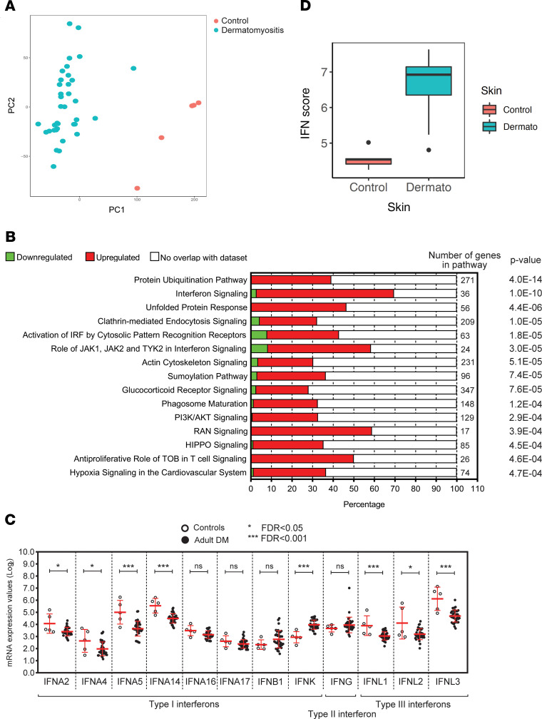
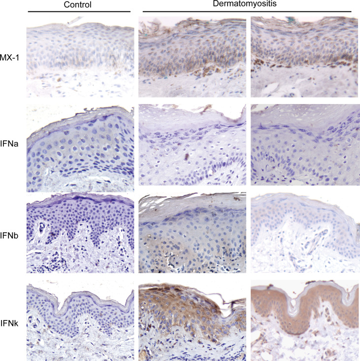
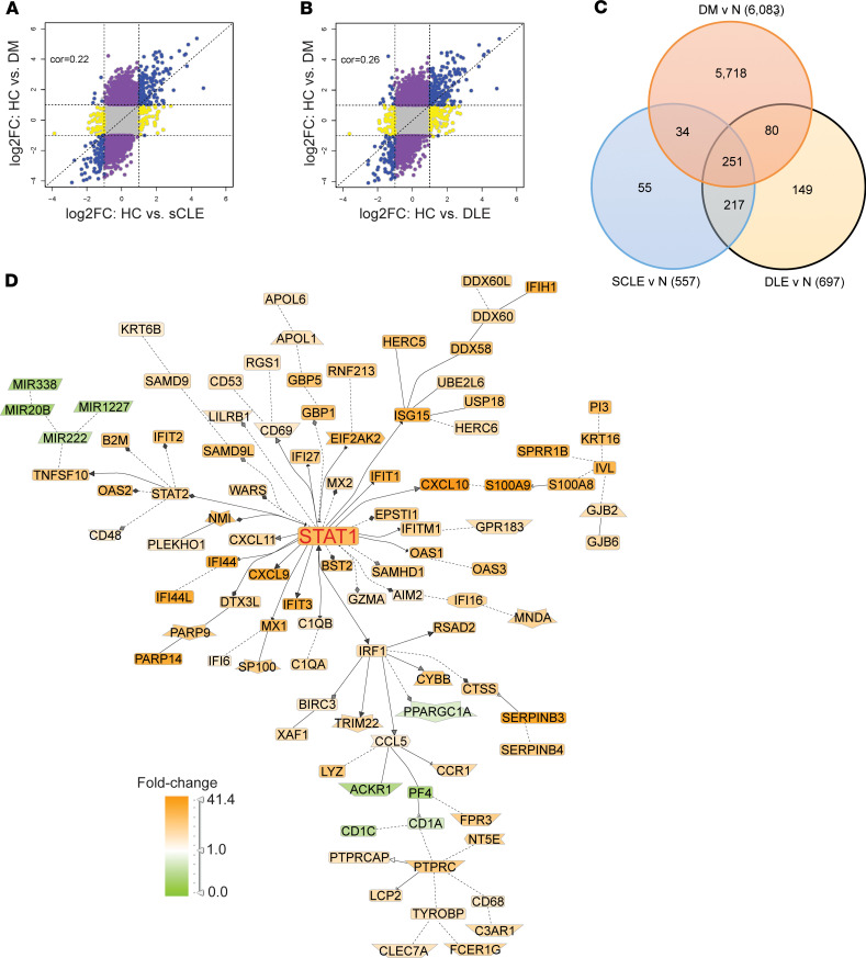
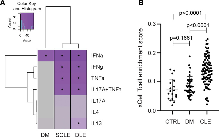
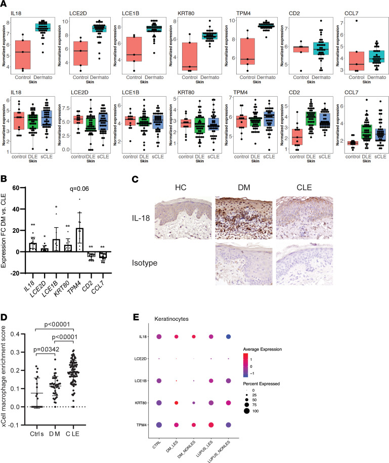
Skin lesions in dermatomyositis (DM) are common, are frequently refractory, and have prognostic significance. Histologically, DM lesions appear similar to cutaneous lupus erythematosus (CLE) lesions and frequently cannot be differentiated. We thus compared the transcriptional profile of DM biopsies with CLE lesions to identify unique features. Type I IFN signaling, including IFN-κ upregulation, was a common pathway in both DM and CLE; however, CLE also exhibited other inflammatory pathways. Notably, DM lesions could be distinguished from CLE by a 5-gene biomarker panel that included IL18 upregulation. Using single-cell RNA-sequencing, we further identified keratinocytes as the main source of increased IL-18 in DM skin. This study identifies a potentially novel molecular signature, with significant clinical implications for differentiating DM from CLE lesions, and highlights the potential role for IL-18 in the pathophysiology of DM skin disease.
Keywords: Autoimmune diseases; Autoimmunity; Cytokines; Dermatology; Skin.
Conflict of interest statement
Figures
Similar articles
-
Proteome study of cutaneous lupus erythematosus (CLE) and dermatomyositis skin lesions reveals IL-16 is differentially upregulated in CLE.Arthritis Res Ther. 2021 Apr 30;23(1):132. doi: 10.1186/s13075-021-02511-0. Arthritis Res Ther. 2021. PMID: 33931094 Free PMC article.
-
The phenotypic profile of dermatomyositis and lupus erythematosus: a comparative analysis.J Cutan Pathol. 2010 Jun;37(6):659-71. doi: 10.1111/j.1600-0560.2009.01443.x. Epub 2009 Nov 4. J Cutan Pathol. 2010. PMID: 19891658
-
Plasmacytoid dendritic cells are present in cutaneous dermatomyositis lesions in a pattern distinct from lupus erythematosus.J Cutan Pathol. 2008 May;35(5):452-6. doi: 10.1111/j.1600-0560.2007.00848.x. Epub 2007 Nov 12. J Cutan Pathol. 2008. PMID: 18005168
-
Immunologic and genetic considerations of cutaneous lupus erythematosus: a comprehensive review.J Autoimmun. 2013 Mar;41:34-45. doi: 10.1016/j.jaut.2013.01.007. Epub 2013 Feb 1. J Autoimmun. 2013. PMID: 23380467 Review.
-
The role of cytokines in the pathogenesis of cutaneous lupus erythematosus.Cytokine. 2015 Jun;73(2):326-34. doi: 10.1016/j.cyto.2015.01.031. Epub 2015 Mar 9. Cytokine. 2015. PMID: 25767072 Review.
Cited by
-
Refractory alopecia universalis associated with dermatomyositis successfully treated with tofacitinib.Mod Rheumatol Case Rep. 2022 Jun 24;6(2):199-202. doi: 10.1093/mrcr/rxac012. Mod Rheumatol Case Rep. 2022. PMID: 35253877 Free PMC article.
-
Anti- Melanoma Differentiation-Associated Gene 5 Antibody Positive Dermatomyositis: Recent Progress in Pathophysiology and Treatment.Curr Rheumatol Rep. 2025 May 5;27(1):23. doi: 10.1007/s11926-025-01188-7. Curr Rheumatol Rep. 2025. PMID: 40323493 Free PMC article. Review.
-
Dermatomyositis: focus on cutaneous features, etiopathogenetic mechanisms and their implications for treatment.Semin Immunopathol. 2025 Aug 6;47(1):32. doi: 10.1007/s00281-025-01054-9. Semin Immunopathol. 2025. PMID: 40770118 Free PMC article. Review.
-
Polishing the crystal ball: mining multi-omics data in dermatomyositis.Ann Transl Med. 2021 Mar;9(5):435. doi: 10.21037/atm-20-5319. Ann Transl Med. 2021. PMID: 33842656 Free PMC article. Review.
-
Single-Cell Sequencing in Rheumatic Diseases: New Insights from the Perspective of the Cell Type.Aging Dis. 2022 Dec 1;13(6):1633-1651. doi: 10.14336/AD.2022.0323. eCollection 2022 Dec 1. Aging Dis. 2022. PMID: 36465169 Free PMC article.
References
-
- Braunstein I, Klein R, Okawa J, Werth VP. The interferon-regulated gene signature is elevated in subacute cutaneous lupus erythematosus and discoid lupus erythematosus and correlates with the cutaneous lupus area and severity index score. Br J Dermatol. 2012;166(5):971–975. doi: 10.1111/j.1365-2133.2012.10825.x. - DOI - PMC - PubMed
Publication types
MeSH terms
Substances
Grants and funding
LinkOut - more resources
Full Text Sources
Molecular Biology Databases
Miscellaneous
