Review
. 2020 Sep 15:257:118056.
doi: 10.1016/j.lfs.2020.118056. Epub 2020 Jul 6.

Structural features of coronavirus SARS-CoV-2 spike protein: Targets for vaccination

Affiliations
Review

Structural features of coronavirus SARS-CoV-2 spike protein: Targets for vaccination

Ariane Sternberg et al. Life Sci. .

Abstract

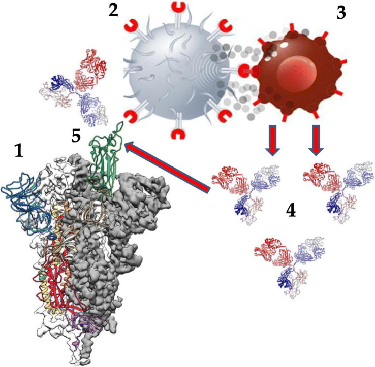
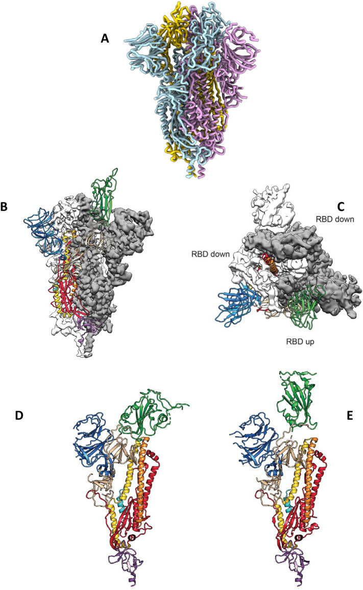
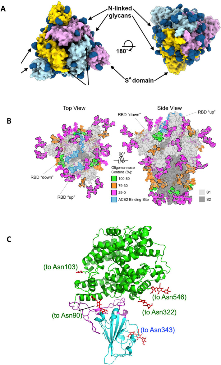
Various human pathogenic viruses employ envelope glycoproteins for host cell receptor recognition and binding, membrane fusion and viral entry. The spike (S) glycoprotein of betacoronavirus SARS-CoV-2 is a homotrimeric class I fusion protein that exists in a metastable conformation for cleavage by host cell proteases furin and TMPRSS2, thereby undergoing substantial structural rearrangement for ACE2 host cell receptor binding and subsequent viral entry by membrane fusion. The S protein is densely decorated with N-linked glycans protruding from the trimer surface that affect S protein folding, processing by host cell proteases and the elicitation of humoral immune response. Deep insight into the sophisticated structure of SARS-CoV-2 S protein may provide a blueprint for vaccination strategies, as reviewed herein.

Keywords: ACE2; Coronavirus SARS-CoV-2; Immune response; Protein structure; Receptor binding domain; Spike protein; Vaccination; Viral fusion protein.


Conflict of interest statement

Declaration of competing interest None.

Figures

Unlabelled Image
Graphical abstract
Fig. 1
Fig. 1
Structural features of the SARS-CoV-2 spike (S) protein. (A) Ribbon diagram of the homotrimeric S, adopted from [19], (with permission from Elsevier Inc.). (B) Side view of the prefusion structure of S, with a single RBD in open (“up”) conformation (green), adopted from [18], (with permission from Science.org). (C) Top view of the prefusion structure of S, with two single RBDs in closed (“down”) conformation (white and grey) and one single RBD in open (“up”) conformation (green), adopted from [18], (with permission from Science.org). (D) Single monomer of S, with the RBD in closed (“down”) conformation (green), adopted from [18], (with permission from Science.org). (E) Single monomer of S, with the RBD in open (“up”) conformation (green), adopted from [18], (with permission from Science.org). (For interpretation of the references to colour in this figure legend, the reader is referred to the web version of this article.)
Fig. 2
Fig. 2
N-linked glycosylation of S, and the RBD of S binding to ACE2. (A) left: top view of the S1 homotrimer, with N-linked glycans as dark blue spheres; (A) right: bottom view of the S2 homotrimer, with N-linked glycans as dark blue spheres. Adopted from [19], (with permission from Elsevier Inc.). (B) Left: top view of the S1 homotrimer, with N-linked glycans colored according to their oligomannose content (green to pink), with the ACE2 binding site in light blue. (B) Right: side view of the S homotrimer, with N-linked glycans colored according to their oligomannose content (green to pink), with the ACE2 binding site in light blue, and the S1 (light grey) and S2 (dark grey) subunits. Adopted from [36], (with permission from Science.org). (C) The RBD (light blue), with its twisted five-stranded antiparallel β sheet of β1, β2, β3, β4 and β7 strands, binding to the bottom side of the small lobe of ACE2 (green helices). Adopted from [34], (with permission from Nature.com). (For interpretation of the references to colour in this figure legend, the reader is referred to the web version of this article.)

References

    1. Liu J., Zheng X., Tong Q., Li W., Wang B., Sutter K., Trilling M., Lu M., Dittmer U., Yang D. Overlapping and dicrete aspects of the pathology and pathogenesis of the emerging human pathogenic coronaviruses SARS-CoV, MERS-CoV, and 2019-nCoV. J. Med. Virol. 2020;92:491–494. - PMC - PubMed
    1. Jiang C., Yao X., Zhao Y., Wu J., Huang P., Pan C., Liu S., Pan C. Comparative review of respiratory diseases caused by coronaviruses and influenza a viruses during epidemic season. Microbes. Infect. May. 2020;13 doi: 10.1016/j.micinf.2020.05.005. - DOI - PMC - PubMed
    1. Yang Y., Peng F., Wang R., Guan K., Jiang T., Xu G., Sun J., Chang C. The deadly coronaviruses: the 2003 SARS pandemic and the 2020 novel coronavirus epidemic in China. J. Autoimmun. 2020;109 - PMC - PubMed
    1. Greber U.F. Virus and host mechanics support membrane penetration and cell entry. J. Virol. 2016;90:3802–3805. - PMC - PubMed
    1. Tong Y., Lavillette D., Li Q., Zhong J. Role of hepatitis C virus envelope glycoprotein E1 in virus entry and assembly. Front. Immunol. 2018;9:1411. - PMC - PubMed

MeSH terms