. 2020 Jul 7;13(7):144.
doi: 10.3390/ph13070144.

Loss of miR-101-3p Promotes Transmigration of Metastatic Breast Cancer Cells through the Brain Endothelium by Inducing COX-2/MMP1 Signaling

Affiliations

Loss of miR-101-3p Promotes Transmigration of Metastatic Breast Cancer Cells through the Brain Endothelium by Inducing COX-2/MMP1 Signaling

Rania Harati et al. Pharmaceuticals (Basel). .

Abstract

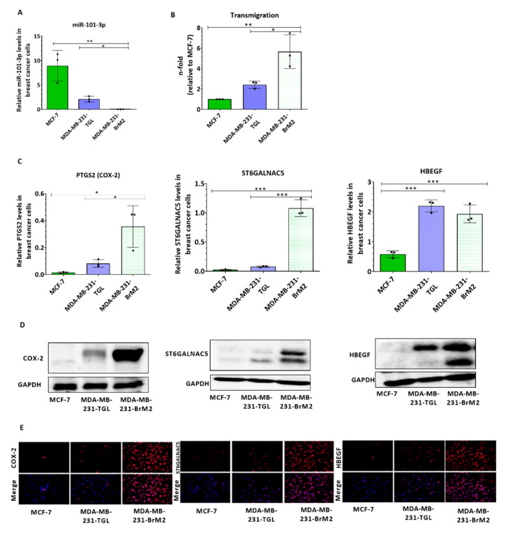
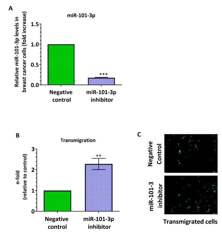
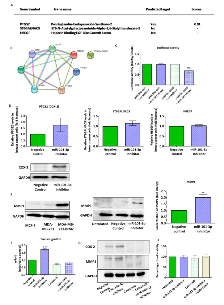
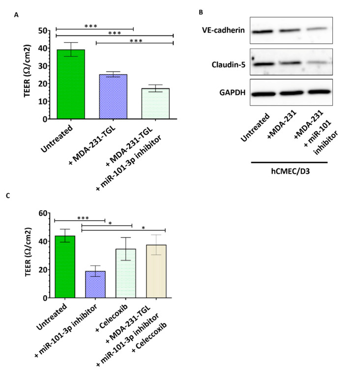
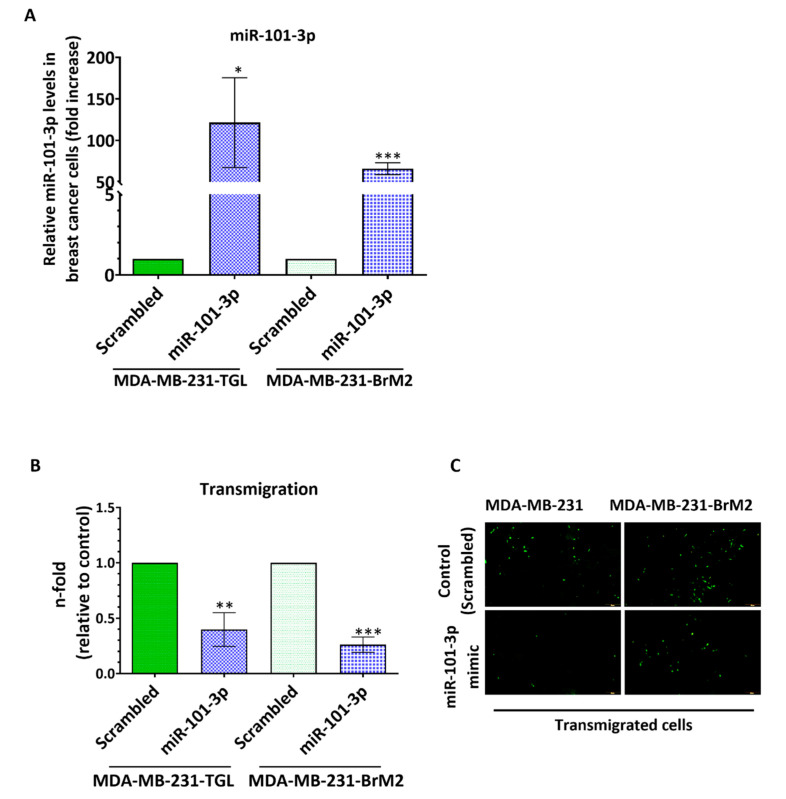
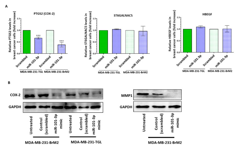
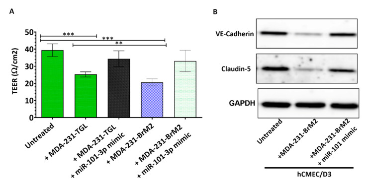
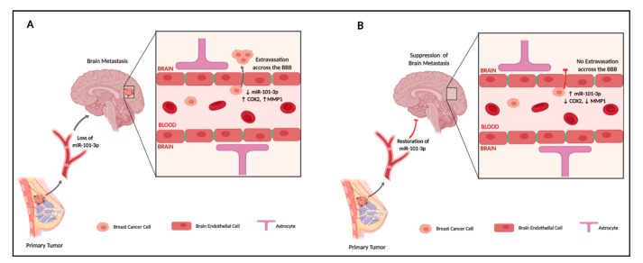
Brain metastases represent one of the incurable end stages in breast cancer (BC). Developing effective or preventive treatments is hampered by a lack of knowledge on the molecular mechanisms driving brain metastasis. Transmigration of BC cells through the brain endothelium is a key event in the pathogenesis of brain metastasis. In this study, we identified miR-101-3p as a critical micro-RNA able to reduce transmigration of BC cells through the brain endothelium. Our results revealed that miR-101-3p expression is downregulated in brain metastatic BC cells compared to less invasive variants, and varies inversely compared to the brain metastatic propensity of BC cells. Using a loss-and-gain of function approach, we found that miR-101-3p downregulation increased transmigration of BC cells through the brain endothelium in vitro by inducing COX-2 expression in cancer cells, whereas ectopic restoration of miR-101-3p exerted a metastasis-reducing effect. In regulatory experiments, we found that miR-101-3p mediated its effect by modulating COX-2-MMP1 signaling capable of degrading the inter-endothelial junctions (claudin-5 and VE-cadherin), key components of the brain endothelium. These findings suggest that miR-101-3p plays a critical role in the transmigration of breast cancer cells through the brain endothelium by modulating the COX-2-MMP1 signaling and thus may serve as a therapeutic target that can be exploited to prevent or suppress brain metastasis in human breast cancer.

Keywords: blood–brain barrier; brain metastasis; breast cancer; micro-RNA.


Conflict of interest statement

The authors declare no conflict of interest. The funders had no role in the design of the study; in the collection, analyses, or interpretation of data; in the writing of the manuscript, or in the decision to publish the results.

Figures

Figure 1
Figure 1
miR-101-3p levels are downregulated in brain metastatic breast cancer cells and vary inversely with their brain metastatic ability. (A) The expression profile of miR-101-3p was examined by real-time PCR in three breast cancer cell lines with different brain metastatic propensities (MCF-7, MDA-MB-231-TGL and MDA-MB-BrM2). Relative miR-101-3p level expression was normalized against the U6 small nuclear RNA levels. (B) The transmigration abilities of the different BC cells were examined by trans-endothelial migration assay. The amount of transmigrated cells was determined by fluorescence measurements.(C) The relative mRNA expression profile of three pro-metastasis genes known to mediate brain trans-endothelial migration of BC cells (PTGS2 coding for COX-2, ST6GALNAC5 and HBEGF) was examined in the three BC cell lines using real time PCR and normalized against GAPDH mRNA levels. (D,E) Western Blot analysis (D) and immunofluorescence images (E) of COX-2, ST6GALNAC5 and HBEGF in MCF-7, MDA-MB-231-TGL and MDA-MB-BrM2 cell lines. Experiments were carried out three times. Data are mean ±SD from three independent experiments. * p < 0.05, ** p < 0.01, *** p < 0.001.
Figure 2
Figure 2
Ectopic downregulation of miR-101-3p in breast cancer cells promotes their transmigration through the brain endothelium. MDA-MB-231-TGL cells were transfected with miR-101-3p inhibitor (30 nM final concentration) or with negative control. (A) Relative miR-101-3p level expression was measured by real time PCR from total RNA. The small nuclear RNA U6 was used as internal control. (B) The transmigration abilities of the different BC cells were examined by trans-endothelial migration assay. (C) Representative images of transmigrated fluorescently labelled MDA-MB-231-TGL (small green cells) in control and anti-miR-101-3p transfected cells. Data are mean ±SD from three independent experiments. * p < 0.05, ** p < 0.01, *** p < 0.001.
Figure 3
Figure 3
Ectopic downregulation of miR-101-3p in breast cancer cells promotes trans-endothelial migration through induction of COX-2-MMP1 expression. (A) List of has-miR-101-3p transendothelial migration-related predicted target genes in silico. (B) Protein–protein interaction network analysis retrieved from STRING (http://www.string-db.org/, version 9.1) showing the link between the different molecules involved in transmigration of breast cancer cells across the blood–brain barrier (BBB) (HBEGF, PTGS2 and ST6GALNAC5). Different colors of the lines represent the types of evidence for association: green line, neighborhood evidence; red line, fusion evidence; purple line, experimental evidence; light blue line, database evidence; black line, coexpression evidence; blue line, co-occurrence evidence; and yellow line, text-mining evidence. (C) Relative luciferase activity expressed as Firefly/Renilla luciferase activity. (D) Relative PTGS2 (COX2), ST6GALNAC5 and HBEGF mRNA and protein expression measured by real time PCR and western blot in control and anti-miR-101-3p transfected cells. (E) Western Blot analysis of MMP1 in the different BC cell lines and in control and anti-miR-101-3p transfected MDA-MB-231-TGL cells. MMP-1 levels released in the culture media were quantified by ELISA. (F) The transmigration ability of MDA-MB-231-TGL cells treated with miR-101-3p inhibitor and/or celecoxib was examined by trans-endothelial migration assay. (G) Western Blot analysis of COX-2 and MMP1 in MDA-MB-231-TGL cells treated with miR-101-3p inhibitor and/or celecoxib. (H) Cell viability of MDA-MB-231-TGL cells treated with miR-101-3p inhibitor and/or celecoxib was measured by MTT assay. Data are mean ±SD from three independent experiments. ** p < 0.01, *** p < 0.001.
Figure 4
Figure 4
Ectopic downregulation of miR-101-3p in breast cancer cells interferes with brain intercellular junctions.(A,C) Transendothelial electrical resistance (TEER) was measured for brain endothelial cells co-cultured with MDA-MB-231-TGL cells treated with negative control, miR-101-3p inhibitor and/or celecoxib (B) Western Blot analysis of VE-cadherin and claudin-5 protein expression was measured by Western blot in hCMEC/D3 co-cultured with MDA-MB-231-TGL cells treated with miR-101-3p inhibitor or control. Data are mean ±SD from three to six independent experiments. * p < 0.05, *** p < 0.001.
Figure 5
Figure 5
Ectopic upregulation of miR-101-3p in low expressing breast cancer cells reduces BC trans-endothelial migration.MDA-MB-231-TGL and MDA-MB-231-BrM2 cells were transfected with miR-101-3p mimic (5 nM final concentration) or with scrambled control. (A) Relative miR-101-3p level expression was measured by real time PCR from total RNA. The small nuclear RNA U6 was used as internal control. (B) The transmigration abilities of the different BC cells were examined by the trans-endothelial migration assay. (C) Representative images of transmigrated fluorescently labelled MDA-MB-231-TGL and MDA-MB-231-BrM2 (small green cells) in control and miR-101-3p mimic transfected cells. * p < 0.05, ** p < 0.01, *** p < 0.001.
Figure 6
Figure 6
Ectopic upregulation of miR-101-3p in breast cancer cells attenuates their transmigration through the brain endothelium by reducing COX-2-MMP1 expression.MDA-MB-231-TGL and MDA-MB-231-BrM2 cells were transfected with miR-101-3p mimic (5 nM final concentration) or with scrambled control. (A) Relative PTGS2, ST6GALNAC5 and HBEGF mRNA measured by real time PCR in control and miR-101-3p mimic transfected cells. (B) Western Blot analysis of COX-2 and MMP1 in control and anti-miR-101-3p transfected MDA-MB-BrM2 cells. Data are mean ±SD from three independent experiments. *** p < 0.001.
Figure 7
Figure 7
Ectopic upregulation of miR-101-3p in breast cancer cells preserves the brain endothelial inter-cellular junctions. (A) Transendothelial electrical resistance (TEER) was measured for brain endothelial cells co-cultured with MDA-MB-231-TGL and MDA-MB-BrM2 cells treated with miR-101-3p mimic or controls. (B) Western Blot analysis of VE-cadherin and claudin-5 protein expression was measured by Western blot in hCMEC/D3 co-cultured with MDA-MB-231-TGL and MDA-MB-BrM2 cells treated with miR-101-3p mimic or controls. Data are mean ±SD from three to six independent experiments. ** p < 0.01, *** p < 0.001.
Figure 8
Figure 8
A proposed model for the pathological role of miR-101-3p in transmigration of metastatic breast cancer cells through the brain endothelium (created with BioRender).

References

    1. Kodack D.P., Askoxylakis V., Ferraro G.B., Fukumura D., Jain R.K. Emerging strategies for treating brain metastases from breast cancer. Cancer Cell. 2015;27:163–175. doi: 10.1016/j.ccell.2015.01.001. - DOI - PMC - PubMed
    1. Weidle U.H., Niewöhner J., Tiefenthaler G. The Blood-Brain Barrier Challenge for the Treatment of Brain Cancer, Secondary Brain Metastases, and Neurological Diseases. Cancer Genom. Proteom. 2015;12:167–177. - PubMed
    1. Pedrosa R.M., Mustafa D.A., Soffietti R., Kros J.M. Breast cancer brain metastasis: Molecular mechanisms and directions for treatment. Neuro Oncol. 2018;20:1439–1449. doi: 10.1093/neuonc/noy044. - DOI - PMC - PubMed
    1. Aversa C., Rossi V., Geuna E., Martinello R., Milani A., Redana S., Valabrega G., Aglietta M., Montemurro F. Metastatic breast cancer subtypes and central nervous system metastases. Breast. 2014;23:623–628. doi: 10.1016/j.breast.2014.06.009. - DOI - PubMed
    1. Anderson R., Balasas T., Callaghan J., Coombes R.C., Evans J., Hall J.A., Kinrade S., Jones D., Jones P.S., Jones R., et al. A framework for the development of effective anti-metastatic agents. Nat. Rev. Clin. Oncol. 2019;16:185–204. doi: 10.1038/s41571-018-0134-8. - DOI - PMC - PubMed