Review
. 2020 Jul 7;7(3):72.
doi: 10.3390/bioengineering7030072.

Polyphenols in Dental Applications

Affiliations
Review

Polyphenols in Dental Applications

Naji Kharouf et al. Bioengineering (Basel). .

Abstract

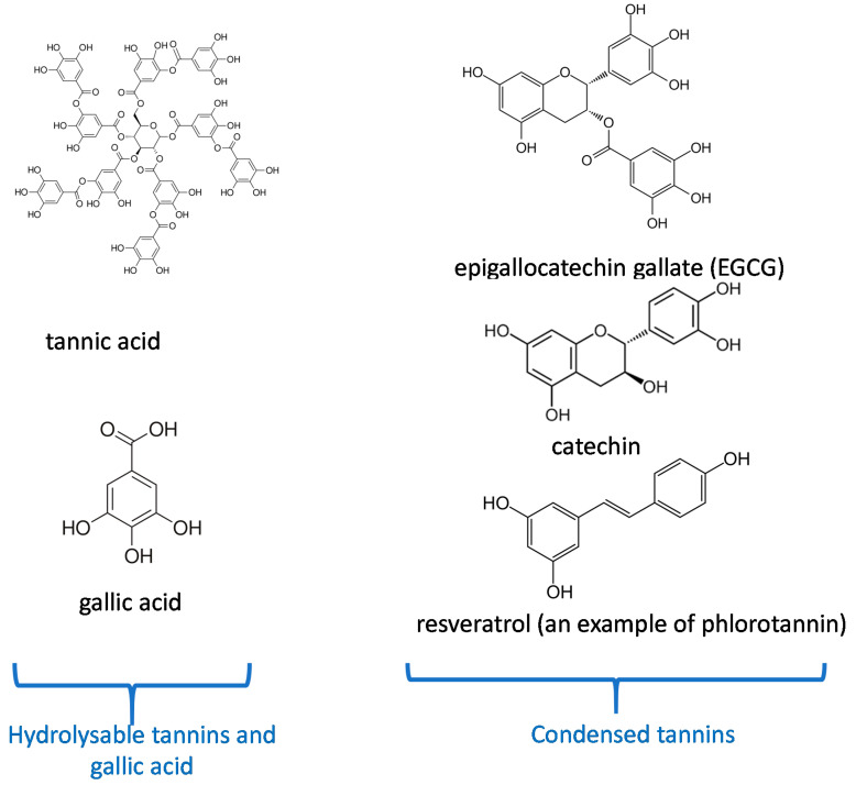
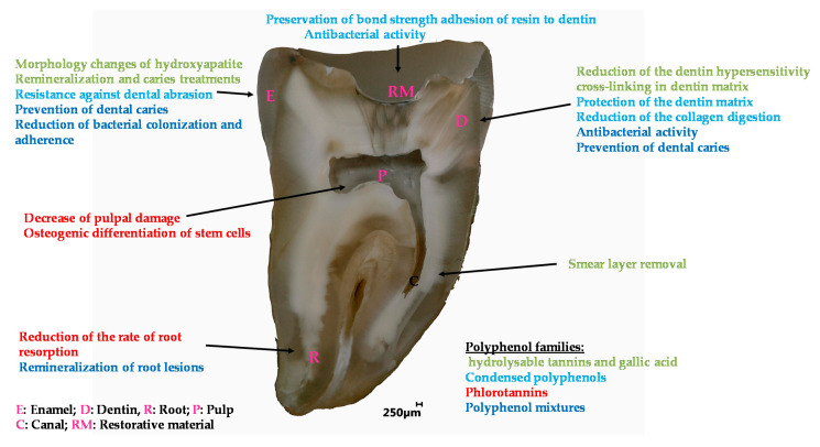
(1) Background: polyphenols are a broad class of molecules extracted from plants and have a large repertoire of biological activities. Biomimetic inspiration from the effects of tea or red wine on the surface of cups or glass lead to the emergence of versatile surface chemistry with polyphenols. Owing to their hydrogen bonding abilities, coordination chemistry with metallic cations and redox properties, polyphenols are able to interact, covalently or not, with a large repertoire of chemical moieties, and can hence be used to modify the surface chemistry of almost all classes of materials. (2) Methods: the use of polyphenols to modify the surface properties of dental materials, mostly enamel and dentin, to afford them with better adhesion to resins and improved biological properties, such as antimicrobial activity, started more than 20 years ago, but no general overview has been written to our knowledge. (3) Results: the present review is aimed to show that molecules from all the major classes of polyphenolics allow for low coast improvements of dental materials and engineering of dental tissues.

Keywords: antibacterial activity; dental resins; dentin; enamel; interactions with collagen; polyphenols.


Conflict of interest statement

The authors declare no conflict of interest.

Figures

Figure 1
Figure 1
Chemical structure of the most important polyphenols used in dentistry according to their classification [10].
Figure 2
Figure 2
Observation of root resorption. (a) SEM image of the tooth, (b) root resorption area (black) and the total surface area (grey), (c) representative SEM pictures of the control group, (d) 5 mg/kg/d resveratrol group, (e) 10 mg/kg/d resveratrol group, (f) the resorption ratio of the three groups were calculated by dividing the surface of black area by the grey area. (** p < 0.01, n = 6). Modified from ref. [54] with authorization.
Figure 3
Figure 3
Scanning electron microscopy of S. mutans. (a) Control; (b) untreated S. mutans cells after 4 days; (c) S. mutans cells treated with 250 µg/mL of epigallocatechin-3-gallate-stearate after 4 days. Modified from Ref. [59] with authorization.
Figure 4
Figure 4
Scanning electron microscopy of S. mutans accumulation on specimens. (a) Adhesive resin surface without epigallocatechin-3-gallate (ECGC); (b) ECGC 100 µg/mL; (c) ECGC 200 µg/mL, (d) ECGC 300 µg/mL. Biofilms accumulated on (c) and (d) were not compact after 24 h incubation. Modified from ref. [63] with authorization.
Figure 5
Figure 5
(a,b) Scanning electron micrographs of dentin hypersensitivity, (c,d) scanning electron micrographs of dentin treated with fluoride-tannin acid-lanthanum-apatite. Modified from ref. [79] with authorization.
Figure 6
Figure 6
Morphology of hydroxyapatite (1) and hydroxyapatite + gallic acid (2) crystals at different times; (a) = 3 h, (b) = 12 h, (c) = 24 h, (d) = 3 days, (e) = 7 days, (f) = 14 days at 80,000× magnification. Modified from ref. [102] with authorization.
Figure 7
Figure 7
Laser scanning confocal microscopy micrographs. (a) Subsurface lesion, (b) subsurface lesion after Galla chinensis extract treatment. Modified from ref. [107] with authorization.
Figure 8
Figure 8
Morphology of the biofilm on orthodontic polyvinyl chloride using scanning electron microscopy at 200×, 1000× and 5000× magnifications, respectively. (ac) Untreated group, (df) green tea polyphenols, (gi) green tea, and Padma Hepaten polyphenols. Modified from ref. [148] with authorization.
Figure 9
Figure 9
Overview of the applications of polyphenols in the engineering of dental materials.

References

    1. Pérez-Jiménez J., Neveu V., Vos F., Scalbert A. Identification of the 100 richest dietary sources of polyphenols: An application of the Phenol-Explorer database. Eur. J. Clin. Nutr. 2010;64(Suppl. 3):S112–S120. - PubMed
    1. Handique J.G., Baruah J.B. Polyphenolic compounds: An overview. React. Funct. Polym. 2002;52:163–188. doi: 10.1016/S1381-5148(02)00091-3. - DOI
    1. Reitzer F., Allais M., Ball V., Meyer F. Polyphenols at interfaces. Adv. Colloid Interface Sci. 2018;257:31–41. doi: 10.1016/j.cis.2018.06.001. - DOI - PubMed
    1. Petti S., Scully C. Polyphenols, oral health and disease: A review. J. Dent. 2009;37:413–423. doi: 10.1016/j.jdent.2009.02.003. - DOI - PubMed
    1. Sánchez M.C., Ribeiro-Vidal H., Esteban-Fernández A., Bartolomé B., Figuero E., Moreno-Arribas M.V., Sanz M., Herrera D. Antimicrobial activity of red wine and oenological extracts against periodontal pathogens in a validated oral biofilm model. BMC Complement. Altern. Med. 2019;19:145. doi: 10.1186/s12906-019-2533-5. - DOI - PMC - PubMed

LinkOut - more resources