. 2020 Jul 9;18(1):278.
doi: 10.1186/s12967-020-02448-z.

Evolving geographic diversity in SARS-CoV2 and in silico analysis of replicating enzyme 3CLpro targeting repurposed drug candidates

Affiliations

Evolving geographic diversity in SARS-CoV2 and in silico analysis of replicating enzyme 3CLpro targeting repurposed drug candidates

Nitin Chitranshi et al. J Transl Med. .

Abstract

Background: Severe acute respiratory syndrome (SARS) has been initiating pandemics since the beginning of the century. In December 2019, the world was hit again by a devastating SARS episode that has so far infected almost four million individuals worldwide, with over 200,000 fatalities having already occurred by mid-April 2020, and the infection rate continues to grow exponentially. SARS coronavirus 2 (SARS-CoV-2) is a single stranded RNA pathogen which is characterised by a high mutation rate. It is vital to explore the mutagenic capability of the viral genome that enables SARS-CoV-2 to rapidly jump from one host immunity to another and adapt to the genetic pool of local populations.

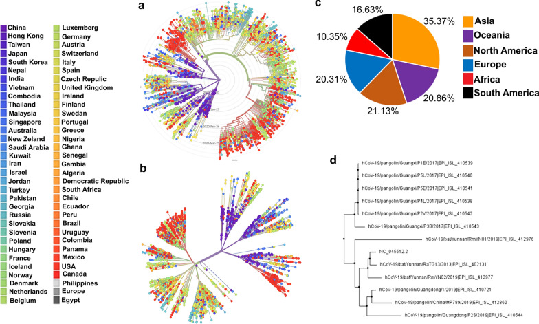
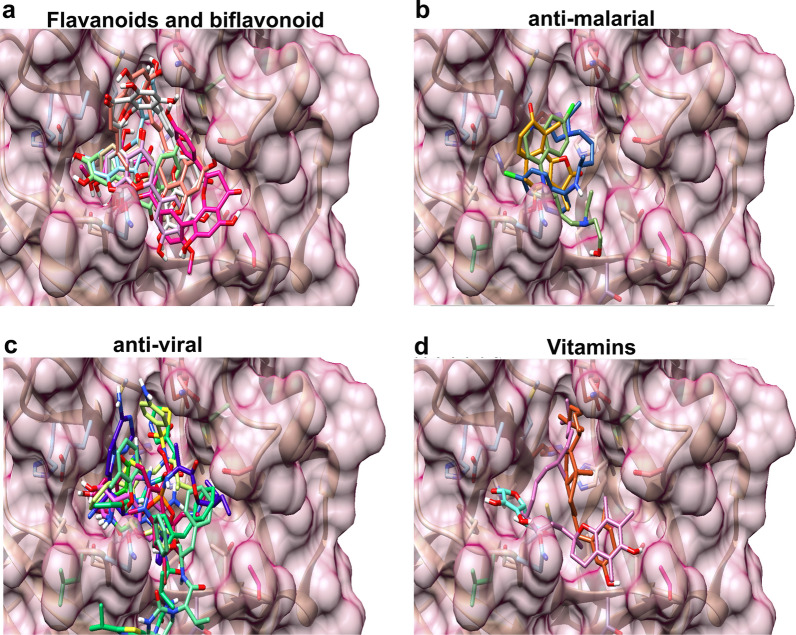
Methods: For this study, we analysed 2301 complete viral sequences reported from SARS-CoV-2 infected patients. SARS-CoV-2 host genomes were collected from The Global Initiative on Sharing All Influenza Data (GISAID) database containing 9 genomes from pangolin-CoV origin and 3 genomes from bat-CoV origin, Wuhan SARS-CoV2 reference genome was collected from GeneBank database. The Multiple sequence alignment tool, Clustal Omega was used for genomic sequence alignment. The viral replicating enzyme, 3-chymotrypsin-like cysteine protease (3CLpro) that plays a key role in its pathogenicity was used to assess its affinity with pharmacological inhibitors and repurposed drugs such as anti-viral flavones, biflavanoids, anti-malarial drugs and vitamin supplements.

Results: Our results demonstrate that bat-CoV shares > 96% similar identity, while pangolin-CoV shares 85.98% identity with Wuhan SARS-CoV-2 genome. This in-depth analysis has identified 12 novel recurrent mutations in South American and African viral genomes out of which 3 were unique in South America, 4 unique in Africa and 5 were present in-patient isolates from both populations. Using state of the art in silico approaches, this study further investigates the interaction of repurposed drugs with the SARS-CoV-2 3CLpro enzyme, which regulates viral replication machinery.

Conclusions: Overall, this study provides insights into the evolving mutations, with implications to understand viral pathogenicity and possible new strategies for repurposing compounds to combat the nCovid-19 pandemic.


Conflict of interest statement

The authors declare that they have no competing interests.

Figures

Fig. 1
Fig. 1
Schematic representation of SARS-CoV-2 structure showing single stranded RNA viral genomic assembly of 29,674 nucleotide base pair which encodes open reading frame 1a (ORF1a, nt 266–13,468), yellow colour, open reading frame 1b (ORF1b, nt 13,468–21,563), blue colour, Spike (S, nt 21,563–25,384), Envelope (E, nt 26,245–26,472), Membrane (M, nt 26,523–27,191) and Nucleocapsid (N, nt 28,274–29,533) proteins in green. ORF1a gene encodes papain-like protease and 3CL protease, ORF1b gene encodes RNA-dependent RNA polymerase, helicase and endo ribo-nuclease, S, E, M and N gene encodes spike, membrane glycoprotein and nucleocapsid phosphoprotein respectively. Three-dimensional crystal structure of 3CL-protese, endoribonuclease and SARS-Cov-2 spike protein receptor binding domain (RBD) engaged human angiotensin converting enzyme 2 (ACE2) receptor were collected from protein data bank
Fig. 2
Fig. 2
(a, b) Phylogenetic evolutionary relationships of SARS-CoV-2 virus showing an initial emergence in Wuhan, China, in Nov-Dec 2019 followed by continued human-to-human transmission. SARS-CoV-2 patient genome sequences deposited in GISAID database from more than 60 different countries (a) radial and (b) unrooted phylogeny created by Nextstrain program [66] (c) Pie chart representation of number of SARS-CoV-2 patient genomes deposited in GISAID till 15th April 2020 from six different regions; Asia (orange, 35.37%), Oceania (purple, 20.86%), North America (brown, 21.13%), Europe (blue, 20.31%), Africa (red, 10.35%) and South America (black, 16.63%). (d) Phylogenetic relationship of CoVs based on whole genome nucleotide sequences from bat, pangolin, and Wuhan SARS-CoV-2 (NC_045512.2) confirms that SARS-CoV-2 share > 90% similarity with bat SARS-CoV while pangolin could be the closest ancestral
Fig. 3
Fig. 3
Graphical representation of SARS-CoV-2 mutation frequency in South American and African patient isolates. a Five novel recurrent hotspots mutations (namely 14,805, 25,563, 26,144, 28,882 and 28,883) were subdivided into 2 geographical areas: South America (n = 307) and Africa (n = 191). Previously confirmed mutations at positions nt3036, nt8782, nt11083, nt14408, nt23403, nt28144 and nt28881 were also present in South American and African populations. We normalize the mutation frequency percentage by estimating the frequency of genomes carrying mutation and comparing it with the overall number of collected genomes per geographical area. The graph shows the cumulative mutation frequency of all given mutations present in South American and African regions. Mutation localisation in viral genes are reported in the legend as well as the proteins (i.e. non-structural protein, nsp) presenting these mutations. b It is also evident that South American and African clusters show a differential pattern of novel mutations: mutation 1059 (black), 9477 (pink), 28,657 (green) and 28,878 (red) in South American, whereas mutation 1059 (black), 15,324 (orange), 28,878 (yellow) and 29,742 (magenta) are present with greater frequency in African patients
Fig. 4
Fig. 4
SARS-CoV-2, Main proteinase 3CLpro analysis. a Cartoon representation structure of the SARS-CoV-2 3CLpro homodimer with inhibitor (green) in greyish black colour. Variant positions of amino acids in 3CLpro (Thr35Val, Ala46Ser, Ser65Asn, Leu86Val, Arg88Lys, Ser94Ala, His134Phe, Lys180Asn, Leu202Val, Ala267Ser, Thr285Ala and Ile286Leu) are shown in yellow colours. b Multiple sequence alignment between SARS-CoV and SARS-CoV-2 3CLpro from Wuhan (Wu) and United States of America (US) patients sharing more than 90% sequence identity. c Surface view representation of SARS-CoV-2 3CLpro (PDB ID: 6Y2G) showing muted amino acid residues in yellow and alpha-ketoamide inhibitor (green) in the substrate binding region. Images are generated by UCSF Chimera software
Fig. 5
Fig. 5
a Cartoon representation of superimposed structures from SARS-CoV 3CLPro (PDB ID: 3TNT, grey) and SARS-CoV-2 3CLpro (PDB ID: 6Y2G, cyan) showing 94.44% sequence identity. Two different 3CLpro inhibitors represented in red and green colour in the substrate binding region (a’) magnified view of substrate binding region. b The residues of the catalytic dyad (His41 and Cys145) are shown in surface view. Autodock 4.2 docking protocol was validated by re-docking of O6K inhibitor in SARS-CoV-2 3CLpro, original O6K inhibitor is shown in red colour and re-docked pose in cyan colour c three-dimensional and d surface view representation
Fig. 6
Fig. 6
Binding modes of different repurposed drugs in the substrate binding region of SARS-CoV-2 3CLpro (a) flavanoids and biflavonoid (7,8 DHF (purple), apigenin (pink), luteolin (orange), quercitin (green), amentoflavone (grey), bilobetin (brown) and ginkgetin (white) (b) anti-malarial (chloroquine (green), hydroxychloroquine (golden yellow) and artemisinin (blue) (c) anti-viral remdesivir (yellow), darunavir (blue), lopinavir (red), galidesivir (dark pink), favipiravir (light blue), ritonavir (light pink) and umifenovir (green) and d vitamins [vitamin C (cyan), vitamin D (golden orange) and vitamin E (pink)]
Fig. 7
Fig. 7
The binding model of repurposed drugs against SARS-CoV-2 3CLpro. a Binding mode of the Amentaflavone drug (red) in SARS-CoV-2 3CLpro (green) substrate binding pocket. b Artemisinin drug (blue) binding mode in SARS-CoV-2 3CLpro (pink) substrate binding pocket. c Binding mode of the Ritonavir drug (magenta) in SARS-CoV-2 3CLpro (cyan) substrate binding pocket. d Binding mode of the Vitamin D (green) in SARS-CoV-2 3CLpro (red) substrate binding pocket. Protein–ligand interaction (hydrogen bond) are shown with red dotted lines

References

    1. Eurosurveillance Editorial T. Note from the editors: World Health Organization declares novel coronavirus (2019-nCoV) sixth public health emergency of international concern. Euro Surveill. 2020;25:200131e. - PMC - PubMed
    1. Coronaviridae Study Group of the International Committee on Taxonomy of V The species Severe acute respiratory syndrome-related coronavirus: classifying 2019-nCoV and naming it SARS-CoV-2. Nat Microbiol. 2020;5:536–544. - PMC - PubMed
    1. To KK, Hung IF, Chan JF, Yuen KY. From SARS coronavirus to novel animal and human coronaviruses. J Thorac Dis. 2013;5(Suppl 2):S103–S108. - PMC - PubMed
    1. Pillaiyar T, Manickam M, Namasivayam V, Hayashi Y, Jung SH. An overview of severe acute respiratory syndrome-coronavirus (SARS-CoV) 3CL protease inhibitors: peptidomimetics and small molecule chemotherapy. J Med Chem. 2016;59:6595–6628. - PMC - PubMed
    1. Seah I, Agrawal R. Can the coronavirus disease 2019 (COVID-19) affect the eyes? A review of coronaviruses and ocular implications in humans and animals. Ocul Immunol Inflamm. 2020;28:391–395. - PMC - PubMed

MeSH terms

Substances

LinkOut - more resources