. 2020 Aug 21;295(34):12143-12152.
doi: 10.1074/jbc.RA120.014168. Epub 2020 Jul 9.

Structure-activity analysis of truncated albumin-binding domains suggests new lead constructs for potential therapeutic delivery

Affiliations

Structure-activity analysis of truncated albumin-binding domains suggests new lead constructs for potential therapeutic delivery

Conan K Wang et al. J Biol Chem. .

Abstract

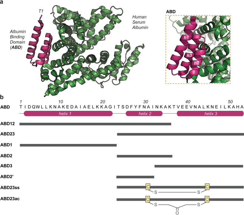
Rapid clearance by renal filtration is a major impediment to the translation of small bioactive biologics into drugs. To extend serum t1/2, a commonly used approach is to attach drug leads to the G-related albumin-binding domain (ABD) to bind albumin and evade clearance. Despite the success of this approach in extending half-lives of a wide range of biologics, it is unclear whether the existing constructs are optimized for binding and size; any improvements along these lines could lead to improved drugs. Characterization of the biophysics of binding of an ABD to albumin in solution could shed light on this question. Here, we examine the binding of an ABD to human serum albumin using isothermal titration calorimetry and assess the structural integrity of the ABD using CD, NMR, and molecular dynamics. A structure-activity analysis of truncations of the ABD suggests that downsized variants could replace the full-length domain. Reducing size could have the benefit of reducing potential immunogenicity problems. We further showed that one of these variants could be used to design a bifunctional molecule with affinity for albumin and a serum protein involved in cholesterol metabolism, PCSK9, demonstrating the potential utility of these fragments in the design of cholesterol-lowering drugs. Future work could extend these in vitro binding studies to other ABD variants to develop therapeutics. Our study presents new understanding of the solution structural and binding properties of ABDs, which has implications for the design of next-generation long-lasting therapeutics.

Keywords: albumin; peptide chemical synthesis; peptide conformation; peptide interaction; peptides.


Conflict of interest statement

Conflict of interest—The authors declare that they have no conflicts of interest with the contents of this article.

Figures

Figure 1.
Figure 1.
Structure and sequence of ABD and truncation variants. a, crystal structure of a three-helix albumin binding domain (ABD; pink) bound to human serum albumin (green) obtained from the PDB (entry 1TF0). Both proteins are largely composed of helices. The first residue of ABD (T1) is labeled for reference. The inset focuses on the contact region between ABD and albumin, highlighting the three helices (i.e. h1, h2, and h3) of ABD. b, amino acid sequence of ABD. Every tenth residue is labeled above the sequence. The location of the three helices is shown below the sequence. The sequences of ABD truncation peptides are schematically shown underneath. Each gray bar spans the length of the corresponding peptide. ABD23ss and ABD23ac include a disulfide and an acetone cross-link, respectively, as shown. The sequences of all peptides are listed in Table S1.
Figure 2.
Figure 2.
Structural characterization of ABD truncation variants by CD and NMR spectroscopy and MD simulation. a, CD spectra of ABD peptides, showing molar ellipticity (θ) as a function of wavelength. b, NMR secondary Hα chemical shifts of ABD, ABD23, and ABD2. Secondary shift plots for the other peptides are shown in the supporting information. c, molecular dynamics simulation of ABD, ABD23, and ABD2 in explicit water. Helicities of the peptides over different simulation times are shown as box plots (left). The structures of the peptides after 150-ns simulation (aligned to their respective starting structures, in gray) are shown as representative structures to illustrate overall helical content. The average root mean squared deviation, RMSD (using backbone heavy atoms), and standard deviation of structural alignments over the course of the simulation (2–150 ns at 2-ns intervals) are indicated below each simulated peptide.
Figure 3.
Figure 3.
Binding of ABD truncation variants to human serum albumin. a, binding isotherm of ABD to human serum albumin measured using ITC. Fitted parameters using one-set-of-side model (with the stoichiometry of the interaction fixed to 0.2) is shown in the bottom right. b, structural analysis of the surface properties of ABD, ABD23, and ABD2. Positively charged (Arg and Lys), negatively charged (Asp and Glu), polar (Gln, Asn, His, Ser, and Thr), and nonpolar (Ala, Ile, Leu, Met, Phe, Val, Pro, Gly, Trp, Tyr, and Met) residues are colored according to the key as indicated. Both cartoon and surface representations are shown.
Figure 4.
Figure 4.
Structure and binding of peptide conjugates. a, schematic illustration of the sequences of ABD peptide conjugates, showing albumin binding peptides (ABD2 or ABD3) linked using a peptide linker to a protein binding peptide (PBP01 or PBP02) that bind to protein convertase subtilisin/kexin type 9 (PCSK9). b, CD spectra of peptide conjugates, showing molar ellipticity (θ) as a function of wavelength. c, 1D 1H NMR spectra of PBP02 and PBP02-ABD2. Similarity in the two spectra indicates the attachment of ABD2 to PBP02 has no significant effect on the structure of PBP02. d, binding isotherm of PBP02-ABD2 to human serum albumin, measured using ITC. e, binding of PBP02-ABD2 to immobilized PCSK9 monitored by SPR. Sensorgrams for varying concentrations of (ranging from 0.31–10.00 μm) are overlaid. f, binding of peptide conjugates to PCSK9 and the effect of human serum albumin (HSA) on binding. The response level at 10 μm peptide concentration is shown for various peptide samples with and without HSA.
Figure 5.
Figure 5.
Structure of a peptide conjugate bound to human serum albumin or protein convertase subtilisin/kexin type 9. a, structure of PBP02-ABD2 (which comprises PBP02 in gray and ABD2 in pink) bound to human serum albumin (green) predicted using homology modeling. b and c, focus on selected interactions between residues of PBP02-ABD2 and human serum albumin. d, structure of PBP02-ABD2 bound to protein convertase subtilisin/kexin type 9 (orange) predicted using homology modeling.

References

    1. Craik D. J., Fairlie D. P., Liras S., and Price D. (2013) The future of peptide-based drugs. Chem. Biol. Drug Des. 81, 136–147 10.1111/cbdd.12055 - DOI - PubMed
    1. Walsh G. (2014) Biopharmaceutical benchmarks 2014. Nat. Biotechnol. 32, 992–1000 10.1038/nbt.3040 - DOI - PubMed
    1. Wang C. K., and Craik D. J. (2018) Designing macrocyclic disulfide-rich peptides for biotechnological applications. Nat. Chem. Biol. 14, 417–427 10.1038/s41589-018-0039-y - DOI - PubMed
    1. Wang C. K., Stalmans S., De Spiegeleer B., and Craik D. J. (2016) Biodistribution of the cyclotide MCoTI-II, a cyclic disulfide-rich peptide drug scaffold. J. Pept. Sci. 22, 305–310 10.1002/psc.2862 - DOI - PubMed
    1. Kontermann R. E. (2016) Half-life extended biotherapeutics. Expert Opin. Biol. Ther. 16, 903–915 10.1517/14712598.2016.1165661 - DOI - PubMed

Publication types

LinkOut - more resources