. 2020 Aug 21;295(34):12181-12187.
doi: 10.1074/jbc.RA120.013547. Epub 2020 Jul 9.

Lysines in the lyase active site of DNA polymerase β destabilize nonspecific DNA binding, facilitating searching and DNA gap recognition

Affiliations

Lysines in the lyase active site of DNA polymerase β destabilize nonspecific DNA binding, facilitating searching and DNA gap recognition

Michael J Howard et al. J Biol Chem. .

Abstract

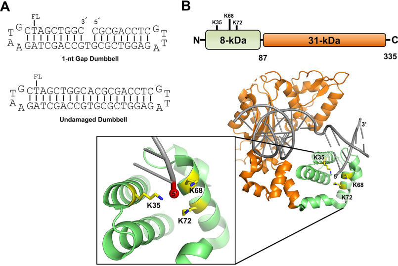
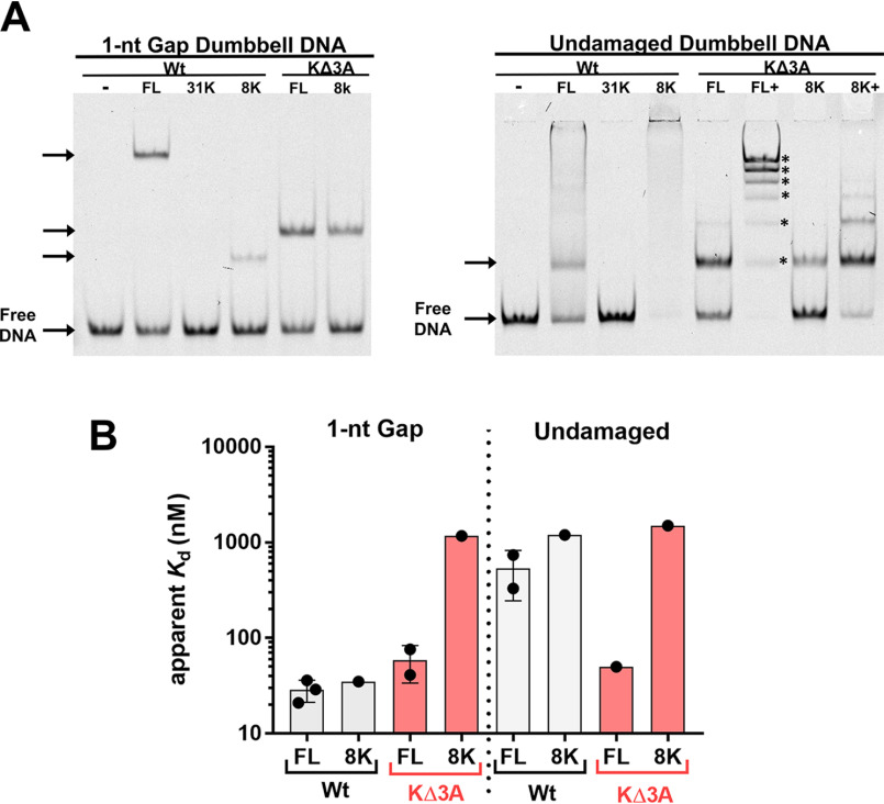
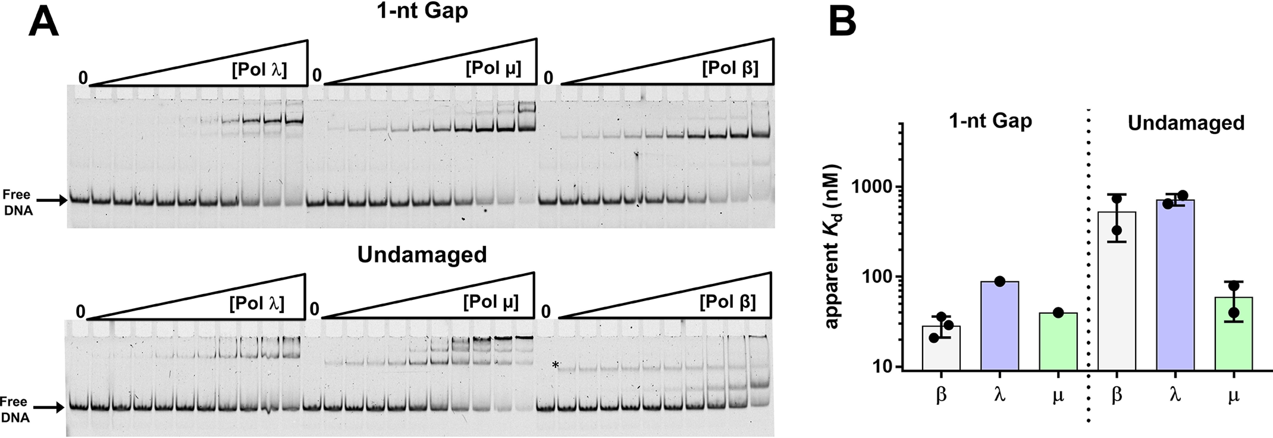
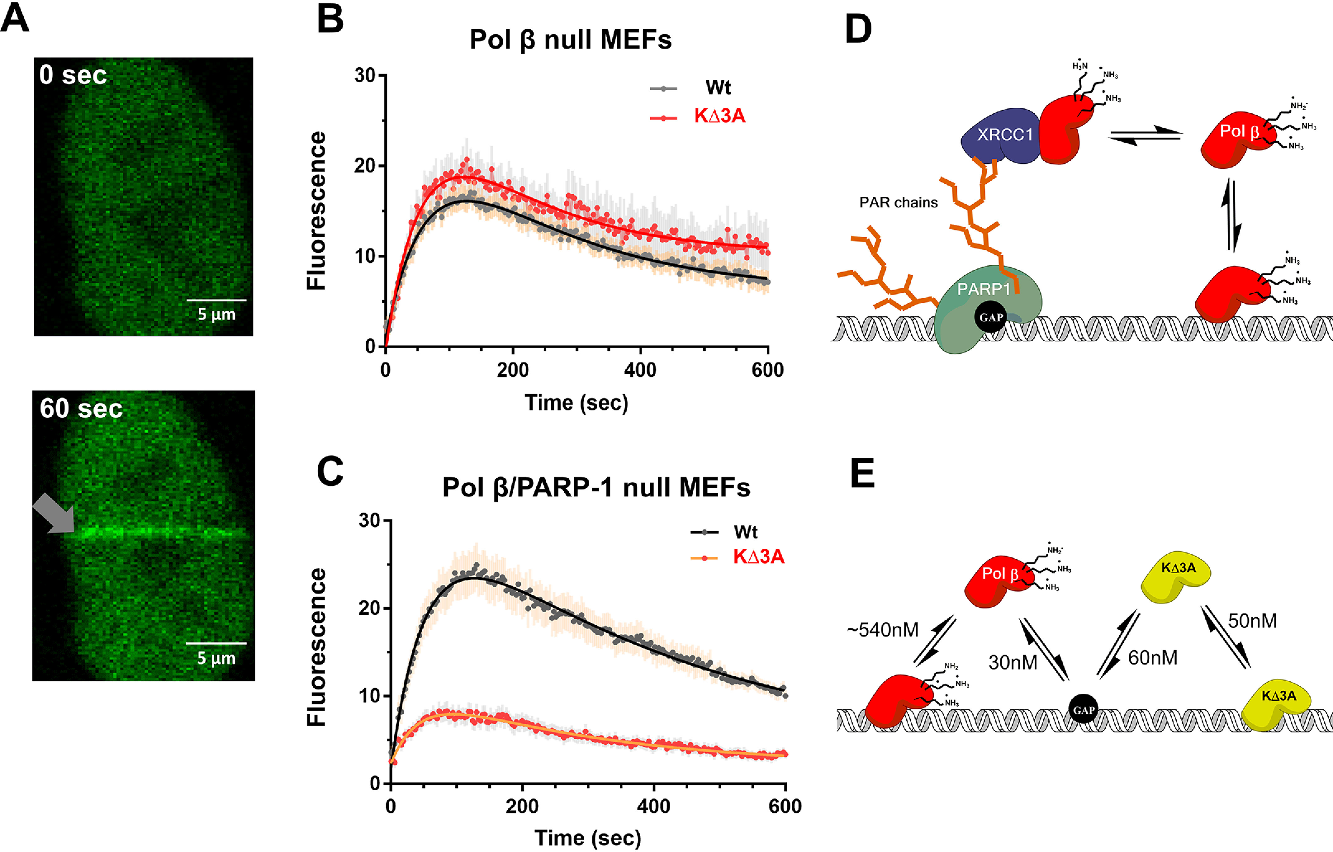
DNA polymerase (pol) β catalyzes two reactions at DNA gaps generated during base excision repair, gap-filling DNA synthesis and lyase-dependent 5´-end deoxyribose phosphate removal. The lyase domain of pol β has been proposed to function in DNA gap recognition and to facilitate DNA scanning during substrate search. However, the mechanisms and molecular interactions used by pol β for substrate search and recognition are not clear. To provide insight into this process, a comparison was made of the DNA binding affinities of WT pol β, pol λ, and pol μ, and several variants of pol β, for 1-nt-gap-containing and undamaged DNA. Surprisingly, this analysis revealed that mutation of three lysine residues in the lyase active site of pol β, 35, 68, and 72, to alanine (pol β KΔ3A) increased the binding affinity for nonspecific DNA ∼11-fold compared with that of the WT. WT pol μ, lacking homologous lysines, displayed nonspecific DNA binding behavior similar to that of pol β KΔ3A, in line with previous data demonstrating both enzymes were deficient in processive searching. In fluorescent microscopy experiments using mouse fibroblasts deficient in PARP-1, the ability of pol β KΔ3A to localize to sites of laser-induced DNA damage was strongly decreased compared with that of WT pol β. These data suggest that the three lysines in the lyase active site destabilize pol β when bound to DNA nonspecifically, promoting DNA scanning and providing binding specificity for gapped DNA.

Keywords: DNA binding protein; DNA binding proteins; DNA damage; DNA polymerase; DNA repair; DNA–protein interaction; base excision repair (BER); facilitated diffusion; nonspecific DNA binding; processive search.


Conflict of interest statement

Conflict of interest—The authors declare that they have no conflicts of interest with the contents of this article.

Figures

Figure 1.
Figure 1.
A, representation of the DNA substrates used in this study. Substrates contain a fluorescein moiety (FL) covalently attached to the designated thymine base. The 1-nt-gap dumbbell DNA substrate (used to evaluate specific binding) contains a 5´-phosphate (not shown). The undamaged DNA substrate (used to evaluate nonspecific binding) was produced as described in Materials and methods. B, cartoon schematic of the DNA pol β domain organization (top) and the crystal structure of pol β bound to 1-nt-gap DNA (PDB entry 3isb). DNA pol β comprises two domains: the 8-kDa (8K) N-terminal dRP lyase domain, which catalyzes 5´-dRP removal, and the 31-kDa (31K) polymerase domain, which catalyzes gap-filling DNA polymerization during BER. The pol β mutations in the 8K lyase domain studied here, K35, K68, and K72, are illustrated; these lysine side chains are proximal to the 5´-phosphate in the 1-nt-gap.
Figure 2.
Figure 2.
A, EMSA representing the different protein-DNA complex mobilities of WT pol β, the WT 8K domain, and two corresponding mutant enzymes, as designated, with 3 lysine to alanine substitutions (KΔ3A). The left panel represents mobilities of complexes bound to 1-nt-gap dumbbell DNA, and the right panel represents mobilities of complexes bound to undamaged dumbbell DNA. The KΔ3A mutants used here included substitutions in full-length (FL) WT and the 8K, as illustrated in red. The concentrations of proteins used were based on Kd measurements, such that about one-half the DNA would be bound: 20 nm FL WT, 1.5 μm 31K, 20 nm 8K WT, 100 nm FL KΔ3A, and 1.5 μm 8K KΔ3A for 1-nt-gap-containing DNA and 500 nm FL WT, 1.5 μm 31K, 1.5 μm 8K WT, 100 nm FL KΔ3A, 1.25 μm, 8K KΔ3A, and 5 μm 8K KΔ3A for undamaged DNA. In panel A, FL+ and 8K+ indicate binding reactions where additional protein was added. This was performed to illustrate that multiple distinct complexes can be detected with these mutants (up to 6 discrete bands [*] were detected by EMSA). Gel quantification is reported in Table S3. B, apparent binding affinities as determined by EMSA for indicated protein and DNA. No binding was observed for 31K with either DNA construct under the concentration range examined (Kd > 3 μm). See the supporting information for gel images and binding isotherms. Data from 3 and 2 independent experiments are shown for FL WT and FL KΔ3A binding, respectively.
Figure 3.
Figure 3.
A, EMSA illustrating binding of pols γ, µ, and β to 1-nt-gap (top) and undamaged (bottom) DNA. Experiments were performed as described in Materials and methods. The asterisk in the reactions performed with pol β and undamaged DNA indicates a band that was not included in the quantification of the gel. Its intensity remains about the same with increasing enzyme concentration and was not observed in other assays under similar conditions (Fig. 2B). B, summary of apparent binding constants of pols to the 1-nt-gap DNA and undamaged DNA. Data in panel A were plotted and fit to a hyperbolic binding equation (Fig. S5 and Table S2).
Figure 4.
Figure 4.
In vivo recruitment of pol β to DNA damage. A, representative confocal fluorescent images of nuclear localized GFP-WT pol β expressed in MEFs at time zero and 60 s after laser-induced DNA damage (gray arrow). B, recruitment of WT (n = 25) and the KΔ3A mutant (n = 17) to laser-induced DNA damage in the cell background of pol β deficiency by virtue of pol β gene deletion. Error bars represent the standard error of the mean. The resulting observed rate constants from a double exponential fit are kobs1 = 0.015 s−1 and kobs2 = 0.005 s−1 as well as kobs1 = 0.017 s−1 and kobs2 = 0.006 s−1 for WT and KΔ3A, respectively. C, recruitment of WT (16 cells) and KΔ3A mutant (21 cells) to laser-induced DNA damage in the cell background of deficiency of both pol β and PARP-1. Error bars represent the standard error of the mean. The resulting observed rate constants from a double exponential fit are kobs1 = 0.02 s−1 and kobs2 = 0.002 s−1 as well as kobs1 = 0.02 s−1 and kobs2 = 0.004 s−1 for WT and KΔ3A, respectively. D, working model explaining in vivo results on pol β localization to DNA damage as shown in panel B. Pol β may localize to DNA damage through an indirect pathway that relies on PARP-1 recognition of the gap, PARylation, and protein–protein interactions with such scaffolding factors as XRCC1; in this model, PARylation signals recruitment and binding of XRCC1, which in turn is a binding partner of pol β, facilitating its recruitment. E, proposed model for results shown in panel C. In the direct model, pol β interacts directly with DNA. The lyase domain lysines destabilize nonspecific DNA binding, allowing the polymerase to scan DNA for damage and provide binding specificity for gapped DNA. The absence of these lysines (KΔ3A) leads to tight nonspecific DNA binding. The black circle on the DNA represents a 1-nt gap. WT pol β is shown in red and KΔ3A in yellow.

References

    1. Krokan H. E., and Bjoras M. (2013) Base excision repair. Cold Spring Harb. Perspect. Biol. 5, a012583 10.1101/cshperspect.a012583 - DOI - PMC - PubMed
    1. Beard W. A., Horton J. K., Prasad R., and Wilson S. H. (2019) Eukaryotic base excision repair: new approaches shine light on mechanism. Annu. Rev. Biochem. 88, 137–162 10.1146/annurev-biochem-013118-111315 - DOI - PMC - PubMed
    1. Wilson S. H., and Kunkel T. A. (2000) Passing the baton in base excision repair. Nat. Struct. Biol. 7, 176–178 10.1038/73260 - DOI - PubMed
    1. Prasad R., Shock D. D., Beard W. A., and Wilson S. H. (2010) Substrate channeling in mammalian base excision repair pathways: passing the baton. J. Biol. Chem. 285, 40479–40488 10.1074/jbc.M110.155267 - DOI - PMC - PubMed
    1. Howard M. J., and Wilson S. H. (2018) DNA scanning by base excision repair enzymes and implications for pathway coordination. DNA Repair 71, 101–107 10.1016/j.dnarep.2018.08.013 - DOI - PMC - PubMed

Publication types

Associated data