. 2020 Jul 28;117(30):17854-17863.
doi: 10.1073/pnas.1920469117. Epub 2020 Jul 9.

Prototypical pacemaker neurons interact with the resident microbiota

Affiliations

Prototypical pacemaker neurons interact with the resident microbiota

Alexander Klimovich et al. Proc Natl Acad Sci U S A. .

Abstract

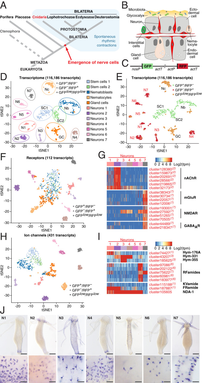
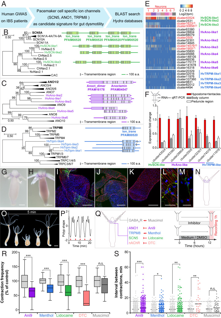
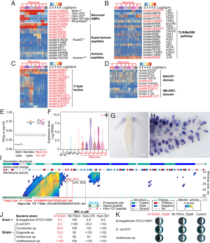
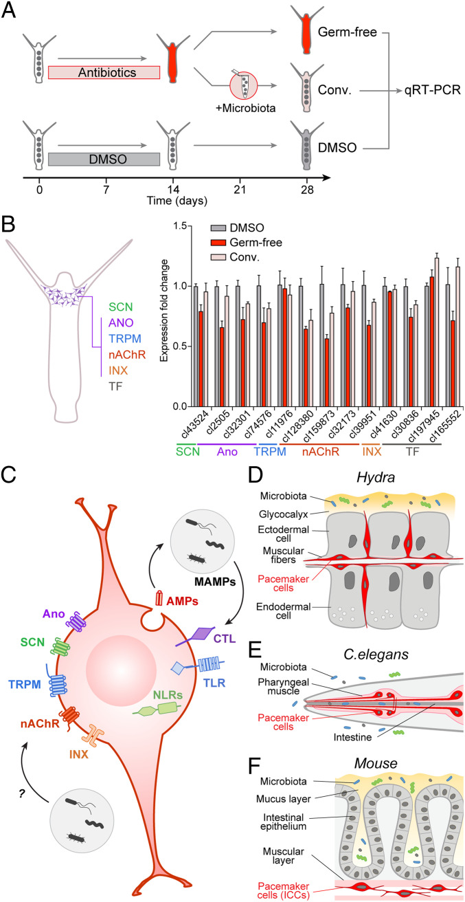
Pacemaker neurons exert control over neuronal circuit function by their intrinsic ability to generate rhythmic bursts of action potential. Recent work has identified rhythmic gut contractions in human, mice, and hydra to be dependent on both neurons and the resident microbiota. However, little is known about the evolutionary origin of these neurons and their interaction with microbes. In this study, we identified and functionally characterized prototypical ANO/SCN/TRPM ion channel-expressing pacemaker cells in the basal metazoan Hydra by using a combination of single-cell transcriptomics, immunochemistry, and functional experiments. Unexpectedly, these prototypical pacemaker neurons express a rich set of immune-related genes mediating their interaction with the microbial environment. Furthermore, functional experiments gave a strong support to a model of the evolutionary emergence of pacemaker cells as neurons using components of innate immunity to interact with the microbial environment and ion channels to generate rhythmic contractions.

Keywords: Hydra; antimicrobial peptide; ion channel; microbiome; pacemaker neuron.


Conflict of interest statement

The authors declare no competing interest.

Figures

Fig. 1.
Fig. 1.
Single-cell transcriptome profiling uncovers the molecular anatomy of Hydra nervous system. (A) Emergence of the first nerve cells preceded the divergence of Cnidaria and Bilateria. Cnidarians possess structurally simple nervous systems and offer a great potential to reveal the fundamental structural and functional principles of neural circuits. Spontaneous rhythmic contractions are ubiquitously observed in Eumetazoa. (B) The Hydra body is made of three cell lineages: The ectodermal and endodermal epithelia separated by the extracellular matrix and the lineage of interstitial cells. The outer surface of the ectoderm is covered by a glycocalyx that serves as a habitat for symbiotic bacteria. The endoderm lining the gastric cavity is free of glycocalyx and stable microbiota. Two nerve nets made of sensory and ganglion neurons are embedded within both epithelia. (C) Genetic construct used to generate transgenic Hydra polyps and differentially label cells within the interstitial lineage by a combination of two fluorescent proteins: GFP expressed under a stem cell-specific nanos promoter (nosP), and RFP driven by the actin promoter (actP) active in terminally differentiated neurons. Both cassettes are flanked by the actin terminator (actT). (D) t-Distributed stochastic neighbor embedding (t-SNE) map constructed by dimensionality-reduction principal component analysis defined by highly covariable genes (Materials and Methods). A total of 928 cells were partitioned in 12 clusters and colored by their cell-type identities inferred from expressed proliferation and cell-type–specific marker genes (Datasets S5 and S6). (E) t-SNE map based on analysis of the entire transcriptome made of 116,186 transcripts segregates 12 clusters, including 7 subpopulations of neurons. Cells are color-coded by their phenotype captured by FACS upon sorting. (F) t-SNE map based on expression analysis of 112 transcripts coding for putative neurotransmitter receptors (Dataset S7). Seven neuronal populations are clearly segregated, indicating that each neuronal population is characterized by a specific set of receptors. (G) Heatmap illustrating expression of genes coding for putative nAChR, mGluR and NMDAR, and GABAAR within seven neuronal populations. Expression within the entire interstitial lineage is presented in SI Appendix, Fig. S3. Transcripts specifically up-regulated in the neurons are labeled red; superscript numbers indicate the nerve cell cluster (N1–N7) where the transcripts are significantly (adjusted P < 0.05) enriched. (H) t-SNE map constructed by expression analysis of 431 transcripts coding for putative ion channels (Dataset S8). Seven neuronal populations are clearly segregated, suggesting that each neuronal population is characterized by a specific set of channels. (I) Heatmap illustrates expression of 11 genes coding for main known neuropeptides in Hydra. Each neuronal population expresses a unique combination of neuropeptides. (J) In situ hybridization for marker genes strongly enriched in each of seven nerve cells clusters (N1–N7) (SI Appendix, Fig. S6) reveals that seven neuronal subpopulations reside in spatially restricted domains along the body column of Hydra (Scale bars, 100 µm upper panel, 25 µm lower panel.)
Fig. 2.
Fig. 2.
Identification of Hydra pacemaker cells using orthologs of human ion channels. (A) Genome-wide association studies on patients with gut motility disorders, such as IBS identified ion channels SCN5, ANO1, and TRPM8 that are expressed in human pacemaker cells (ICCs) and found to be essential for gut motility control. A BLAST search was used to identify the homologous genes in Hydra. (BD) Pacemaker-specific ion channels are highly conserved in Hydra. Phylogenetic tree and domain structure of human SCN (B), ANO1 (C), and TRPM (D) channels and the orthologs from Hydra (Hv). Additionally, sequences from other cnidarians, Nematostella vectensis (Nv) and Clytia hemisphaerica (Ch) are included into the phylogenetic analysis. Noncollapsed trees are presented in SI Appendix, Figs. S8–S10. The topology and domain structure of three Hydra SCN-like sodium channels, six ANO1-like chloride channels and four homologs of TRPM-like cation channels are remarkably similar to their human counterparts. (E) Expression of genes encoding SCN-, ANO1-, and TRPM-like channels in Hydra single-cell dataset is overall very weak and restricted to only few cells. However, several transcripts coding for SCN and ANO1 homologs are significantly up-regulated in neurons (red) with some of the transcripts specifically enriched in the neuronal subpopulation N2 (red superscript). (F) Expression levels of most transcripts coding for SCN-, ANO1-, and TRPM-like channels are highest in the head region of Hydra, including the hypostome and tentacles, as revealed by real-time PCR analysis. Mean ± SEM, n = 3 to 6. (G and H) In situ hybridization with two probes specific for the transcripts cluster2505 (coding for an ANO1-like channel) and cluster30856 (SCN-like), confirms their localized expression in the base of tentacles. (Scale bars, 100 µm.) (IM) Immunohistochemical analysis confirmed the presence of ANO1-like (I and L) and SCN-like (J and M) channels in the neurons of the subpopulation N2 at the base of tentacles. No signal can be detected in the peduncle region of Hydra (K). (Scale bars, 20 µm in L and M and 50 µm in I, J, and K.) (N) Taken together, the expression of gut dysmotility-associated ion channels identifies the nerve cell population N2 resident at the base of tentacles as putative pacemakers in Hydra. Pharmacological interference experiments corroborate the essential role of ANO-, SCN-, and TRPM-like channels and nicotinic acetylcholine receptors expressed in the neuronal population N2 in pacemaker activity in Hydra. (O) Hydra demonstrates spontaneous rhythmic contractions followed by body extensions that occur on average every 5 min. (Scale bar, 200 µm.) (P) The contraction pattern was video-recorded and transformed into a diagram of polyp shape to assess the contraction frequency, defined as the number of full body contractions (red arrowheads) that occurred within 1 h, and the time intervals between consecutive contractions. (Q) Polyps were exposed to chemicals specifically modulating the activity of the ANO1-, SCN-, and TRPM-like ion channels and nAChR expressed in the neuronal population N2. Experimental design: Hydra polyps were treated for 12 h prior to a 1-h recording of contractions. Polyps incubated in 0.16% DMSO-supplemented (for Ani9) or pure (other chemicals) Hydra medium served as control. (R) Contraction frequency is reduced in the presence of all chemicals targeting the channels expressed on the pacemaker population N2, but not affected in the presence of muscimol, which likely interferes with the population N7. (S) Modulating the activity of the pacemaker-specific ANO1-, SCN-, and TRPM-like channels and nAChRs in Hydra also disturbs the rhythmicity of spontaneous contractions, since the intervals between contractions become longer and less regular. Sampling size: n = 10 to 49 animals (contraction frequency), n = 25 to 283 intervals (interval length), *P < 0.05; **P < 0.005; ***P < 0.0005; n.s., P > 0.05.
Fig. 3.
Fig. 3.
Neurons in Hydra are immunocompetent cells. (A) Neurons express a rich set of peptides that have been previously characterized as antimicrobial peptides or their homologs. *ref. ; **ref. ; ***ref. . (B) Heatmap illustrates expression of transcripts coding for components of the TLR/MyD88-dependent immune pathway. Most components are present in the neurons, and five of them are significantly enriched in the neuronal population (red). Superscript numbers indicate the nerve cell cluster (N1 to N7), where the transcripts are significantly (adjusted P < 0.05) enriched. (C) Heatmap illustrates expression of some transcripts coding for NACHT and NB-ARC domain-containing NOD-like receptors that have immune function. (D) Multiple C-type lectin receptors that might recognize bacterial products are strongly expressed in the neurons. (E) Over 70% of top 100 transcripts specifically expressed in each of seven neuronal subpopulations (N1 to N7) is represented by genes that have no homologs outside of Cnidaria, and thus are considered as TRGs. In contrast, among the top 100 transcripts specifically enriched in the interstitial stem cells, only 15% are identified as TRGs. (F) Transcripts of TRG cluster62692 are strongly up-regulated in the neuronal subpopulation N7, weakly expressed in other neurons, and absent from nonneuronal cells of the interstitial lineage. (G and H) In situ hybridization provides evidence that the TRG cluster62692 is expressed exclusively in the sensory neurons of the tentacles. (Scale bars, 100 µm in G and 10 µm in H.) (I) Moving-window small-peptide scan prediction map for the peptide encoded by TRG cluster62692 with residue charge and secondary structure annotations. The heat map reflects the peptide’s probability (σ-score) of being membrane active as predicted by the machine learning classifier (49). High σ-scores (yellow) suggest that cluster62692 peptide is a potent antimicrobial peptide. N-terminal signal peptide, putative proteolysis sites, and a sequence identical to a previously described peptide Hym-121 (50) are found within the cluster62692 peptide, providing evidence that a preprohormone cluster62692 is processed and gives rise to a secreted active peptide. The 17-aa-long peptide corresponding to amino acids 47 to 63 (SPPWNKFGAFVKSKLAK = Hym-121) with high membrane activity score (σ = 2.317) and control peptide amino acids 59 to 75 (SKLAKSKREMSNSDGSE) with no membrane activity (σ = −1.878) were synthesized, C terminally amidated, and tested for antimicrobial activity in a MIC assay. (J) The peptide 47–63 Am is a potent antimicrobial peptide that shows selective growth inhibiting activity against gram-positive and -negative bacteria. Control peptide 59–75 Am demonstrates no antimicrobial activity. Consistently with previous observations (43), dual-function neuropeptides Hym-370 and Hym-357 show some antibacterial activity, yet weaker and more restricted than the peptide 47–63 Am. (K) Representative wells from plates of MIC assay. At concentration 25 µM, the peptide 47–63 Am inhibits growth of Curvibacter sp. and Acidovorax sp. and affects colony morphology of B. megaterium. The growth in the presence of control peptide (59–75 Am, 25 µM) is not different from that in the pure medium (control).
Fig. 4.
Fig. 4.
Prototypical pacemaker neurons interact with microbiota. (A) To test the immune function of the pacemaker neurons in Hydra, GF animals were generated by antibiotic treatment and then recolonized with the natural Hydra microbiota to obtain conventionalized (Conv.) polyps. Polyps treated with DMSO solvent were used as control. Total RNA was extracted from polyps for gene-expression analysis by qRT-PCR. (B) Analysis of expression level of 13 genes specific for the pacemaker neurons in Hydra (scheme, Left) in GF, conventionalized (conv.) and control (DMSO) polyps. Average fold-changes (mean ± SEM; n = 3) are shown. Most of the genes are substantially down-regulated in GF polyps compared to control, and their expression level is restored to values close to control in conventionalized polyps. Thus, expression of pacemaker genes in Hydra is dependent on the presence of symbiotic microbes. (CF) The molecular anatomy of the pacemaker cell. The gene-expression program characteristic for pacemaker cells is highly conserved and is present in the neuronal population N2 that controls spontaneous contractions in Hydra (D), in the neuro-muscular pacemaker complex of C. elegans pharynx (E), and in the ICCs driving the gut motility in mammals (F). This evolutionary conserved signature of a pacemaker cell is composed of ANO-, SCN-, and TRPM-like ion channels, nAChRs, and innexin gap junction. It also includes receptors such as Toll- and NOD-like receptors and C-type lectins that are capable of recognizing bacterial products (MAMPs). Bacteria-derived products may have profound effects onto the gene-expression program of the pacemakers via immune pathways or might directly target the pacemaker ion channels or neuromediator receptors (C).

Comment in

  • Linking neurons to immunity: Lessons from Hydra.
    Obata Y, Pachnis V. Obata Y, et al. Proc Natl Acad Sci U S A. 2020 Aug 18;117(33):19624-19626. doi: 10.1073/pnas.2011637117. Epub 2020 Aug 5. Proc Natl Acad Sci U S A. 2020. PMID: 32759220 Free PMC article. No abstract available.

References

    1. Sasselli V., Pachnis V., Burns A. J., The enteric nervous system. Dev. Biol. 366, 64–73 (2012). - PubMed
    1. Bosch T. C. G. et al. ., Back to the basics: Cnidarians start to fire. Trends Neurosci. 40, 92–105 (2017). - PMC - PubMed
    1. Olsson C., Holmgren S., The control of gut motility. Comp. Biochem. Physiol. A Mol. Integr. Physiol. 128, 481–503 (2001). - PubMed
    1. Barajas-López C., Berezin I., Daniel E. E., Huizinga J. D., Pacemaker activity recorded in interstitial cells of Cajal of the gastrointestinal tract. Am. J. Physiol. 257, C830–C835 (1989). - PubMed
    1. Sanders K. M., Ward S. M., Koh S. D., Interstitial cells: Regulators of smooth muscle function. Physiol. Rev. 94, 859–907 (2014). - PMC - PubMed

Publication types

LinkOut - more resources