. 2020 Jul 9;10(1):11402.
doi: 10.1038/s41598-020-68413-4.

In vivo characterization of target cells for acute elephant endotheliotropic herpesvirus (EEHV) infection in Asian elephants (Elephas maximus)

Affiliations

In vivo characterization of target cells for acute elephant endotheliotropic herpesvirus (EEHV) infection in Asian elephants (Elephas maximus)

Thunyamas Guntawang et al. Sci Rep. .

Abstract

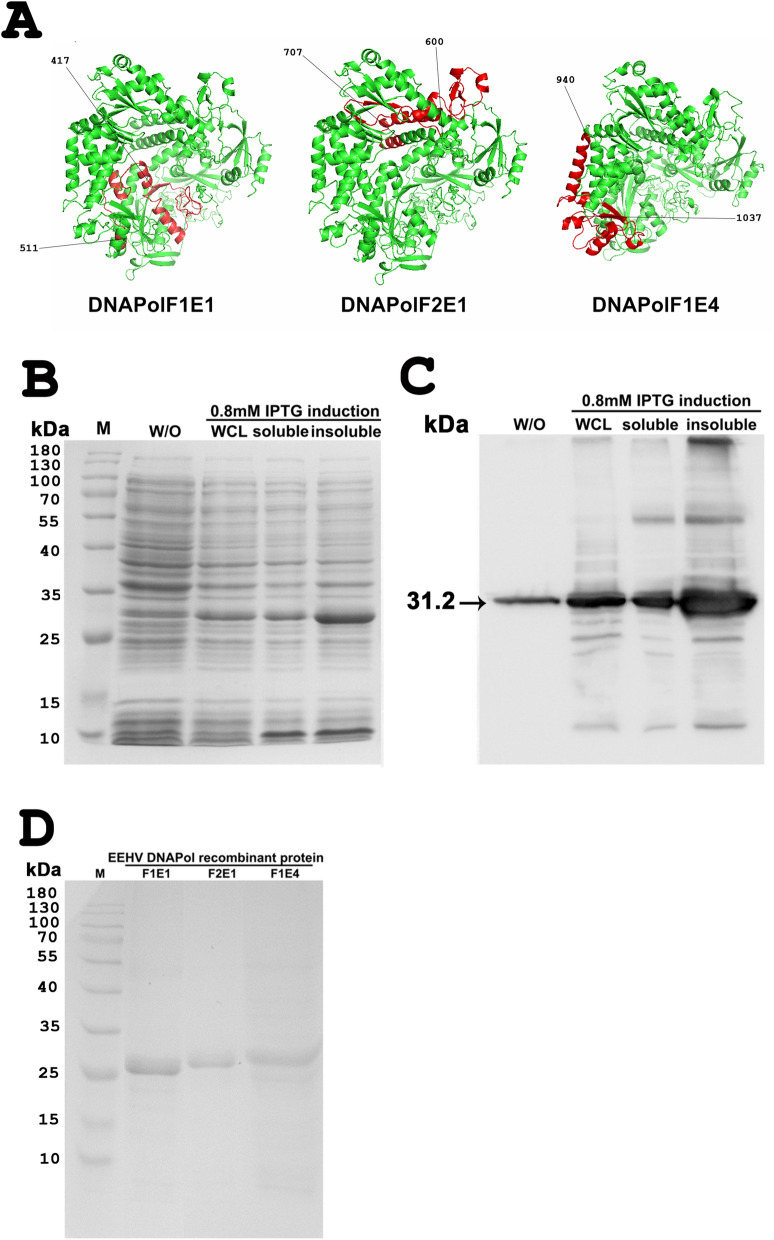
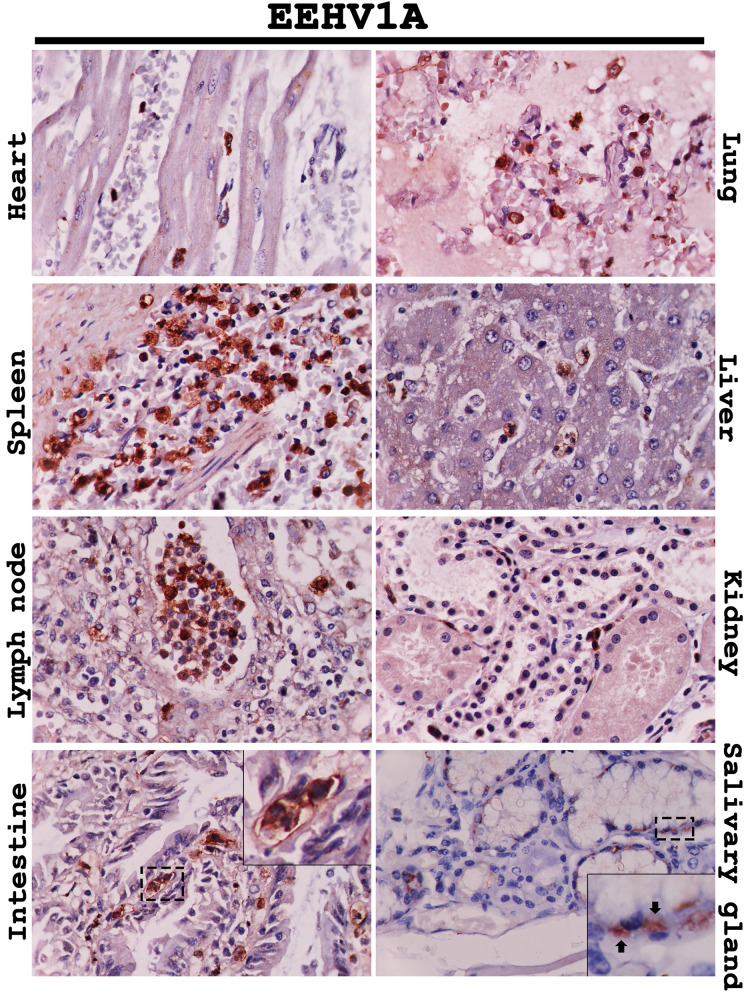
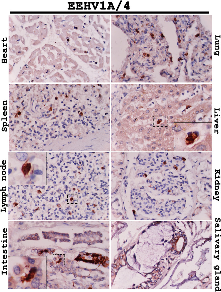
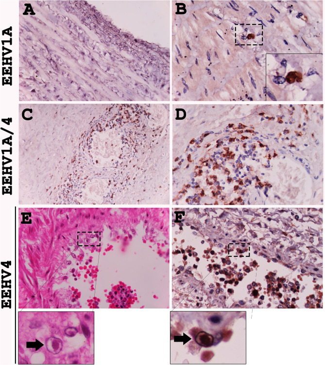
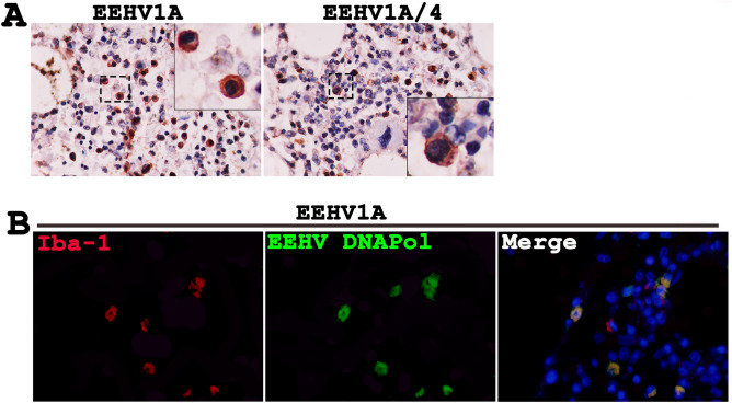
Elephant endotheliotropic herpesvirus-hemorrhagic disease (EEHV-HD) is a dangerous viral infectious disease in young Asian elephants. Despite hypotheses underlying pathogenesis of the disease, it is unclear which cell types the virus targets during acute or persistent infections. This study investigated the tissues and target cells permissive for EEHV infection and replication in vivo. Rabbit polyclonal antibodies against the non-structural proteins of EEHV, DNA polymerase (EEHV DNAPol), were generated and validated. These were used to examine EEHV infection and replication in various tissues of acute EEHV-HD cases and compared to an EEHV-negative control. The results indicated that viral antigens were distributed throughout the epithelia of the alimentary tract and salivary glands, endothelia and smooth muscle cells, and monocytic lineage cells of the EEHV-infected elephants. Moreover, EEHV DNAPol proteins were also found in the bone marrow cells of the EEHV1A-HD and EEHV1A/4-HD cases. This study demonstrated for the first time the target cells that favor in vivo EEHV replication during acute infection, providing a promising foundation for investigating EEHV propagation in vitro.


Conflict of interest statement

The authors declare no competing interests.

Figures

Figure 1
Figure 1
Three dimensional (3D) protein structures and characterization of the EEHV DNAPol recombinant proteins. Predicted regions of DNAPolF1E1, DNAPolF2E1 and DNAPolF1E4 were obtained from the U38 at the amino acid position 417–511 and 600–707 of EEHV1A and 940–1,037 of EEHV4, respectively (A). SDS-PAGE (B) and western blot analysis (C) of DNAPolF2E1 from the BL-21 E. coli induced by 0.8 mM IPTG showed a molecular size of 31.2 kDa, which mainly produced in insoluble form (C). M: Marker, W/O: cells without induction, WCL: Whole cell lysate after induced with 0.8 mM IPTG. SDS-PAGE analysis of the purified EEHV DNAPol histidine-tagged proteins through Ni–NTA column indicated molecular protein sizes of DNAPolF1E1, DNAPolF2E1 and DNAPolF1E4 of 29.9, 31.2 and 30.6 kDa, respectively (D). The 3D structures were generated from digital information available at I-TASSER server and image was modified with Adobe Photoshop CS6 v.13.0.1.
Figure 2
Figure 2
Representative photomicrographs of the EEHV DNAPol proteins in the EEHV1A-HD cases by immunohistochemistry. Immunolabeling positive cells for EEHV DNAPol were observed in the monocytes/macrophages (intestine, inset) of the heart, lung, spleen, liver, lymph node, kidney and intestine. Epithelial cells of the salivary gland were shown to be immunolabeling positive in the cytoplasm of the infected cells (inset, arrow). Image was modified with Adobe Photoshop CS6 v.13.0.1.
Figure 3
Figure 3
Representative photomicrographs of the EEHV DNAPol proteins in the EEHV4-HD cases by immunohistochemistry. Detection of the EEHV DNAPol proteins in the EEHV4-HD calves showed immunolabeling for EEHV DNAPol in the monocytes/macrophages (inset) of the heart, lung, spleen, lymph node and kidney, while immunolabeling positive cells were seen in the endothelia of hepatic sinusoid (inset, arrow), crypt epithelia of the intestine and squamous epithelia of the tongue. Image was modified with Adobe Photoshop CS6 v.13.0.1.
Figure 4
Figure 4
Representative photomicrographs of the EEHV DNAPol proteins in the EEHV1A/4-HD cases by immunohistochemistry. EEHV DNAPol proteins were observed in the monocytes/macrophages (inset) of the heart, lung, spleen, liver, lymph node and kidney. Epithelial cells of the intestinal crypts (inset) and salivary gland were also shown to immunolabel positive for the EEHV DNAPol antibodies. Image was modified with Adobe Photoshop CS6 v.13.0.1.
Figure 5
Figure 5
Immunohistochemical labeling for the EEHV DNAPol in the central nervous system (CNS) and peripheral nervous system (PNS). Spinal cords of the EEHV1A-HD, EEHV4-HD and EEHV1A/4-HD cases were immunostained with rabbit anti-EEHV DNAPol antibodies and indicated that only the infiltrating blood leukocytes were positive (A; inset). Nerve ganglion of the EEHV4-HD case also showed the immunolabeling positive cells of the leukocyte within blood vessels (B; inset). Image was modified with Adobe Photoshop CS6 v.13.0.1.
Figure 6
Figure 6
EEHV preferentially infect certain types of endothelia of the visceral organs. Immunohistochemical staining of the EEHV-HD calves with polyclonal anti-EEHV DNAPol antibodies revealed that endothelia and smooth muscle of the large blood vessel (aorta; A) were not targeted by EEHV, while infiltrating monocytes in the vasa vasorum of tunica adventitia were shown to be infected by EEHV (B). On the other hand, EEHV are prone to infect and replicate in the small blood vessels (artery and vein) of the internal organs (CF). Basophilic intranuclear inclusion bodies (arrows) of the EEHV were observed in the endothelia of small blood vessel (E, inset; H&E) and positive for EEHV DNAPol immunolabeling (F, inset). Image was modified with Adobe Photoshop CS6 v.13.0.1.
Figure 7
Figure 7
Bone marrow cells of the elephant calves were targeted by the EEHV, as determined by immunohistochemistry and double immunofluorescence. Bone marrow of the EEHV1A-HD and EEHV1A/4-HD calves were immunostained with anti-EEHV DNAPol antibodies and showed positive in the cytoplasm and nucleus of the Iba-1 positive cells (A, B), indicating their monocytic phenotypes. Image was modified with Adobe Photoshop CS6 v.13.0.1.

References

    1. Long SY, Latimer EM, Hayward GS. Review of elephant endotheliotropic herpesviruses and acute hemorrhagic disease. ILAR J. 2016;56:283–296. doi: 10.1093/ilar/ilv041. - DOI - PMC - PubMed
    1. Wilkie GS, et al. First fatality associated with elephant endotheliotropic herpesvirus 5 in an Asian elephant: Pathological findings and complete viral genome sequence. Sci. Rep. 2014;4:6299. doi: 10.1038/srep06299. - DOI - PMC - PubMed
    1. Richman LK, et al. Novel endotheliotropic herpesviruses fatal for Asian and African elephants. Science. 1999;283:1171–1176. doi: 10.1126/science.283.5405.1171. - DOI - PubMed
    1. Dastjerdi A, Seilern-Moy K, Darpel K, Steinbach F, Molenaar F. Surviving and fatal Elephant Endotheliotropic Herpesvirus-1A infections in juvenile Asian elephants—Lessons learned and recommendations on anti-herpesviral therapy. BMC Vet. Res. 2016;12:178. doi: 10.1186/s12917-016-0806-5. - DOI - PMC - PubMed
    1. Boonprasert K, et al. Survival analysis of confirmed elephant endotheliotropic herpes virus cases in Thailand from 2006–2018. PLoS ONE. 2019;14:e0219288. doi: 10.1371/journal.pone.0219288PONE-D-18-33945. - DOI - PMC - PubMed

Publication types

MeSH terms