. 2020 Aug;27(8):763-767.
doi: 10.1038/s41594-020-0468-7. Epub 2020 Jul 9.

SARS-CoV-2 and bat RaTG13 spike glycoprotein structures inform on virus evolution and furin-cleavage effects

Affiliations

SARS-CoV-2 and bat RaTG13 spike glycoprotein structures inform on virus evolution and furin-cleavage effects

Antoni G Wrobel et al. Nat Struct Mol Biol. 2020 Aug.

Erratum in

Abstract

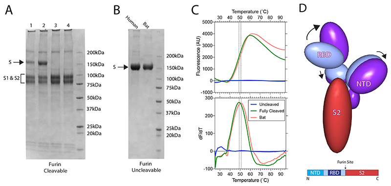
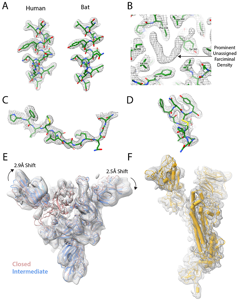
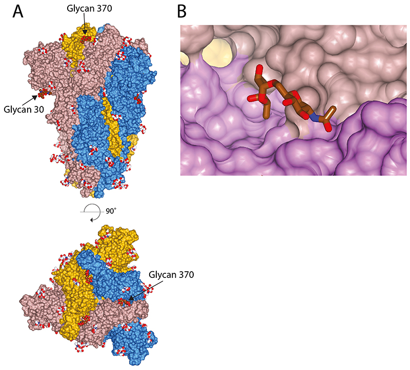
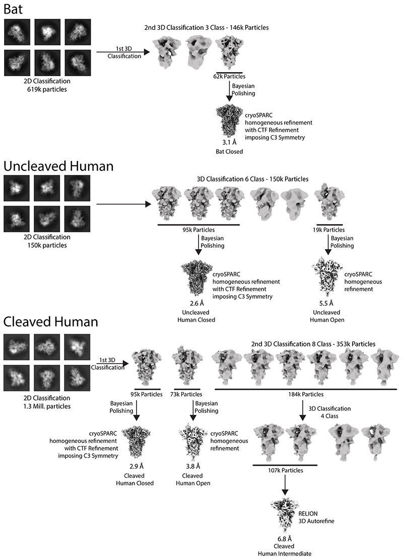
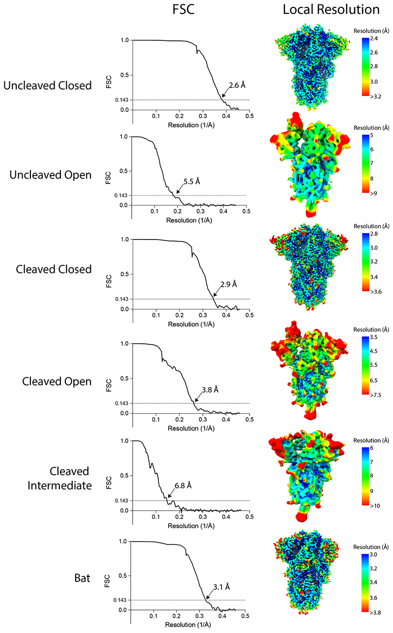
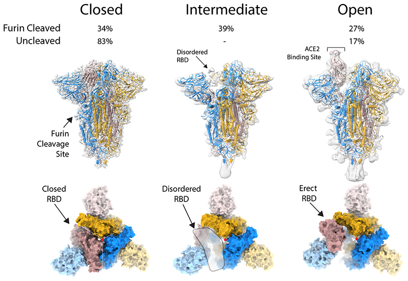
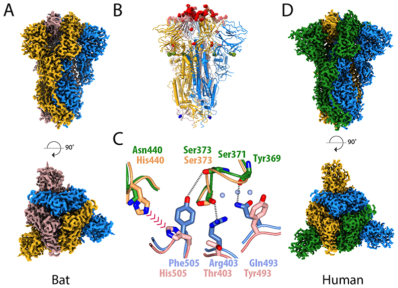
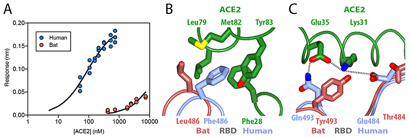
SARS-CoV-2 is thought to have emerged from bats, possibly via a secondary host. Here, we investigate the relationship of spike (S) glycoprotein from SARS-CoV-2 with the S protein of a closely related bat virus, RaTG13. We determined cryo-EM structures for RaTG13 S and for both furin-cleaved and uncleaved SARS-CoV-2 S; we compared these with recently reported structures for uncleaved SARS-CoV-2 S. We also biochemically characterized their relative stabilities and affinities for the SARS-CoV-2 receptor ACE2. Although the overall structures of human and bat virus S proteins are similar, there are key differences in their properties, including a more stable precleavage form of human S and about 1,000-fold tighter binding of SARS-CoV-2 to human receptor. These observations suggest that cleavage at the furin-cleavage site decreases the overall stability of SARS-CoV-2 S and facilitates the adoption of the open conformation that is required for S to bind to the ACE2 receptor.


Conflict of interest statement

Competing interests

The authors declare no competing interests.

Figures

Extended Data Fig. 1
Extended Data Fig. 1. Biochemical analyses of spike proteins.
(A) SDS-PAGE of furin-cleavable SARS-CoV-2 protein. 1: 5 hr furin cleavage; 2: Expressed protein, not cleaved in vitro; 3: 12 hr furin cleavage; 4: 32 hr furin cleavage. (B) SDS-PAGE of uncleavable bat and human virus S proteins. (C) Differential Scanning Fluorimetry measurement of melting temperature for uncleavable human and bat, and fully-furin-cleaved SARS-CoV-2 proteins. (D) The changes in domain orientation, between the closed and open forms, shown schematically, for the monomer that undergoes the most substantive change in the RBD position. The image is produced by the CCP4 MG 'bloboid representation' and is calculated from the shape and centre of mass of the molecular model. Also shown is a bar representation of the domains with the furin cleavage site indicated.
Extended Data Fig. 2
Extended Data Fig. 2. Density features of spike protein cryoEM density.
EM density shown as grey mesh, with built models shown in green. (A) Density for residues 1003-1016 of the uncleaved closed human and bat structures. (B) Unassigned farciminal density seen in maps of the closed conformation of human spike protein. (C+D) Typical density for examples of previously unbuilt external loops in uncleaved closed human structures: residues 174-185 of the NTD (C) and residues 479-489 of the RBD (D). (E) EM density of Intermediate conformation of furin-cleaved spike. The fitted model (blue) is compared to that of the closed conformation (pink). Shifts in the NTDs are indicated with arrows, with values calculated from translations of their centres of mass. (F) Example EM density of a monomer of the Intermediate conformation map with the built structure shown in yellow.
Extended Data Fig. 3
Extended Data Fig. 3. N-linked glycosylation of bat spike protein.
(A) All buildable glycans shown with bat-specific glycosylations, attached to Asn-30 and Asn-370, highlighted in brown (B) Glycan at Asn-370, shown in brown, which is inserted in the cleft between RBDs, shown in different hues of pink.
Extended Data Fig. 4
Extended Data Fig. 4. Fourier Shell Correlations (FSCs) and local resolution estimates for calculated maps.
Extended Data Fig. 5
Extended Data Fig. 5. Processing schemes for cryo-EM data processing.
Figure 1
Figure 1. Structure of protease-cleaved SARS-CoV-2 spike glycoprotein.
Three structures are calculated from micrographs of furin-cleaved material: closed, intermediate and open forms of approximately equal proportions. In the uncleaved material, most of the population represents the closed form with a small proportion in the open conformation. Density maps for the three types of particles, overlaid with a ribbon representation of the built molecular models, as viewed with the long axis of the trimer vertical (middle panel); the three monomers are coloured blue, yellow and brown. An orthogonal view (lower panel) looking down the long axis (indicated by a red dot), the colouring is as in the middle panel with the NTDs in a lighter hue.
Figure 2
Figure 2. Structural comparison of the spike glycoprotein from the bat virus RaTG13 and SARS-CoV-2.
(A) The density map for the bat virus trimeric S is shown with the long axis vertical in the top panel and viewed orthogonally in the bottom panel. All of the particles are in the closed conformation likely because of the cross-linking of the material. The three monomers are coloured blue, yellow and brown. (B) Molecular model of the bat virus S protein, coloured as in (A), with substitutions between the bat virus and SARS-CoV-2 highlighted. Most of the changes are in the RBD and coloured red, there are four substitutions in S1 outside of the RBD, which are shown in green, and a single substitution in S2 shown in blue. (C) Overlay of the molecular structure of a portion of the RBD–RBD interface; the two bat virus S monomers are coloured gold (upper) and pink (lower) while the two superposed SARS-CoV-2 S RBD chains are shown in green (upper) and blue (lower). Analysis suggests that the residues at the interface of SARS-CoV-2 S RBD chains support several additional stabilising interactions and avoid potential steric repulsion between His505 and His440 seen in the bat virus structure. (D) The density map for the uncleaved SARS-CoV-2 S protein, in the closed conformation, shown in the same orientation as (A) with the subunits coloured blue, green and yellow. This sample gave the best quality maps and enabled the most extensive build of the polypeptide chain.
Figure 3
Figure 3. Binding of ACE2 receptor to bat virus and SARS-CoV-2 spike protein.
(A) Plot of surface biolayer amplitude measurement as a function of ACE2 concentration with the data for spike from SARS-CoV-2 (blue, Kd calculated as 91 ± 18 nM) and from the bat virus (red, Kd estimated to be >40 μM). The equilibrium dissociation constant for SARS-CoV-2 protein calculated from kinetic constants (koff = 0.0105 s-1 and kon = 1.56 x 10 M-1s-1) was 67.5 +/- 9 nM. (B & C) Ribbon representation of modelled molecular interactions between ACE2 (green) with RBD from spike in SARS-CoV-2 (blue) (both PDB 6VW1) and bat virus (brown, this study). (B) Details of a hydrophobic pocket on ACE2 that accommodates a phenylalanine residue from the SARS-CoV-2 S RBD. (C) Shows two salt bridges and a charged hydrogen bond linking SARS-CoV-2 S RBD to ACE2, while the interface with bat virus S RBD is not able to make these interactions and presents a potential steric clash between SARS-CoV-2 RBD Tyr493 with ACE2 Lys31.

References

    1. Lai M, Perlman S, Anderson L. Coronaviridae. Fields Virology. 2013:1305–1336.
    1. Zhou P, et al. A pneumonia outbreak associated with a new coronavirus of probable bat origin. Nature. 2020;579:270–273. - PMC - PubMed
    1. Wu F, et al. A new coronavirus associated with human respiratory disease in China. Nature. 2020;579:265–269. - PMC - PubMed
    1. Li W, et al. Bats are natural reservoirs of SARS-like coronaviruses. Science (80- ) 2005;310:676–679. - PubMed
    1. Yang L, et al. Novel SARS-like betacoronaviruses in bats, China, 2011. Emerg Infect Dis. 2013;19:989–991. - PMC - PubMed

Publication types

MeSH terms