. 2019 Oct;4(10):651-668.
doi: 10.1038/s41578-019-0135-y. Epub 2019 Aug 16.

Dissecting and rebuilding the glioblastoma microenvironment with engineered materials

Affiliations

Dissecting and rebuilding the glioblastoma microenvironment with engineered materials

Kayla J Wolf et al. Nat Rev Mater. 2019 Oct.

Abstract

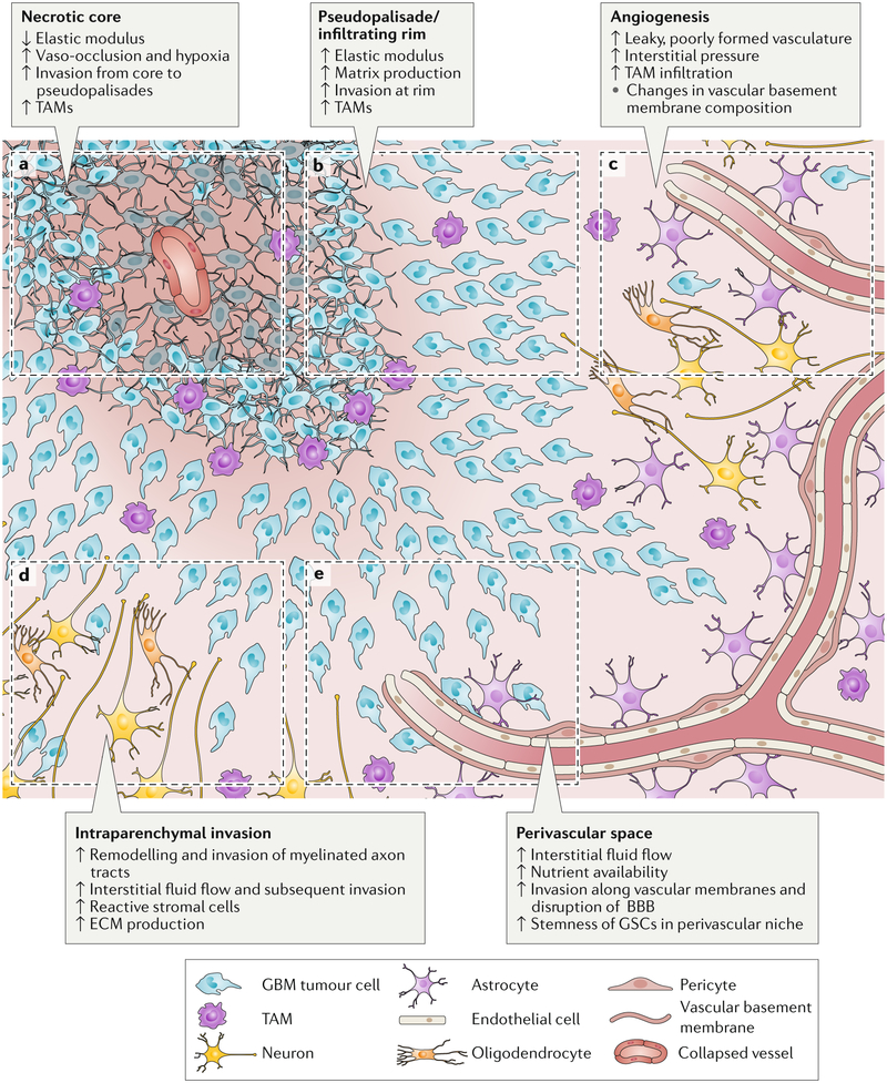
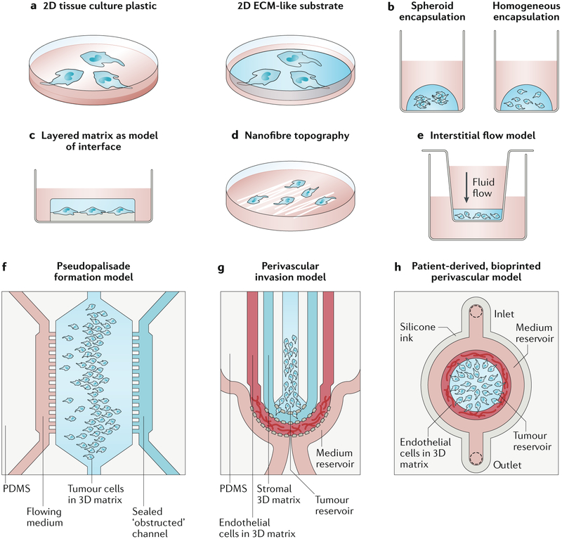
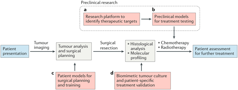
Glioblastoma (GBM) is the most aggressive and common form of primary brain cancer. Several decades of research have provided great insight into GBM progression; however, the prognosis remains poor with a median patient survival time of ~ 15 months. The tumour microenvironment (TME) of GBM plays a crucial role in mediating tumour progression and thus is being explored as a therapeutic target. Progress in the development of treatments targeting the TME is currently limited by a lack of model systems that can accurately recreate the distinct extracellular matrix composition and anatomic features of the brain, such as the blood-brain barrier and axonal tracts. Biomaterials can be applied to develop synthetic models of the GBM TME to mimic physiological and pathophysiological features of the brain, including cellular and ECM composition, mechanical properties, and topography. In this Review, we summarize key features of the GBM microenvironment and discuss different strategies for the engineering of GBM TME models, including 2D and 3D models featuring chemical and mechanical gradients, interfaces and fluid flow. Finally, we highlight the potential of engineered TME models as platforms for mechanistic discovery and drug screening as well as preclinical testing and precision medicine.


Conflict of interest statement

Competing Interest Statements: The authors declare no competing financial interests.

Figures

Figure 1.
Figure 1.. Schematic of gliobastoma (GBM) regions.
This GBM schematic illustrates changes during tumour progression in the different microenvironmental regions. a) The necrotic core is softer than surrounding tissue and is thought to form after increases in cell density beyond a certain threshold or vaso-occlusive events result in hypoxia. b) Psuedopalisades are regions of high cell density thought to form as cells migrate away from hypoxic regions. These zones have an increased elastic modulus and matrix production compared to healthy tissue and necrotic regions. GBM cells invade from the outer edge of the cell-dense tumour into healthy tissue at the infiltrating rim. c) GBM tumours show hypervascularity with increased angiogenesis compared to healthy brain tissue. Tumour-associated vasculature is poorly formed, leaky and leads to an increase in interstitial fluid pressure. d) Tumour cells invading through the parenchyma often follow and remodel the surface of myelinated tracts – a region in which high interstitial fluid flow may also drive invasion. e) Tumour cells rapidly invade the vasculature, where they are exposed to nutrients, high interstitial fluid flow and haptotactic cues in basement membranes. The perivascular niche also supports stemness and survival of glioblastoma stem cells (GSCs). TAM, tumour-associated macrophage; ECM, extracellular matrix; BBB, blood-brain barrier.
Figure 2.
Figure 2.. Engineered glioblastoma models.
a) 2D models often include a matrix layer with tunable mechanical properties and composition. b) In 3D matrices, cells can be encapsulated as spheroids or as single cells. c) Cells can be cultured between extracellular matrix (ECM) layers of distinct composition and mechanics to model cell migration at the interface of the vascular basement membrane and the intraparenchymal matrix. d) Nanofibres with ECM coatings are often used to mimic linear, white matter tracts. e) Media height in a Boyden chamber can be used to generate interstitial flow through matrix-encapsulated cells. f) A microfluidic device with an open (nutrient-rich) and closed (occluded) channel surrounding matrix-encapsulated cells can be used to test how psuedopalisades form. g) A microfluidic model of the perivascular niche (PVN) containing a glioblastoma stem cell (GSC)-rich tumour reservoir, an intraparenchymal region with stromal matrix and a region of matrix-encapsulated endothelial networks can be used to investigate the role of the PVN in GSC tumourigenicity. h) A bioprinted microfluidic model with a matrix-encapsulated endothelial network arranged concentrically around patient-derived tumour cells can be applied for the development of patient-specific engineered tumour microenvironments (TMEs). Panel f adapted from REF. 223. Panel g adapted from REF. 224. Panel h adapted from REF. 195.
Figure 3.
Figure 3.. Glioblastoma microenvironment models in the preclinical and clinical pipeline.
Red dashed lines indicate stages at which engineered models are or could be used. a) Engineered tumour microenvironments (TMEs) have been widely employed as research platforms to investigate the TME and they can be used to identify therapeutic targets. b) With refinement, these platforms can serve as a basis for precision medicine using patient-specific cells and/or matrices. c) Images of tumours from patients can be used to generate mechanically-matched patient-specific models of the tumour and brain anatomy for surgical planning and training. d) After surgical resection, engineered TMEs can aid in maintaining heterogeneity during culture for patient-specific treatment validation. The cells can be selected by molecular profiling and histological analysis.

References

    1. Ostrom QT et al. CBTRUS Statistical Report: Primary Brain and Other Central Nervous System Tumors Diagnosed in the United States in 2011–2015. Neuro. Oncol 20, iv1–iv86 (2018). - PMC - PubMed
    1. Koshy M et al. Improved survival time trends for glioblastoma using the SEER 17 population-based registries. J. Neurooncol 107, 207–212 (2012). - PMC - PubMed
    1. Stupp R et al. Effects of radiotherapy with concomitant and adjuvant temozolomide versus radiotherapy alone on survival in glioblastoma in a randomised phase III study: 5-year analysis of the EORTC-NCIC trial. Lancet. Oncol 10, 459–66 (2009). - PubMed
    1. Watanabe M, Tanaka R & Takeda N Magnetic resonance imaging and histopathology of cerebral gliomas. Neuroradiology 34, 463–469 (1992). - PubMed
    1. Young RM, Jamshidi A, Davis G & Sherman JH Current trends in the surgical management and treatment of adult glioblastoma. Ann. Transl. Med 3, 121 (2015). - PMC - PubMed