. 2019 Jun 7:10:4.
doi: 10.1186/s43008-019-0005-7. eCollection 2019.

A taxonomic summary of Aphelidiaceae

Affiliations

A taxonomic summary of Aphelidiaceae

Peter M Letcher et al. IMA Fungus. .

Abstract

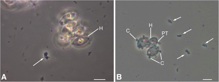
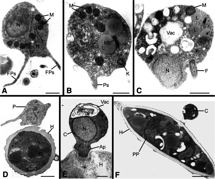
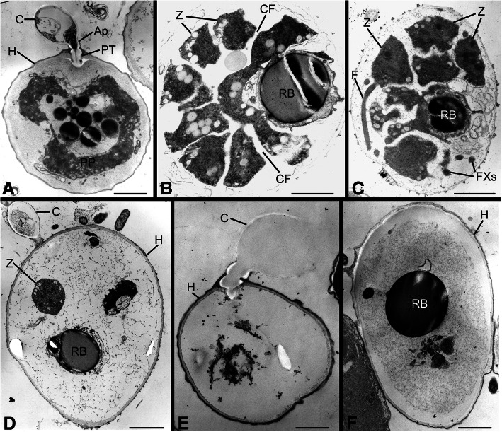
Aphelids are parasitoids of various algae and diatoms, and in a recent classification are contained in family Aphelidiaceae, phylum Aphelidiomycota, kingdom Fungi. Family Aphelidiaceae (the only family in the phylum) is composed of four genera: Aphelidium, Paraphelidium, Amoeboaphelidium, and Pseudaphelidium. All species are known morphologically, and most have been illustrated. Few have been examined ultrastructurally, and even fewer have been sequenced for molecular comparisons. Recent studies in molecular phylogenetics have revealed an abundance of related environmental sequences that indicate unrealized biodiversity within the group. Herein, we briefly summarize the history of aphelids and acknowledge the controversy of placement of the group with related organisms. With light microscopic images and transmission electron micrographs, we illustrate typical life cycle stages for aphelids, provide updated descriptions and taxonomy for all described species, and provide a key to the species.

Keywords: Amoeboaphelidium; Aphelidiomycota; Aphelidium; Basal groups; Paraphelidium; Pseudaphelidium.


Conflict of interest statement

Competing interestsThe authors declare that they have no competing interests.

Figures

Fig. 1
Fig. 1
Light microscopic images of Aphelidium protococcorum (FD 95), representative of Aphelidiaceae. a Motile zoospore (arrow) in vicinity of uninfected host (H) (Scenedesmus) cells. b Motile zoospores (arrows) and host cells to which zoospores have attached, encysted C and penetrated the host via a penetration tube (PT). Bars: A = 5 μm, B = 10 μm
Fig. 2
Fig. 2
Transmission electron micrographs of life stages representative of Aphelidiaceae. a Motile zoospore with filose pseudopodia (FPs) and containing a nucleus (N), multiple mitochondria (M), and dispersed ribosomes (R). b Motile zoospore with a pseudopodium (Ps), multiple lipid globules (L) and an adjacent microbody (Mb), and a kinetosome (K). c Motile zoospore with a posterior flagellum (F) and central vacuole (Vac). d Amoeboid zoospore (P) approaching a host cell. e Encysted zoospore (C) containing an anterior vacuole (Vac) and a nucleus, attached to host via an appressorium (Ap). f An infected host cell, with a zoospore cyst attached and the parasitoid plasmodium (PP) inside the host. Figs. A, B, D, E, F =Am. occidentale (FD 01); Fig. C = Aph. desmodesmi strain FD 104. Bars: A–E = 5 μm, F = 2 μm
Fig. 3
Fig. 3
Transmission electron micrographs of life stages representative of Aphelidiaceae. a Host cell (H) with a remnant cyst (C) attached to host via an appressorium (Ap) and infecting the host with a penetration tube (PT). Parasitoid plasmodium (PP) has engulfed much of the host cytoplasm. b Parasitoid plasmodium cleaved into multiple uninucleate cells (Z) delineated by cleavage furrows (CF); a residual body (RB) remains. c Cleaved zoospores, one with a flagellum (F); flagellar cross-sections (FXs) also visible. d An evacuated sporangium that retained a zoospore and the residual body. e An empty sporangium with ephemeral remnant of zoospore cyst. f An evacuated sporangium with a residual body. Figs. A, E = Am. occidentale (FD 01); Fig. B–D = Aph. desmodesmi (FD 104); Fig. F = Aph. protococcorum (FD 95) Bars: A–D, F = 2 μm, E = 1 μm)

References

    1. Adl SM, Bass D, Lane CE, Lukes J, Schoch CL, Smirnov A, et al. Revisions to the classification, nomenclature, and diversity of eukaryotes. Journal of Eukaryotic Microbiology. 2019;66:4–119. - PMC - PubMed
    1. Cavalier-Smith T. The origin of Fungi and pseudofungi. In: Rayner AD, Brasier CM, Moore D, editors. Evolutionary biology of the Fungi. Cambridge: Cambridge University Press; 1987. pp. 339–353.
    1. Cavalier-Smith T. Early evolution of eukaryote feeding modes, cell structure diversity, and classification of the protozoan phyla Loukozoa, Sulcozoa, and Choanozoa. European Journal of Protistology. 2013;49:115–178. doi: 10.1016/j.ejop.2012.06.001. - DOI - PubMed
    1. Dangeard PA. Mémoire sur quelques maladies des algues et des animaux. Le Botaniste. 1890;2:231–268.
    1. de Bruyne C. Monadines et Chytriacées, parasites des algues du Golfe de Naples. Archives de Biologie (Paris) 1890;10:43–104.

LinkOut - more resources