. 2020 Sep;33(9):e4359.
doi: 10.1002/nbm.4359. Epub 2020 Jul 9.

Nitroxide-enhanced MRI of cardiovascular oxidative stress

Affiliations

Nitroxide-enhanced MRI of cardiovascular oxidative stress

Soham A Shah et al. NMR Biomed. 2020 Sep.

Abstract

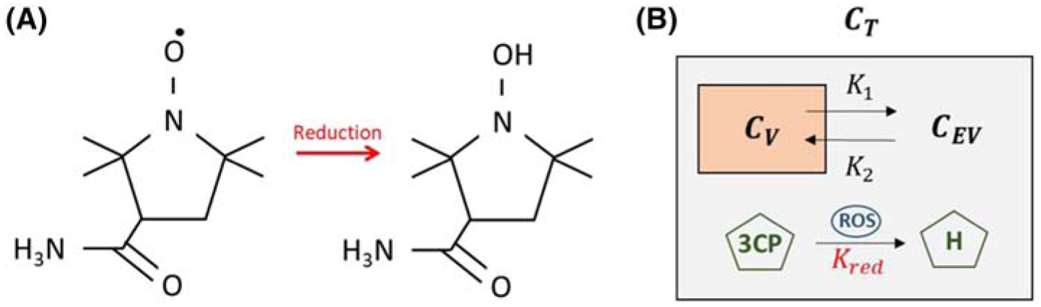
Background: In vivo imaging of oxidative stress can facilitate the understanding and treatment of cardiovascular diseases. We evaluated nitroxide-enhanced MRI with 3-carbamoyl-proxyl (3CP) for the detection of myocardial oxidative stress.

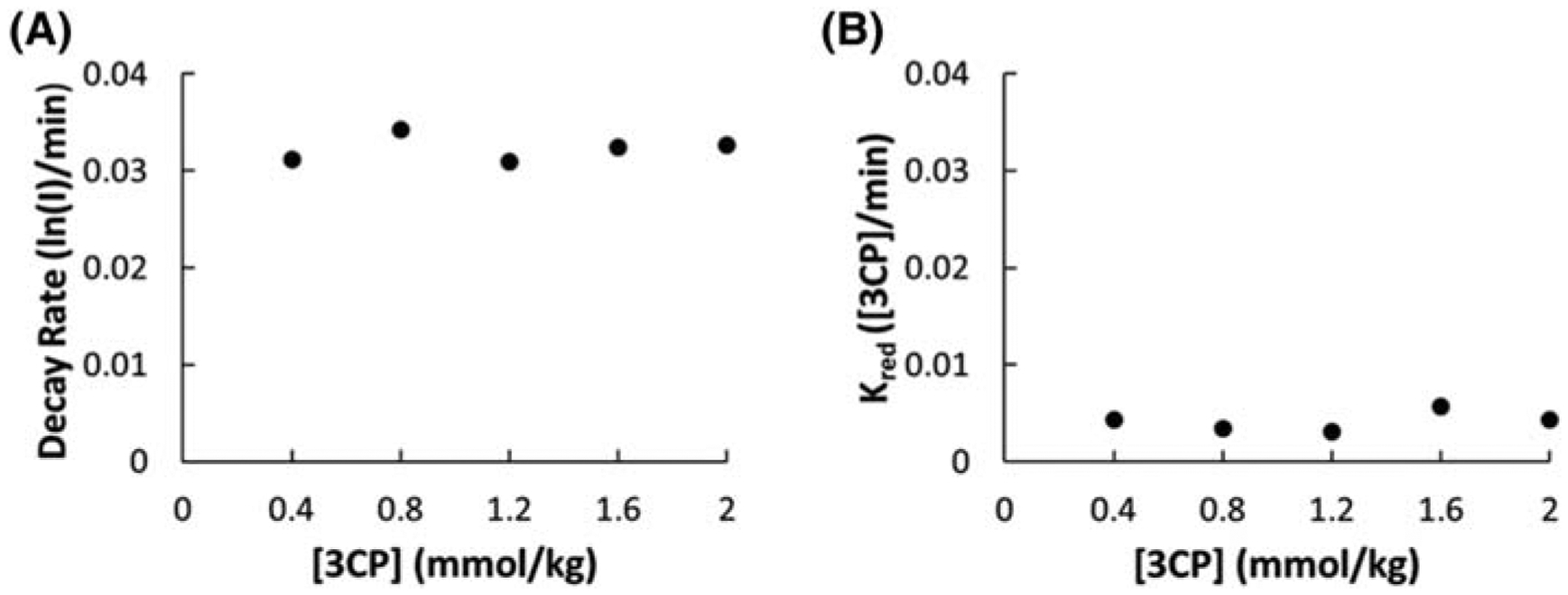
Methods: Three mouse models of cardiac oxidative stress were imaged, namely angiotensin II (Ang II) infusion, myocardial infarction (MI), and high-fat high-sucrose (HFHS) diet-induced obesity (DIO). For the Ang II model, mice underwent MRI at baseline and after 7 days of Ang II (n = 8) or saline infusion (n = 8). For the MI model, mice underwent MRI at baseline (n = 10) and at 1 (n = 8), 4 (n = 9), and 21 (n = 8) days after MI. For the HFHS-DIO model, mice underwent MRI at baseline (n = 20) and 18 weeks (n = 13) after diet initiation. The 3CP reduction rate, Kred , computed using a tracer kinetic model, was used as a metric of oxidative stress. Dihydroethidium (DHE) staining of tissue sections was performed on Day 1 after MI.

Results: For the Ang II model, Kred was higher after 7 days of Ang II versus other groups (p < 0.05). For the MI model, Kred , in the infarct region was significantly elevated on Days 1 and 4 after MI (p < 0.05), whereas Kred in the noninfarcted region did not change after MI. DHE confirmed elevated oxidative stress in the infarct zone on Day 1 after MI. After 18 weeks of HFHS diet, Kred was higher in mice after diet versus baseline (p < 0.05).

Conclusions: Nitroxide-enhanced MRI noninvasively quantifies tissue oxidative stress as one component of a multiparametric preclinical MRI examination. These methods may facilitate investigations of oxidative stress in cardiovascular disease and related therapies.

Keywords: MRI; cardiovascular; heart; nitroxides; oxidative stress; preclinical.


Conflict of interest statement

CONFLICTS OF INTEREST

The authors declare that they have no competing interests.

Figures

FIGURE A1
FIGURE A1
A, The myocardial 3CP decay rate quantified by linear fitting of the natural log of signal intensity (I) decay as a function of the 3CP dose. B, The 2CXRM myocardial 3CP reduction rate, Kred, as a function of the 3CP dose
FIGURE 1
FIGURE 1
A, The molecular structure of the paramagnetic nitroxide 3CP, showing a single unpaired electron, and the molecular structure of 3CP after undergoing a reduction reaction into a diamagnetic hydroxylamine, without an unpaired electron. B, The 2CXRM, which accounts for (1) nitroxide exchange between the vascular and extravascular compartments within the tissue compartment with rate constants K1 and K2, and (2) nitroxide reduction, with rate constant Kred, in the extravascular compartment due to intracellular reactions driving 3CP reduction to hydroxylamine via ROS. Cv, CEV, and CT are the 3CP concentrations in the vascular, extravascular, and tissue compartments, respectively
FIGURE 2
FIGURE 2
A, Example T1-weighted saturation-recovery gradient-echo short-axis images of the mouse heart acquired before and after the injection of 3CP at doses of 0.4, 0.8, 1.2, 1.6, and 2.0 mmol/kg body weight. B, Signal-to-noise ratios (mean ± standard error, n = 3) are shown for each 3CP dose from ROIs in the LV blood pool and myocardial tissue
FIGURE 3
FIGURE 3
A, Example dynamic nitroxide-enhanced MR images before and after injection of 3CP demonstrate signal enhancement kinetics. A proton-density-weighted image (PD) is used to visualize myocardial borders. B, Example ROIs of the LV blood pool and myocardium used to estimate vascular and tissue 3CP concentrations, CV and CT respectively. C, Example CV(t) fit and 2CXRM fitting of CT(t) of a control mouse. Kred was 0.0019 [3CP]/min and R2 was 0.99. D, Kred was significantly elevated in Ang II mice at Week 1 versus Ang II mice at baseline and control mice at Week 1. *p < 0.05 using a two-way ANOVA
FIGURE 4
FIGURE 4
A, An example DENSE circumferential strain map was used to identify infarcted and noninfarcted remote regions of the myocardium in post-MI mice, and these regions were applied to nitroxide-enhanced images acquired at matched locations. B, Example vascular (CV) and 2CXRM remote zone and infarct zone CT fits in a mouse 1 day after infarction. C, Time courses of mean Kred for infarct and remote regions from mice before and for 21 days after MI demonstrate the rise and decline of oxidative stress in the infarct region. D, Example confocal images of DHE-stained myocardium obtained on Day 1 after MI. Specifically, overlays of 488 nm (for oxidation products of DHE) and 405 nm (for DHE) are shown for remote and infarct regions. E, DHE fluorescence in the infarct zone is elevated compared with the remote zone in 1 day post-MI mice, indicative of oxidative stress in infarcted myocardium. In contrast, in control mice DHE fluorescence was equivalent in the septal and lateral walls. *p < 0.05 compared with baseline and Day 21 infarct, and Day 1 remote using a two-way ANOVA. **p < 0.05 compared with baseline and Day 21 infarct, and Day 4 remote using a two-way ANOVA. $p < 0.05 compared with control using a log ratio t test
FIGURE 5
FIGURE 5
The myocardial 3CP reduction rate, Kred, was increased in mice fed an HFHS diet for 18 weeks compared with baseline (0.0327 ± 0.0102 versus 0.0014 ± 0.0009). *p < 0.05 compared with baseline using a t test

References

    1. Münzel T, Camici GG, Maack C, Bonetti NR, Fuster V, Kovacic JC. Impact of oxidative stress on the heart and vasculature: part 2 of a 3-part series. J Am Coll Cardiol. 2017;70(2):212–229. - PMC - PubMed
    1. Duilio C, Ambrosio G, Kuppusamy P, DiPaula A, Becker LC, Zweier JL. Neutrophils are primary source of O2 radicals during reperfusion after prolonged myocardial ischemia. Am J Physiol Heart Circ Physiol. 2001;280(6):H2649–H2657. - PubMed
    1. Furukawa S, Fujita T, Shimabukuro M, et al. Increased oxidative stress in obesity and its impact on metabolic syndrome. J Clin Invest. 2017;114(12): 1752–1761. - PMC - PubMed
    1. Münzel T, Gori T, Keaney JF, Maack C, Daiber A. Pathophysiological role of oxidative stress in systolic and diastolic heart failure and its therapeutic implications. Eur Heart J. 2015;36:2555–2564. - PMC - PubMed
    1. Elas M, Ichikawa K, Halpern HJ. Oxidative stress imaging in live animals with techniques based on electron paramagnetic resonance. Radiat Res. 2012; 177(4):514–523. - PMC - PubMed

Publication types

MeSH terms