Review
. 2020 Aug 10;38(2):167-197.
doi: 10.1016/j.ccell.2020.06.001. Epub 2020 Jul 9.

Oxidative Stress in Cancer

Affiliations
Review

Oxidative Stress in Cancer

John D Hayes et al. Cancer Cell. .

Abstract

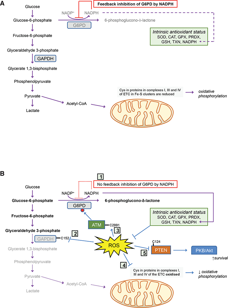
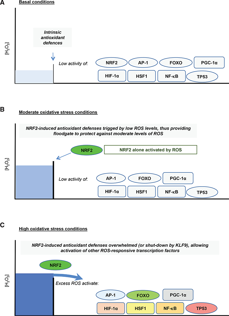
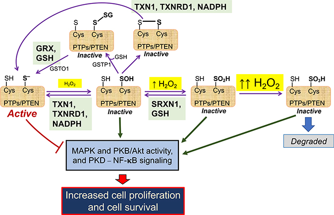
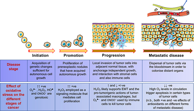
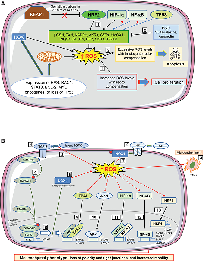
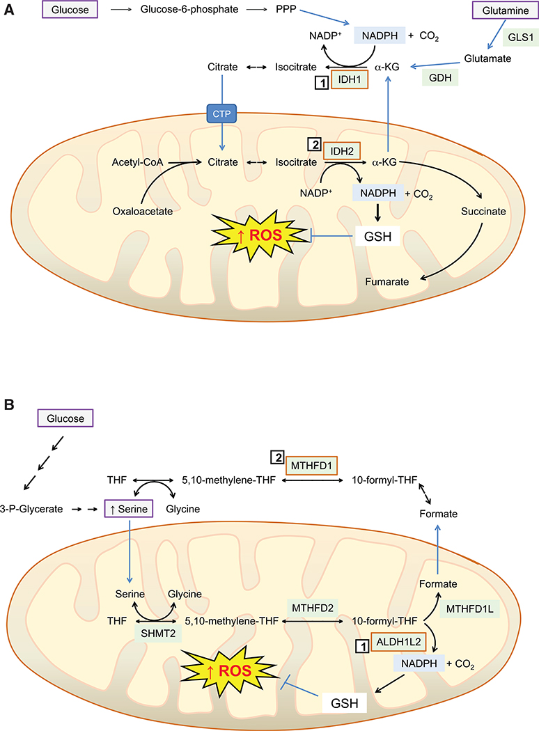
Contingent upon concentration, reactive oxygen species (ROS) influence cancer evolution in apparently contradictory ways, either initiating/stimulating tumorigenesis and supporting transformation/proliferation of cancer cells or causing cell death. To accommodate high ROS levels, tumor cells modify sulfur-based metabolism, NADPH generation, and the activity of antioxidant transcription factors. During initiation, genetic changes enable cell survival under high ROS levels by activating antioxidant transcription factors or increasing NADPH via the pentose phosphate pathway (PPP). During progression and metastasis, tumor cells adapt to oxidative stress by increasing NADPH in various ways, including activation of AMPK, the PPP, and reductive glutamine and folate metabolism.

Keywords: AP-1; BACH1; FOXO; HIF-1alpha; HSF1; NADPH generation; NF-κB; NRF2; PGC-1alpha; TP53; adaptation; antioxidant; dormant cancer cell; folate metabolism; glutathione; initiation; metastasis; oxidative stress; pentose phosphate pathway; progression; reactive oxygen species; recurrent disease; redox signaling; reductive glutamine metabolism; thioredoxin; tumorigenesis.


Conflict of interest statement

Declaration of Interests A.D.K. is on the Scientific Advisory Board of Evgen Pharma and is a consultant for Aclipse Therapeutics and Vividion Therapeutics.

Figures

Figure 1.
Figure 1.. Metabolic Responses to Acute Oxidative Stress
In cells under normal redox homeostatic conditions (A), glucose is principally oxidized by glycolysis to pyruvate, and via acetyl-CoA through the tricarboxylic acid cycle, with G6PD inhibited by NADPH and minimal flux through the PPP. However, upon acute oxidative stress (B), feedback inhibition of G6PD by NADPH is greatly diminished (1) and Cys residues in GAPDH (2), ATM (3), and complexes I, III, and IV of the electron transport chain (4) are oxidized, a combination of circumstances that result in inhibition of glycolysis, phosphorylation of G6PD, and increased metabolism through the PPP. Moreover, oxidation of Cys residues in PTEN (5) causes activation of PKB/Akt, resulting in increased cell survival.
Figure 2.
Figure 2.. NRF2 Provides an Inducible Floodgate Defense against Oxidative Stress
Under non-stressed basal conditions (A), cellular redox homeostasis is maintained by constitutive expression of a battery of antioxidant genes. However, when exposed to acute oxidative stress (B), cells adapt to the increase in ROS levels by inducing genes encoding detoxification, GSH- and TXN-dependent antioxidants, and NADPH-generating enzymes that are regulated by NRF2. Should the capacity of the antioxidant systems that are induced by NRF2 become saturated and therefore insufficient to counter additional oxidative stress (C), or prolonged oxidative stress causes activation of KLF9 and downregulation of NRF2, the excess levels of ROS that are not countered by the NRF2-directed defences then trigger additional redox switches that activate other members of the antioxidant transcription factor network. When NRF2-orchestrated defences become saturated, activation of other members of the antioxidant transcription factor network may occur simultaneously, or they may be activated in a stratified manner with each transcription factor being activated at a distinct ROS threshold, which results in various cellular responses, including metabolic reprogramming, damage repair, cell-cycle arrest, senescence, and apoptosis.
Figure 3.
Figure 3.. Redox Regulation of PTP- and PTEN-Mediated Inhibition of Cell Proliferation and Survival
Protein tyrosine phosphatases PTP1B, PTPN2, and PTPN11 and the lipid phosphatase PTEN suppress MAPK and PKB/Akt activity, as well as NF-κB signaling (bottom left). These phosphatases each possess an active-site Cys residue in a thiolate anion (S) state that is susceptible to oxidation: as depicted across the center of the cartoon, the thiolate form of the active-site Cys can be oxidized to sulfenate (SOH), sulfinate (SO2H), or sulfonate (SO3H) states depending on the levels of H2O2 and duration of exposure to H2O2 (see Box 6). Alternatively, as shown at the top of the cartoon, the active-site Cys may form mixed disulfides by reacting with GSH (S-glutathionylated protein-SSG), which can be catalyzed by GST P1–1, or react with another thiol internally or in another protein to form a disulfide bridge (-S-S-). These oxidative modifications of the phosphatases result in their inactivation and therefore an increase in MAPK and PKB/Akt activity and NF-κB signaling. However, oxidative inactivation to sulfenate or sulfinate states can be reversed by the TXN1 or SRXN1 antioxidant systems (shown in green boxes), thereby allowing rescue of phosphatase activity and suppression of MAPK, PKB/Akt, and NF-κB activities. Similarly, active-site Cys that have been S-glutathionylated or have formed a disulfide bridge can be reversed by the GRX/GSH antioxidant system (green box), or de-glutathionylated by GST O1–1, thereby allowing recovery of phosphatase activity. Oxidation of the active-site Cys to a sulfonate state is irreversible (right), and the protein has to be eliminated.
Figure 4.
Figure 4.. An Ambiguous Role for Oxidative Stress in Tumorigenesis
The cartoon depicts development of malignant disease from initiation through promotion and progression, until it acquires a highly malignant, invasive and metastatic phenotype. The contributions that excess levels of O2●–, H2O2, HO, and ONOO may make to the different stages of the disease are indicated in panel at the bottom.
Figure 5.
Figure 5.. Influence of Oxidative Stress on Cell Fate during Early Stages of Tumorigenesis
The cartoons depict how levels of ROS stimulate proliferation or apoptosis in preneoplastic cells during initiation of tumorigenesis, and how ROS support EMT during progression of tumorigenesis by altering TGF-β signaling and by activation of antioxidant transcription factors that control expression of EMT-TFs. During the earliest stages of tumorigenesis (A), activation of oncogenes, coupled with higher metabolic demands, results in an increase in intracellular ROS levels in early neoplastic lesions/adenomas. To benefit from the proliferative advantages associated with the increase in ROS, without succumbing to apoptosis, cells harboring activated oncogenes augment their antioxidant capacity by increasing transactivation of genes encoding GSH- and TXN-dependent enzymes along with antioxidant/detoxification enzymes (1). Often this readjustment of redox entails loss or blunting of repression of NRF2 by KEAP1 and induction of NRF2 target genes. In addition, HIF-1α increases expression of key metabolic proteins, such as GLUT1, HK2, and MCT4. Also, NF-κB and TP53 probably contribute to this adaptive process, although in lesions harboring mutant TP53 the latter is unlikely. Treatment of animals with BSO, which inhibits synthesis of GSH, before initiation of carcinogenesis will stimulate apoptosis of premalignant cells (2). However, once carcinogenesis has been initiated, stimulation of apoptosis in malignant cells requires inhibition of both the GSH-based (by BSO) and the TXN-based (by sulfasalazine or auranofin) antioxidant systems. During the progression stage of tumorigenesis (B), EMT is triggered by a variety of environmental factors, including those that alter intracellular redox. In this regard, TGF-β signaling (1), growth factor signaling (2), and tumor-associated macrophages (TAMs) in the microenvironment (3) will produce ROS. Binding of TGF-β to its cognate receptor causes phosphorylation of SMAD2/3 (4) and induction of NOX4 gene expression (5), which results in production of H2O2 at the ER (6). NOX4-generated ROS within the tumor cell is augmented by growth factor signaling causing phosphorylation and activation of NOX1 and production of O2●– at the plasma membrane (7), with increased ROS increasing processing of latent TGF-β (8). The increases in ROS from TGF-β and growth factor signaling, along with those generated by TAMs, activate TP53, which combines with SMAD proteins to induce transcription of genes encoding the EMT-TFs SNAIL and TWIST (9). Similarly, increased ROS levels activate AP-1 (10) and HIF-1α (11) and induce SNAIL and TWIST, whereas the activation by ROS of NF-κB (12) leads to induction of genes encoding SNAIL, TWIST, SLUG, ZEB1, and ZEB2. Together, SNAIL, TWIST, SLUG, ZEB1, and ZEB2 positively control expression of mesenchymal-associated genes and negatively control expression of epithelial-associated genes. ROS also activate HSF1 (13), whereas its downregulation decreases TGF-β-mediated expression of SNAIL and SLUG and inhibits EMT, although the precise mechanism(s) is not understood.
Figure 6.
Figure 6.. Reductive Glutamine Metabolism and Serine-Driven Folate Metabolism Suppresses Mitochondrial ROS Accumulation to Support Anchorage-Independent Growth and/or Metastatic Disease
(A) The increase in mitochondrial ROS that occurs when tumor cells are grown as spheroids can be mitigated by the concerted actions of IDH1 (1) and IDH2 (2), located in the cytoplasm and mitochondrion, respectively. Specifically, within the cytoplasm, IDH1 catalyzes the reductive carboxylation of α-ketoglutarate (α-KG), obtained from glutamine (by the sequential actions of GLS1 and GDH, see top right of cartoon), utilizing NADPH provided by the pentose phosphate pathway (PPP) (see top left and center of cartoon), to provide a supply of isocitrate and citrate. In turn, citrate in the cytoplasm is transferred to the mitochondrion via the citrate transporter protein (CPT, SLC25A1), before it is utilized by IDH2 to produce α-KG and NADPH. The latter is required to reduce GSSG and maintain high GSH levels, which ensures mitochondrial ROS levels are restrained and anoikis averted. Thus, IDH1 and IDH2 activities in the different subcellular compartments enable NADPH generated by the PPP, along with high glutamate/glutamine levels, to drive reductive carboxylation of α-KG in the cytoplasm, and so transfer reducing equivalents from the cytoplasm to the mitochondrion. (B) The 10-formyl-tetrahydrofolate (THF) pathway represents a major source of NADPH for a variety of cell lines grown under in vitro cell culture conditions. For melanoma tumor cells to survive the relatively high O2 levels in the bloodstream and then colonize the liver, they increase production of NADPH by augmenting ALDH1L2 protein levels and maintaining levels of MTHFD1; ALDH1L2 (1) and MTHFD1 (2) are located in the mitochondrion and cytoplasm, respectively. The cartoon depicts how high de novo synthesis of serine, derived from glucose and 3-phosphoglycerate (top left-hand side), followed by transport into the mitochondrion, allows donation of a methyl group to THF, yielding 5,10-methylene-THF through a serine hydroxymethyl transferase (SHMT) 2-catalyzed reaction. In turn, 5,10-methylene-THF within the mitochondrion is converted to 10-formyl-THF by MTHFD2, which can in turn be utilized by ALDH1L2 to form NADPH (bottom right-hand side), which maintains GSH levels and prevents excess ROS from accumulating. Alternatively, 10-formyl-THF can be used by MTHFD1L to generate formate, which when transported out of the mitochondrion can be used in reversible MTHFD1-catalyzed reactions to generate 5,10-methylene-THF.

References

    1. Ahn SG, and Thiele DJ (2003). Redox regulation of mammalian heat shock factor 1 is essential for Hsp gene activation and protection from stress. Genes Dev. 17, 516–528. - PMC - PubMed
    1. Akter S, Fu L, Jung Y, Conte ML, Lawson JR, Lowther WT, Sun R, Liu K, Yang J, and Carroll KS (2018). Chemical proteomics reveals new targets of cysteine sulfinic acid reductase. Nat. Chem. Biol. 14, 995–1004. - PMC - PubMed
    1. Alexander MS, Wilkes JG, Schroeder SR, Buettner GR, Wagner BA, Du J, Gibson-Corley K, O’Leary BR, Spitz DR, Buatti JM, et al. (2018). Pharmacologic ascorbate reduces radiation-induced normal tissue toxicity and enhances tumor radiosensitization in pancreatic cancer. Cancer Res. 78, 6838–6851. - PMC - PubMed
    1. Aljagthmi AA, Hill NT, Cooke M, Kazanietz MG, Abba MC, Long W, and Kadakia MP (2019). DeltaNp63alpha suppresses cells invasion by downregulating PKCgamma/Rac1 signaling through miR-320a. Cell Death Dis. 10, 680. - PMC - PubMed
    1. Andersson DA, Gentry C, Moss S, and Bevan S (2008). Transient receptor potential A1 is a sensory receptor for multiple products of oxidative stress. J. Neurosci. 28, 2485–2494. - PMC - PubMed

Publication types