. 2020 Sep 17;27(9):1140-1150.e4.
doi: 10.1016/j.chembiol.2020.06.015. Epub 2020 Jul 9.

In Situ Fucosylation of the Wnt Co-receptor LRP6 Increases Its Endocytosis and Reduces Wnt/β-Catenin Signaling

Affiliations

In Situ Fucosylation of the Wnt Co-receptor LRP6 Increases Its Endocytosis and Reduces Wnt/β-Catenin Signaling

Senlian Hong et al. Cell Chem Biol. .

Abstract

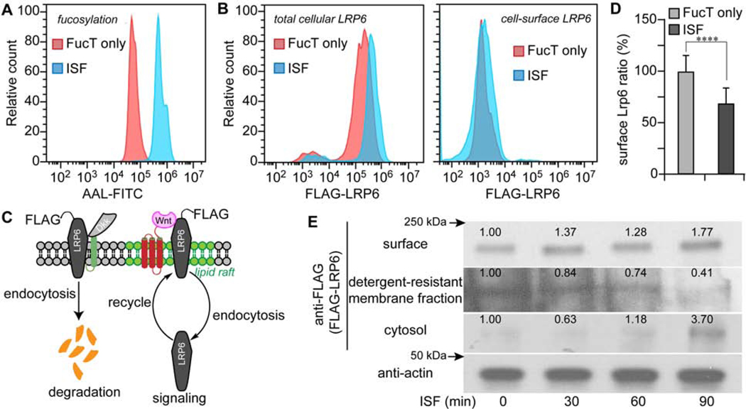
Wnt/β-catenin signaling regulates critical, context-dependent transcription in numerous physiological events. Among the well-documented mechanisms affecting Wnt/β-catenin activity, modification of N-glycans by L-fucose is the newest and the least understood. Using a combination of Chinese hamster ovary cell mutants with different fucosylation levels and cell-surface fucose editing (in situ fucosylation [ISF]), we report that α(1-3)-fucosylation of N-acetylglucosamine (GlcNAc) in the Galβ(1-4)-GlcNAc sequences of complex N-glycans modulates Wnt/β-catenin activity by regulating the endocytosis of low-density lipoprotein receptor-related protein 6 (LRP6). Pulse-chase experiments reveal that ISF elevates endocytosis of lipid-raft-localized LRP6, leading to the suppression of Wnt/β-catenin signaling. Remarkably, Wnt activity decreased by ISF is fully reversed by the exogenously added fucose. The combined data show that in situ cell-surface fucosylation can be exploited to regulate a specific signaling pathway via endocytosis promoted by a fucose-binding protein, thereby linking glycosylation of a receptor with its intracellular signaling.

Keywords: (1–3)-fucosylation; LRP6; Wnt signaling/β-catenin signaling; endocytosis.


Conflict of interest statement

Declaration of Interests The authors declare no competing interest.

Figures

Figure 1.
Figure 1.
Enhanced α(1–3)-fucosylation via ISF inhibits Wnt signaling in zebrafish embryos, CHO parent and Lec2 cells. (A) Relativye mean fluorescence intensity (MFI) of traditional bioorthogonal chemical reporter labeling in the FucT/GDP-Fuc or FucT/GDP-FucAl treated embryos by ImageJ. (B) Flow chart of the impacts of ISF on Wnt signaling activity in a zebrafish Wnt reporter line. At the 8-cell stage, a FucT/GDP-Fuc pre-mixture, or FucT only, were injected into the chorion to perform ISF or act as control, respectively. To further boost the Wnt signaling readout, recombinant WNT8a was injected into the chorion at the 1000-cell stage before the embryos were transferred individually into single wells. Expression of the GFP reporter was quantified at 7 hpf. (C) Comparing the GFP expression level of Wnt reporter embryos treated by ISF or FucT only in the presence or absence of WNT8a. The MFI of GFP was quantified for healthy live embryos at 7 hpf by subtracting background in samples with no GFP expression, and the GFP-MFI of embryos treated with FucT only was set as 100%. The numbers on the bar graph represent the numbers of healthy embryos used in this assay. (D) Typical complex N-glycans present on cell surface glycoproteins of CHO Pro-5 cells, and glycosylation mutants Lec2, LEC30 and Lec8 cells.(North et al., 2010) Pro-5 and Lec2 cells carry LacNAc with no Fuc attached and are excellent substrates for ISF. Lec8 has no LacNAc and LEC30 has many fucosylated LacNAcs. Sugar symbols are from the Symbol Nomenclature for Glycans (SNFG).(Neelamegham et al., 2019) (E) Comparison of Wnt activity in Pro-5 cells compared to LEC30 cells in the presence or absence of WNT8a. (F) Western blot assay for detection of biotinylated glycoproteins in Pro-5 cells generated via GDP-Fuc-biotin-ISF before or after PNGase F-treatment. Equal loading determined by Coomassie blue staining (lower panel). (G) Comparison of Wnt activity in CHO parent and three mutant lines treated with ISF or FucT only. (H) Quantitative flow cytometry analysis of time-dependent incorporation of α(1–3) Fuc onto Pro-5 cell-surface glycans via ISF-treatment. (I) Time-dependent decrease of Wnt activity in Pro-5 cells treated by ISF. Error bars represent the standard deviation of three biological replicates. In A and B, results are from the same set of samples and the signal intensity of sample at 0 min treatment is set as 100%. (J) Time-dependent decrease of the β-catenin level in ISF-treated Pro-5 cells. Western blot of anti-β-catenin (upper panel) and actin (lower panel). In Figure 1J, the numbers indicate the relative quantification of anti-β-catenin level to actin by ImageJ. The 30 min, non-treated sample was set as 100%. Number (in white) indicates the relative β-catenin level normalized to corresponding level of actin. * unpaired, two-tailed Student’s t-test *p<0.05; **p<0.01; ***p<0.001; NS, not significant.
Figure 2.
Figure 2.
ISF treatment relocates LRP6 from detergent-resistant membranes to cytoplasm. (A, B) Flow cytometry histograms of FucT-treated (Red) and ISF-treated (Blue) Pro-5 CHO cells after 90 min incubation. LRP6 in (B) was transiently-expressed FLAG-LRP6. (A) Detection of cell surface N-glycan LacNAc fucosylation by the lectin AAL-FITC. (B) Immunofluorescent staining of total cellular LRP6 and cell-surface LRP6. (C) Scheme of LRP6 location and maintenance regulated by endocytosis. (D) Ratio of LRP6 (surface to total) in Pro-5 cells treated by ISF, or FucT only. Mock control was set as 100%. Error bars show standard error. N=53,664 cells, **** unpaired, two-tailed Student’s t-test (p<0.001). (E) Western blot assay of membrane LRP6, lipid raft LRP6, cytoplasmic LRP6 and actin after different times of ISF treatment. Number indicates the relative LRP6 level normalized to the corresponding level of actin.
Figure 3.
Figure 3.
ISF relocalizes LRP6 away from lipid rafts. (A) A schematic representation (top panel) of the membrane topological location of the lipid-raft marker CTB and LRP6. The colocalization of LRP6 and CTB (white dots) in lipid rafts was calculated from confocal images using ImageJ, based on immunofluorescence of antibody to LRP6 (green) and labeled CTB (red). (B) Comparing the colocalization of LRP6 and CTB showed that LRP6 moved from membrane to cytoplasm in ISF-treated Pro-5 cells. The numbers show the cells counted in this assay. (C, D) Plots of the colocalization of LRP6 and CTB (per cell) in ISF-treated and FucT-treated Lec2 cells (C) or in untreated LEC30 cells (D). Colocalization was measured by ImageJ and is indicated by white dots. Note: ****, unpaired, two-tailed Student’s t-test p<0.001. Scale bar: 20 μm.
Figure 4.
Figure 4.
Tracking LRP6 endocytosis induced by ISF. (A, B) Confocal visualization of LRP6 (FLAG-LRP6, green) in FucT only-treated and ISF-treated Pro-5 cells. Early endosome marker RAB5-mCherry (A, red) and recycling endosome marker RAB11-mCheery (B, red) were transiently expressed in Pro-5 cells. The colocalization of LRP6 with RAB5 or RAB11 is shown in yellow. Scale bar: 20 μm. (C-E) Colocalization of RAB5 (C), RAB7 (D) or RAB11 (E) with LRP6 in FucT-only-treated and ISF-treated Pro-5 cells, is shown as Pearson’s correlation coefficient (PCC). Note: **, two-sided Student’s t test p<0.01; NS, not significant.
Figure 5.
Figure 5.
Free Fuc suppresses ISF-induced Wnt inhibition. (A) Diagram summarizing our new findings and predicting the presence of a Fuc-binding receptor. Increasing cell-surface N-glycan LacNAc α(1–3)-fucosylation via ISF-treatment exacerbates the endocytosis of the lipid-raft localized Wnt-receptor LRP6 which, in turn, leads to reduced Wnt/β-catenin signaling. The majority of endocytosed cell-surface LRP6 enters the recycling pathway rather than the degradation pathway following endocytosis induced by ISF treatment. (B) The chemical structure of four monosaccharaides in a vertebrate N-glycan, including L-fucose (Fuc), D-glucose (Glc), D-galactose (Gal) and D-mannose (Man). (C, D) Wnt signaling activity in Pro-5 cells treated by ISF or FucT only, in the presence or absence of a free monosaccharaide. The bars represent standard error of three biological replicates. * unpaired, two-tailed Student’s t-test p<0.05; ** unpaired, two-tailed Student’s t-test p<0.01; NS, not significant.
Figure 6.
Figure 6.
Cell-surface ISF suppresses Wnt/β-catenin signaling in breast cancer cells. (A) Time-dependent decrease of the β-catenin level in ISF-treated MDA-MB-231. Western blot of anti-β-catenin (upper panel) and anti-actin (lower panel). (B) Immunoprecipitation of LRP6 from indicated MDA-MB-231 cells and detection of LRP6 and phosphorylated LRP6 by immunoblot. ISF-dependent decrease of LRP-6 phosphorylation. (C) Wound healing assay results from MDA-MB-231 cells treated with ISF or not. (D) Time-dependent decrease of the β-catenin level in ISF-treated MCF-7. (E) Wound healing of MCF-7 cells treated with ISF or not. (F) Time-dependent gap closure of MDA-MB-231 and MCF-7 cells treated with ISF or not, in wound healing assays. * one-way ANOVA p<0.05; ** one-way ANOVA p<0.01. In figure A, B and D, the number indicates the relative protein level normalized to corresponding level of actin.

Comment in

Similar articles

Cited by

References

    1. Anastas JN, Moon RT, 2013. WNT signalling pathways as therapeutic targets in cancer. Nat. Rev. Cancer 13, 11–26. doi:10.1038/nrc3419 - DOI - PubMed
    1. Bennett CN, Ross SE, Longo KA, Bajnok L, Hemati N, Johnson KW, Harrison SD, MacDougald OA, 2002. Regulation of Wnt signaling during adipogenesis. J. Biol. Chem 277, 30998–31004. doi:10.1074/jbc.M204527200 - DOI - PubMed
    1. Bienz M, Clevers H, 2000. Linking colorectal cancer to Wnt signaling. Cell 103, 311–320. - PubMed
    1. Bilic J, Huang YL, Davidson G, Zimmermann T, Cruciat C-M, Bienz M, Niehrs C, 2007. Wnt induces LRP6 signalosomes and promotes dishevelled-dependent LRP6 phosphorylation. Science 316, 1619–1622. doi:10.1126/science.1137065 - DOI - PubMed
    1. Brown DA, 2006. Lipid rafts, detergent-resistant membranes, and raft targeting signals. Physiology (Bethesda) 21, 430–439. doi:10.1152/physiol.00032.2006 - DOI - PubMed

Publication types

MeSH terms

LinkOut - more resources