. 2020 Aug 20;27(8):1084-1096.e4.
doi: 10.1016/j.chembiol.2020.06.014. Epub 2020 Jul 9.

Parallel Chemoselective Profiling for Mapping Protein Structure

Affiliations

Parallel Chemoselective Profiling for Mapping Protein Structure

Zachary E Potter et al. Cell Chem Biol. .

Abstract

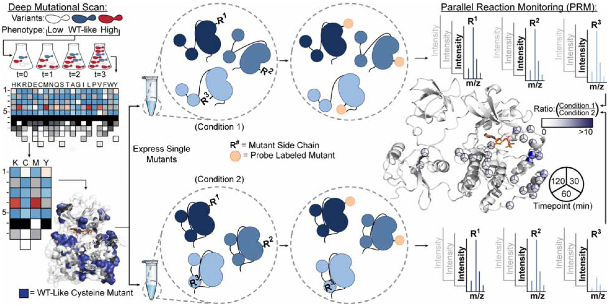
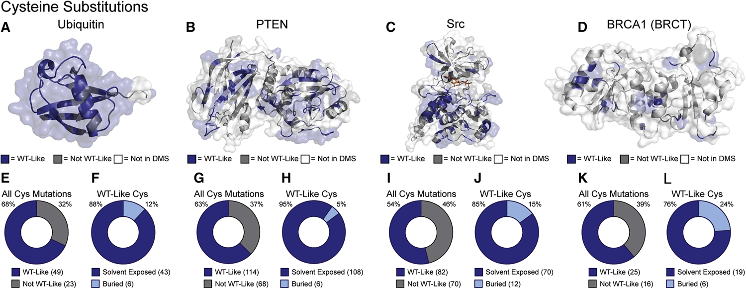
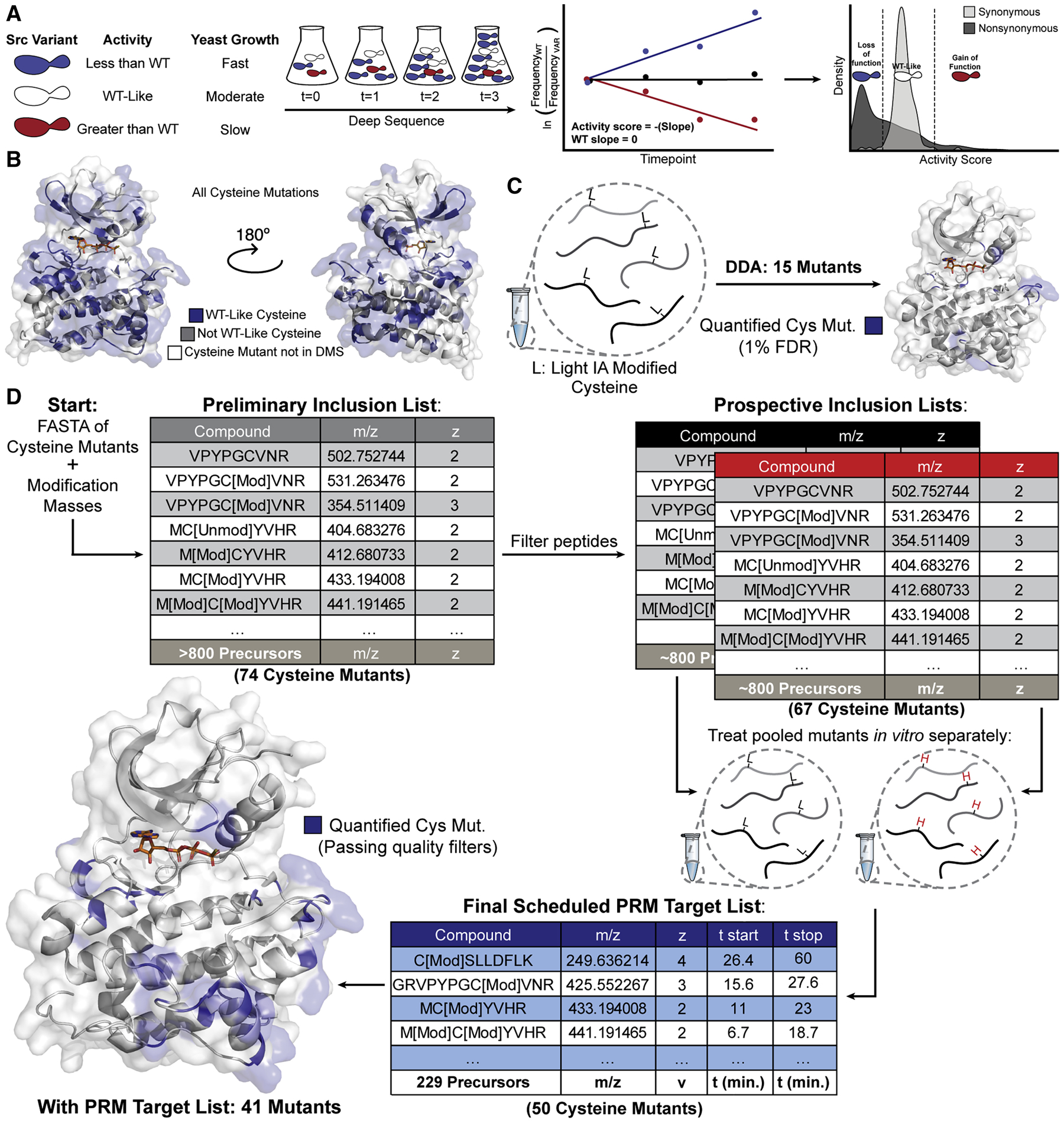
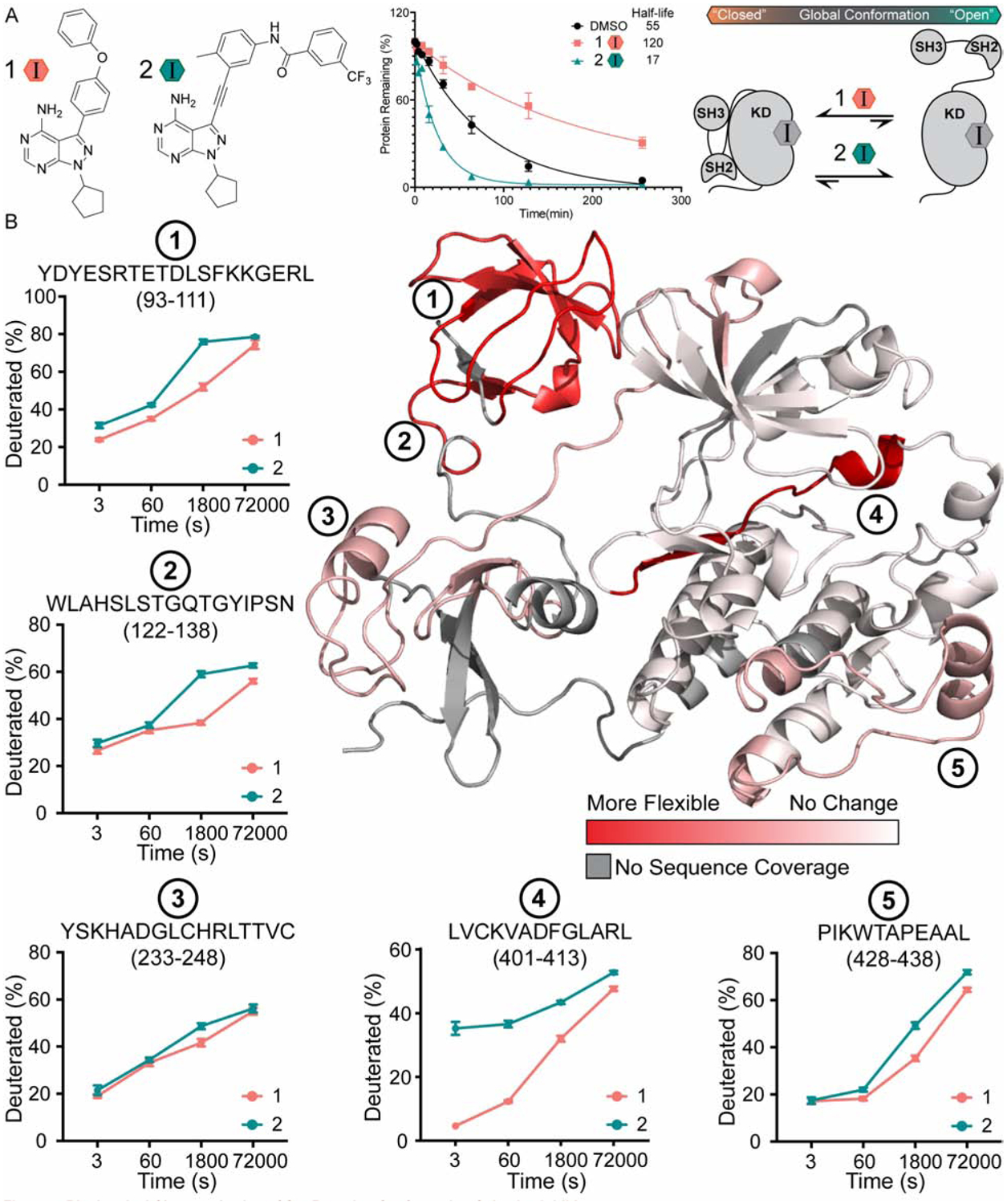
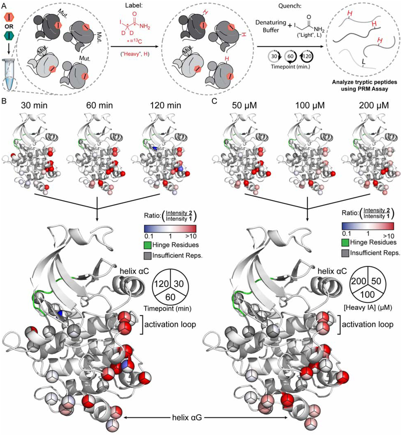
Solution-based structural techniques complement high-resolution structural data by providing insight into the oft-missed links between protein structure and dynamics. Here, we present Parallel Chemoselective Profiling, a solution-based structural method for characterizing protein structure and dynamics. Our method utilizes deep mutational scanning saturation mutagenesis data to install amino acid residues with specific chemistries at defined positions on the solvent-exposed surface of a protein. Differences in the extent of labeling of installed mutant residues are quantified using targeted mass spectrometry, reporting on each residue's local environment and structural dynamics. Using our method, we studied how conformation-selective, ATP-competitive inhibitors affect the local and global structure and dynamics of full-length Src kinase. Our results highlight how parallel chemoselective profiling can be used to study a dynamic multi-domain protein, and suggest that our method will be a useful addition to the relatively small toolkit of existing protein footprinting techniques.

Keywords: mass spectrometry; molecular dynamics; parallel chemoselective profiling; protein structure; structural proteomics.


Conflict of interest statement

Declaration of Interests The authors declare no competing interests.

Figures

Figure 1.
Figure 1.. General Schematic for Parallel Chemoselective Profiling
Deep Mutational Scanning is used to determine positions where chemically reactive reporter amino acids can be installed on a protein of interest without perturbing protein function. Individual mutants are expressed and purified in a pooled format and are treated under comparative conditions in vitro. Following treatment, solvent exposed mutant residues are labeled with a chemoselective reagent. Unreacted mutant residues are then capped under denaturing conditions, protein is digested, and labeled residues are quantified using Parallel Reaction Monitoring targeted mass spectrometry. Ratios of labeled residues are compared across timepoints or labeling reagent conditions, which can be visualized and used to infer changes to the local environment of each mutant residue.
Figure 2.
Figure 2.. Tolerance and Solvent Accessibility of Cys Substitutions
(A-D) Mutational effect scores for Cys substitutions (Table 1) mapped on the crystal structures of ubiquitin, PTEN, Src kinase domain, and BRCA1 BRCT domain (PDB: 1UBQ, 1D5R, 2SRC and 1T29, respectively). (E,G,I,K) Fractions of all Cys mutants classified as WT-like. (F,H,J,L) Fraction of WT-like Cys mutants whose endogenous amino acid contains a solvent-exposed sidechain.
Figure 3.
Figure 3.. Parallel Reaction Monitoring Assay Development
(A) Schematic of the yeast growth-based DMS of Src’s kinase domain (Ahler et al. 2019). (B) Binned activity scores for all Cys substitutions mapped onto Src’s kinase domain (PDB: 3DQW). (C) Initial attempt to identify Cys mutants without enrichment or targeting yielded 15 mutant IDs with mutant peptide FDR<1%. (D) Schematic of PRM assay development. Candidate precursors were identified for all mutant Cys residues by inputting mutant FASTA sequences and modification masses of heavy and light IA into Skyline. Precursors were filtered by removing potentially problematic peptides resulting in prospective inclusion lists for each mass label . Each prospective DDA inclusion list was run in triplicate and resulting spectra were aggregated using Skyline to generate a spectral library and a corresponding Scheduled PRM Target List.
Figure 4.
Figure 4.. Biochemical Characterization of Inhibitor-Bound Src 3D Complexes
(A) Structures of inhibitors 1 and 2 (Krishnamurty et al., 2013; Fang et al., 2020). Quantification of the limited proteolysis experiments performed with inhibitor-bound complexes of Src 3D. Points represent mean ± SEM. (B) HDX-MS analysis of inhibitor-bound Src 3D complexes. Deuteration differences between the inhibitor 1- and 2-Src 3D complexes are plotted on the crystal structure of Src 3D (PDB: 2SRC) from no change (white) to the largest change (red). Deuterium uptake plots for peptides at regions of interest are shown on the periphery. Points represent mean ± SEM.
Figure 5.
Figure 5.. Parallel Chemoselective Profiling of Inhibitor-Bound Src FL Complexes
(A) Schematic of the Parallel Chemoselective Profiling experiment. Conformation-selective inhibitors 1 or 2 were added to the SrcCysLib at saturating concentrations to form inhibitor-Src FL complexes. After complexation, heavy IA was added to a final concentration of 50, 100 or 200 μM. At the indicated timepoints, aliquots of the reaction were removed and quenched. Protein was then digested and peptides were analyzed using our PRM assay. (B-C) 2-SrcCysLib/1-SrcCysLib intensity ratios from select conditions (B: 50 μM heavy IA timecourse; C: 60 min timepoint for each heavy IA concentration) mapped on the kinase domain of Src (residues 264–536, human numbering) extracted from PDB ID: 2SRC. Crystal structures are superimposed to generate pie charts.
Figure 6.
Figure 6.. Parallel Chemoselective Profiling Beyond Cys
(A-D) Mutational effect scores for Cys substitutions (Table 1) mapped on the crystal structures of ubiquitin, PTEN, Src kinase domain, and BRCA1 BRCT domain (PDB: 1UBQ, 1D5R, 2SRC and 1T29, respectively). (E,G,I,K) Fractions of all Met mutants classified as WT-like. (F,H,J,L) Fraction of all WT-like Met substitutions that are solvent exposed in each crystal structure. (M) All single amino acid substitutions from the DMS of Src kinase domain and the fraction of those that result in WT-like substitutions. (N) Chemoselective labeling reagents for Met, Tyr, and Lys. (O) WT-like Met, Lys or Tyr residues mapped with WT-like Cys residues.

References

    1. Abo M, Li C, Weerapana E. (2018). Isotopically-labeled iodoacetamide-alkyne probes for quantitative cysteine-reactivity profiling. Mol Pharm. 15, 743–749. - PMC - PubMed
    1. Agius MP, Ko KS, Johnson TK, Kwarcinski FE, Phadke S, Lachacz EJ, Soellner MB (2019). Selective proteolysis to study the global conformation and regulatory mechanisms of c-Src kinase. ACS Chem Biol 14 7, 1556–1563. - PMC - PubMed
    1. Ahler E Register AC, Chakraborty S, Fang L, Dieter EM, Sitko KA, Vidadala RSR, Trevillian BM, Golkowski M, Gelman H, Stephany JJ, Rubin AF, Merritt EA, Fowler DM, Maly DJ (2019). A combined approach reveals a regulatory mechanism coupling Src’s kinase activity, localization and phosphotransferase-independent functions. Mol. Cell74, 2, 393–408 - PMC - PubMed
    1. Azam M, Seeliger MA, Gray N, Kuriyan J, Daley GQ (2008). c-Src kinase domain Thr338Ile mutant in complex with ATPgS Nat. Struct. Mol. Biol 15: 1109–1118 - PMC - PubMed
    1. Baker PR, Chalkley RJ (2014). MS-viewer: A web-based spectral viewer for proteomics results. Mol. Cell. Proteomics 13(5) 1392–6. - PMC - PubMed

Publication types

MeSH terms

LinkOut - more resources