. 2020 Jul;8(7):726-737.
doi: 10.1016/S2213-2600(20)30168-5.

Interstitial lung abnormalities detected incidentally on CT: a Position Paper from the Fleischner Society

Affiliations

Interstitial lung abnormalities detected incidentally on CT: a Position Paper from the Fleischner Society

Hiroto Hatabu et al. Lancet Respir Med. 2020 Jul.

Abstract

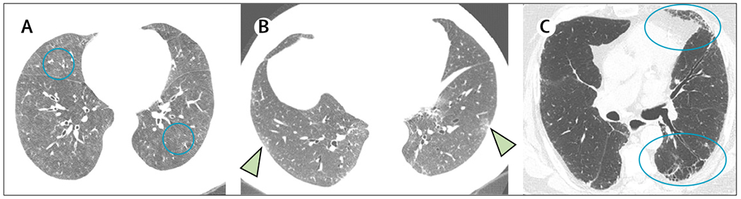
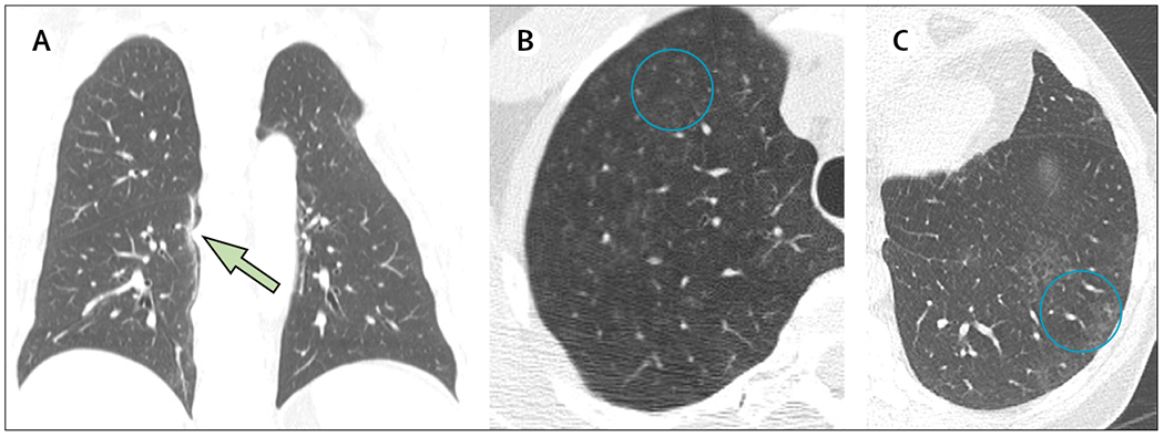
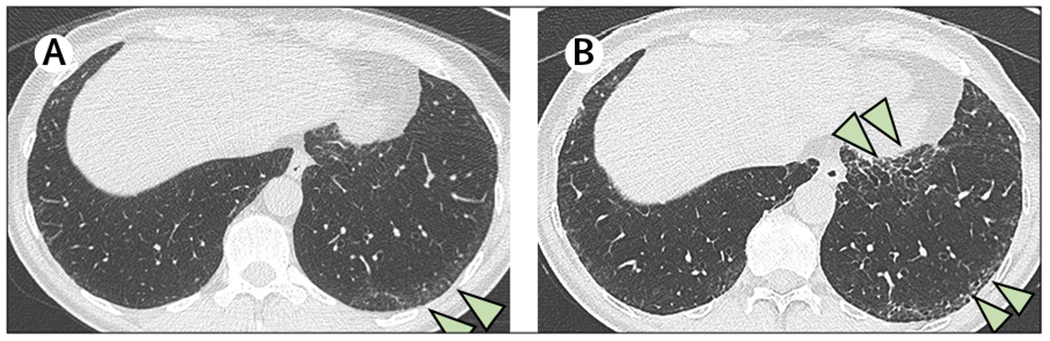
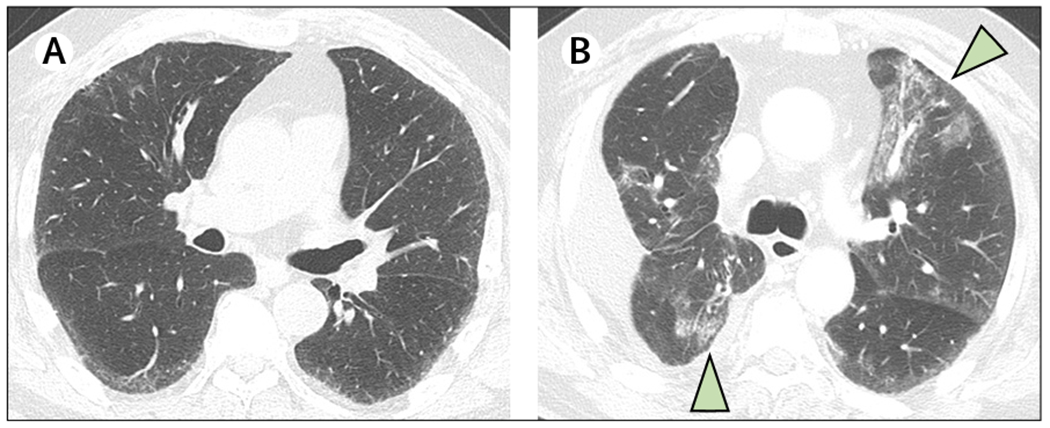
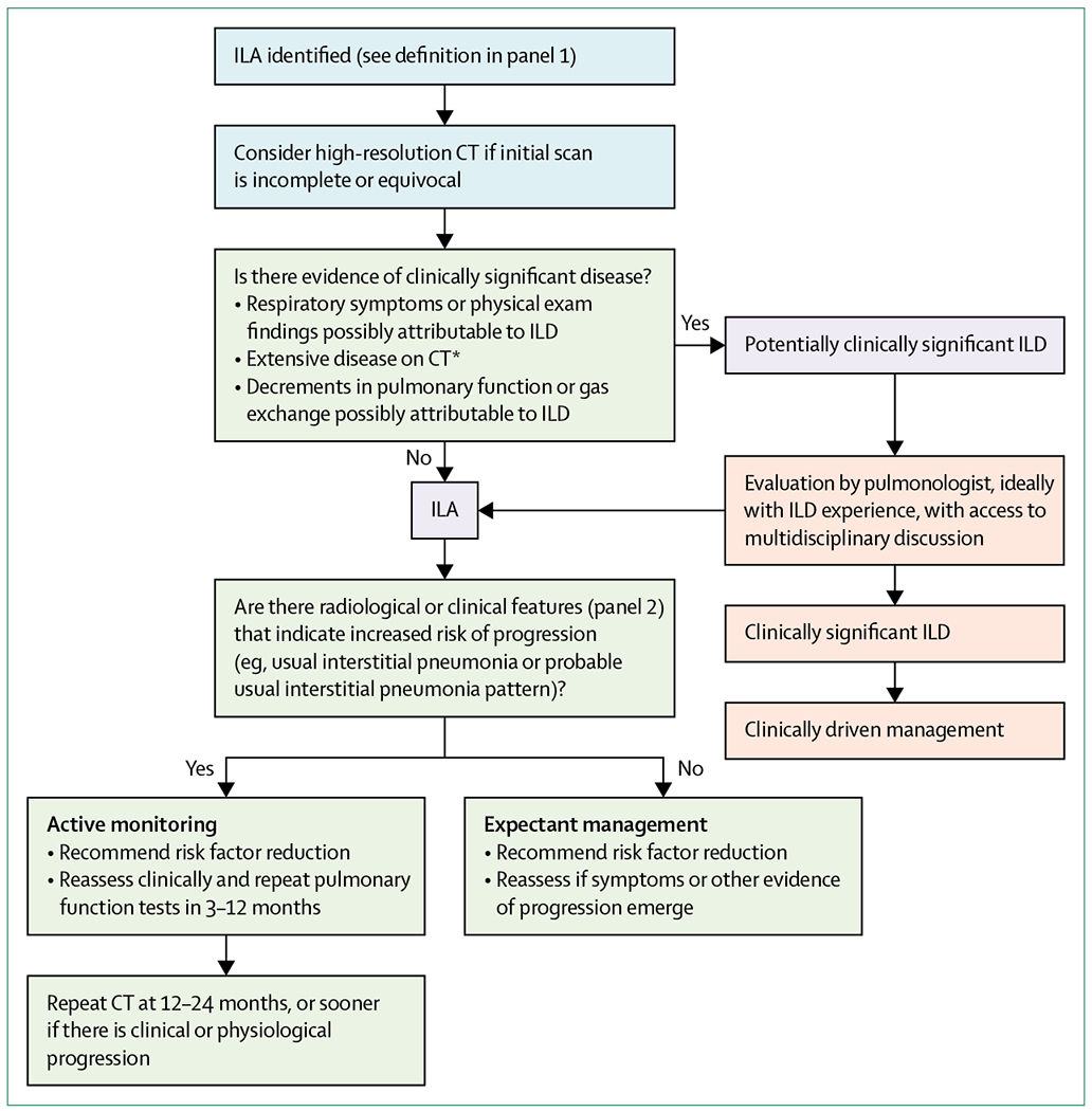
The term interstitial lung abnormalities refers to specific CT findings that are potentially compatible with interstitial lung disease in patients without clinical suspicion of the disease. Interstitial lung abnormalities are increasingly recognised as a common feature on CT of the lung in older individuals, occurring in 4-9% of smokers and 2-7% of non-smokers. Identification of interstitial lung abnormalities will increase with implementation of lung cancer screening, along with increased use of CT for other diagnostic purposes. These abnormalities are associated with radiological progression, increased mortality, and the risk of complications from medical interventions, such as chemotherapy and surgery. Management requires distinguishing interstitial lung abnormalities that represent clinically significant interstitial lung disease from those that are subclinical. In particular, it is important to identify the subpleural fibrotic subtype, which is more likely to progress and to be associated with mortality. This multidisciplinary Position Paper by the Fleischner Society addresses important issues regarding interstitial lung abnormalities, including standardisation of the definition and terminology; predisposing risk factors; clinical outcomes; options for initial evaluation, monitoring, and management; the role of quantitative evaluation; and future research needs.


Figures

Figure 1:
Figure 1:. Subcategories of interstitial lung abnormalities
(A) Non-subpleural and non-fibrotic. CT shows widespread ground-glass abnormality with central predominance (circled). (B) Subpleural non-fibrotic. CT shows predominantly subpleural ground-glass and linear abnormality without evidence of fibrosis (arrows). (C) Subpleural fibrotic. Traction bronchiectasis and architectural distortion are indicated by the ovals in the lingula and left lower lobe. This pattern would correspond to a probable usual interstitial pneumonia pattern.,
Figure 2:
Figure 2:. Imaging abnormalities that do not represent interstitial lung abnormalities
(A) Focal paraspinal fibrosis. Coronal CT shows a linear fibrotic band in the medial right lower lobe, closely related to osteophytes (arrow). (B) Centrilobular nodularity in a heavy smoker. CT shows numerous poorly defined ground-glass centrilobular nodules without other findings of interstitial abnormality (circled). (C) Unilateral mild focal abnormality. CT shows patchy ground-glass abnormality in the left lower lobe that is thought to be due to aspiration (circled).
Figure 3:
Figure 3:. Progression from subpleural non-fibrotic to subpleural fibrotic interstitial lung abnormality
High-resolution CT examination obtained in an asymptomatic, 61-year-old ex-smoker referred for suspicion of radiographic abnormality. (A) Prone high-resolution CT section at the lung bases shows subpleural lung abnormality, primarily ground-glass opacity (arrows). (B) Prone high-resolution CT section obtained 7 years later shows increased severity and extent of abnormality, with new traction bronchiectasis and honeycomb cysts in the anterior and posterior left lower lobe, indicating the interim development of lung fibrosis (arrows). This patient was still asymptomatic at the time of this follow-up CT examination.
Figure 4:
Figure 4:. Interstitial lung abnormalities after surgery and chemotherapy for lung cancer
High-resolution CT examination in a 79-year-old man with lung cancer. (A) A preoperative CT showed mild subpleural interstitial abnormality without evident fibrosis (subpleural non-fibrotic). The patient developed shortness of breath following a fourth cycle of pemetrexed. (B) Subsequent CT showed bilateral peribronchovascular ground-glass abnormality compatible with drug-related pneumonitis, as well as a postoperative effusion (arrows).
Figure 5:
Figure 5:. Proposed schema for management of interstitial lung abnormalities detected on chest CT
Action items for the radiologist are in blue, action items for the treating physician or pulmonologist are in green, and action items for a pulmonologist, ideally with ILD experience, are in orange. ILA=interstitital lung abnormality. ILD=interstitial lung disease. *Non-trivial abnormalities present in three or more lung zones (above the bottom of the aortic arch, between the aortic arch and top of the inferior pulmonary vein, and below the inferior pulmonary vein).
Figure 6:
Figure 6:. Computer-based classification of interstitial lung abnormalities with the histogram approach
(A) CT images with subpleural non-fibrotic interstitial lung abnormalities and emphysema in a participant in the COPDGene study. (B) CT image overlays of computer-based classification of interstitial lung abnormalities using artificial intelligence, showing objective quantification of different injury patterns. Regions of interstitial lung abnormality are shown in blue. Normal parenchyma (red), emphysema (green), and paraseptal emphysema (yellow) are also subtyped.
Figure 7:
Figure 7:. Quantification of progression for interstitial lung abnormalities with data-driven texture analysis
(A) A baseline CT scan shows subpleural non-fibrotic interstitial lung abnormalities with fibrotic changes. (B) CT 5 years later shows clear progression. (C) Baseline data-driven textural analysis shows overall extent of fibrosis as 1·5% (red). (D) Data-driven textural analysis of follow-up scan at 5 years shows that the extent of fibrosis increased to 4·6% (red).

References

    1. Schwarz MI, King TE Jr. Interstitial lung disease, 5th edn. Shelton, CT: People’s Medical Publishing House, 2011.
    1. Hatabu H, Hunninghake GM, Lynch DA. Interstitial lung abnormality: recognition and perspectives. Radiology 2019; 291: 1–3. - PubMed
    1. Washko GR, Lynch DA, Matsuoka S, et al. Identification of early interstitial lung disease in smokers from the COPDGene Study. Acad Radiol 2010; 17: 48–53. - PMC - PubMed
    1. Washko GR, Hunninghake GM, Fernandez IE, et al. Lung volumes and emphysema in smokers with interstitial lung abnormalities. N Engl J Med 2011; 364: 897–906. - PMC - PubMed
    1. Seibold MA, Wise AL, Speer MC, et al. A common MUC5B promoter polymorphism and pulmonary fibrosis. N Engl J Med 2011; 364: 1503–12. - PMC - PubMed

MeSH terms