. 2020 Jul 8;9(7):1640.
doi: 10.3390/cells9071640.

Inflammation Differentially Modulates the Biological Features of Adult Derived Human Liver Stem/Progenitor Cells

Affiliations

Inflammation Differentially Modulates the Biological Features of Adult Derived Human Liver Stem/Progenitor Cells

Hoda El-Kehdy et al. Cells. .

Abstract

The progression of mesenchymal stem cell-based therapy from concept to cure closely depends on the optimization of conditions that allow a better survival and favor the cells to achieve efficient liver regeneration. We have previously demonstrated that adult-derived human liver stem/progenitor cells (ADHLSC) display significant features that support their clinical development. The current work aims at studying the impact of a sustained pro-inflammatory environment on the principal biological features of ADHLSC in vitro.

Methods: ADHLSC from passages 4-7 were exposed to a cocktail of inflammatory cytokines for 24 h and 9 days and subsequently analyzed for their viability, expression, and secretion profiles by using flow cytometry, RT-qPCR, and antibody array assay. The impact of inflammation on the hepatocytic differentiation potential of ADHLSC was also evaluated.

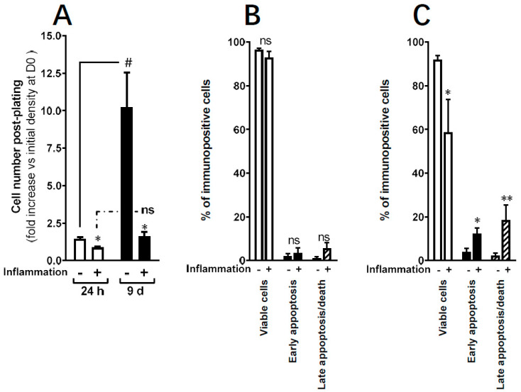
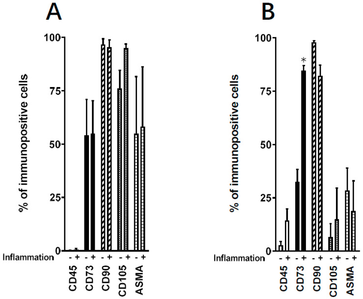
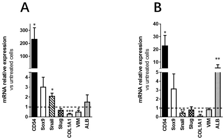
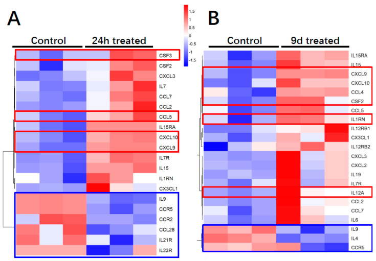
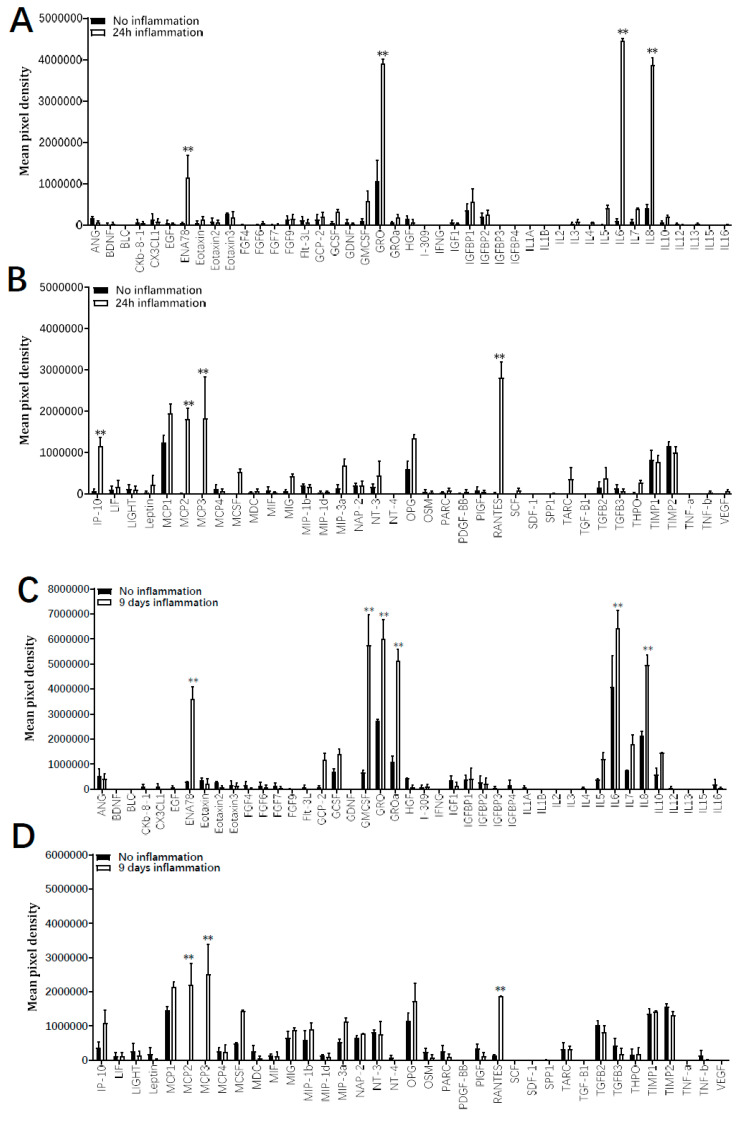
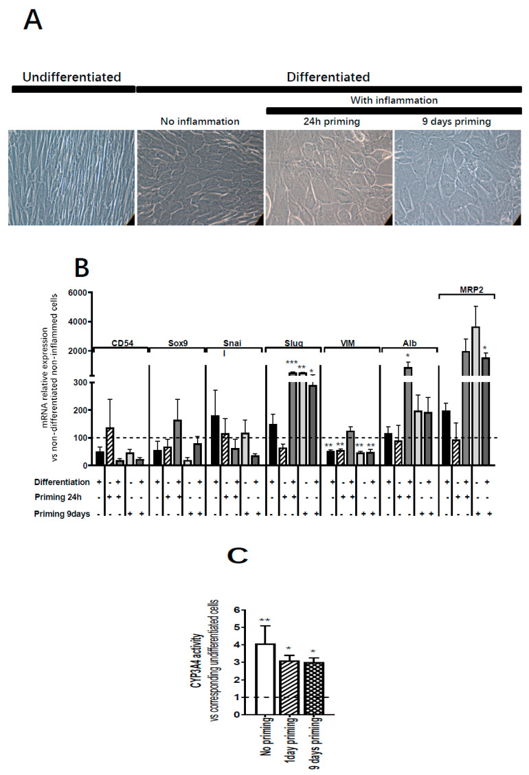
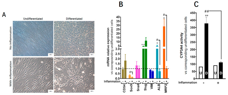
Results: ADHLSC treated with a pro-inflammatory cocktail displayed significant decrease of cell yield at both times of treatment while cell mortality was observed at 9 days post-priming. After 24 h, no significant changes in the immuno-phenotype of ADHLSC expression profile could be noticed while after 9 days, the expression profile of relevant markers has changed both in the basal conditions and after inflammation treatment. Inflammation cocktail enhanced the release of IL-6, IL-8, CCL5, monocyte-chemo-attractant protein-2 and 3, CXCL1/GRO, and CXCL5/ENA78. Furthermore, while IP-10 secretion was increased after 24 h priming, granulocyte macrophage colony-stimulating factor enhanced secretion was noticed after 9 days treatment. Finally, priming of ADHLSC did not affect their potential to differentiate into hepatocyte-like cells.

Conclusion: These results indicate that ADHLSCs are highly sensitive to inflammation and respond to such signals by adjusting their gene and protein expression. Accordingly, monitoring the inflammatory status of patients at the time of cell transplantation, will certainly help in enhancing ADHLSC safety and efficiency.

Keywords: immuno-biology; inflammation; liver; liver stem/progenitor cells.


Conflict of interest statement

E.M.S. and M.N. (Mustapha Najimi) are founders and scientific advisors for Promethera Biosciences and have founding shares and/or stock options. All other authors declare that they have no competing financial interests.

Figures

Figure 1
Figure 1
Effect of inflammation on ADHLSC culture. Morphology of ADHLSC observed microscopically after different times post-treatment with the inflammation cocktail (n = 6 samples from different donors). Magnification: 100× and 200×.
Figure 2
Figure 2
Effect of inflammation on ADHLSC viability in culture. (A) Significant decrease in adherent ADHLSC number after 24 h and 9 days treatment with the inflammation cocktail (n = 4 samples from different donors for each timepoint). Results are expressed as mean ± standard error of the mean (SEM). * p value < 0.05. # p < 0.05 control-9-day inflammation vs. control-24 h inflammation, one-way ANOVA followed by Dunnett post hoc test. (B) Following Annexin V–DAPI staining, no significant difference in cell death induction was noticed after 24 h treatment with the inflammation cocktail. (C) In contrast, maintaining the treatment for 9 days significantly decreases ADHLSC viability in correlation to an increase in cell apoptosis. Results are expressed as mean ± standard error of the mean (SEM) (n = 4). ** denotes a p value < 0.01; * p < 0.05 vs. corresponding control, paired Student’s t-test.
Figure 3
Figure 3
Effect of inflammation on ADHLSC mesenchymal expression profile. Positive expression of mesenchymal cell surface (CD105, CD90, and CD73) and intracellular markers (alpha-smooth muscle actin—ASMA) was evaluated using validated corresponding primary antibodies and flow cytometry. Negative expression of CD45 was also analyzed. (A) No changes in the mesenchymal expression profile were noticed after 24 h treatment. (B) After 9 days post-treatment with the inflammation cocktail, a significant increase was only observed for CD73 expression. Results are expressed as mean ± standard error of the mean (SEM) (n = 3 samples from different donors). * denotes a p value p < 0.05 vs. corresponding control, paired Student’s t-test.
Figure 4
Figure 4
Effect of inflammation on ADHLSC mRNA expression profile. (A) Differential modulation of ADHLSC gene expression profile by 24 h inflammation. (B) Differential modulation of ADHLSC gene expression profile by 9-days inflammation. RT-qPCR gene expression analysis demonstrated that inflammation differentially modulated the mRNA expression pattern of several ADHLSC genes. Differences in the upregulated and downregulated genes are observed between 24 h and 9 days of inflammatory treatment. For the ADHLSC treated group, results are expressed as mRNA relative expression versus untreated cells. CD54 (Intercellular Adhesion Molecule 1; ICAM-1), Sox9 (SRY-Related HMG-Box 9 encoding gene), Snail (SNAI1), Slug (SNAI2), COL1alpha1 (collagen type 1 alpha 1), VIM (Vimentin), and ALB (Albumin). Data shown are the mean ± SEM of three independent experiments (three samples from different donors). *** denotes a p value < 0.001; ** p < 0.01; * p < 0.05 vs. corresponding control, paired Student’s t-test.
Figure 5
Figure 5
Effect of inflammation on ADHLSC cytokine and cytokine receptors transcriptome profile. (A,B) Heat map presenting the differential cytokine upregulated and downregulated mRNA expression profile after 24 h and 9 days of inflammation priming (n = 3 samples from different donors).
Figure 6
Figure 6
Effect of inflammation on ADHLSC secretion profile. Secretion profile was evaluated on supernatants from ADHLSC untreated and treated with the inflammation cocktail using the antibody array assay. The data of each antibody array was normalized against the average of six positive control spots which are present on each antibody array and detect all proteins present in the sample. This means that the expression of each protein was first normalized against the total protein present in the individual sample before comparing expression levels between samples. (A,B) After 24 h, the inflammation cocktail significantly increased the secretion of ENA-78, GRO, IL-6, Il-8, IP-10, MCP 2 and 3, and RANTES by ADHLSC (n = 3 samples from different donors). (C,D) After 9 days of treatment, increased levels of ENA-78, GRO, IL-6, Il-8, MCP 2 and 3, and RANTES were maintained, whereas GRO-alpha and GM-CSF were additionally augmented (n = 3 samples from different donors). Results are expressed as mean ± standard error of the mean (SEM) of calculated pixel densities in stimulated ADHLSC versus untreated cells. ** denotes a p value < 0.01 vs. corresponding control, paired Student’s t-test.
Figure 7
Figure 7
Effect of inflammation on ADHLSC hepatocytic differentiation potential. (A) Primed ADHLSC acquired polygonal shape with granular cytoplasm after in vitro hepatocytic differentiation similar to the standard condition. Magnification: 200×. (B) RT-qPCR gene expression analysis demonstrated that inflammation did not alter the mRNA expression of mesenchymal markers except for slug and vimentin. Slug expression remains upregulated in differentiated 24 h-primed cells and both undifferentiated and differentiated 9-day primed cells while the expression of vimentin remains downregulated in non-differentiated 24 h and 9-day primed cells as well as differentiated 9-day primed cells. Furthermore, inflammation did not impact the upregulation of hepatocytic markers that normally occurs after hepatocytic differentiation (Albumin expression in differentiated 24 h primed cells and MRP2 expression in 9-day primed and differentiated cells) which is correlated with the morphological changes described above in Figure 7A. For treated and untreated differentiated cell groups, results are expressed as fold change in differentiated versus undifferentiated ADHLSC. *** denotes a p value < 0.001, ** p < 0.01, * p < 0.05 vs. non-primed and non-differentiated control, one-way ANOVA followed by Dunnett post-hoc test. CD54 (Intercellular Adhesion Molecule 1; ICAM-1), SOX9 (SRY-Related HMG-Box 9 encoding gene), Snail (SNAI1), Slug (SNAI2), VIM (Vimentin), ALB (Albumin), MRP2 (multi-drug resistance-associated protein-2 encoding gene). (C) Undifferentiated and differentiated ADHLSC from untreated and treated groups were incubated with IPA substrate and luciferase activity was measured. Results are expressed as the % of relative luminescence unit detected in the differentiated ADHLSC versus undifferentiated counterparts. Data shown are the mean ± SEM of three independent experiments (three different samples from different donors).
Figure 8
Figure 8
Effect of 30 days-inflammation on ADHLSC hepatogenic differentiation potential. (A) alteration of the typical morphological changes noted after in vitro hepatogenic differentiation of ADHLSC when the inflammation cocktail is added. Magnification: 200×. (B) RT-qPCR gene expression analysis demonstrated the inflammation does not alter the mRNA expression of mesenchymal markers expect for slug whose expression remains upregulated. However, inflammation significantly inhibits the upregulation of hepatocytic markers that normally occurs after hepatogenic differentiation which is in correlation with the morphological changes. For treated and untreated groups, results are expressed as fold change in differentiated versus corresponding undifferentiated ADHLSC. CD54 (Intercellular Adhesion Molecule 1; ICAM-1), Sox9 (SRY-Related HMG-Box 9 encoding gene), Snail (SNAI1), Slug (SNAI2), VIM (Vimentin), ALB (Albumin), MRP2 (multi-drug resistance-associated protein-2 encoding gene). *** denotes a p value < 0.001; * p < 0.05 vs. non-primed and non-differentiated control, # denotes a p < 0.05 vs primed and differentiated cells, One-way ANOVA followed by Dunnett post-hoc test. (C) Undifferentiated and differentiated ADHLSC from untreated and treated groups were incubated with IPA substrate and luciferase activity was measured. Results are expressed as the relative luminescence unit detected in the differentiated ADHLSC versus undifferentiated counterparts. Data shown are the mean ± SEM of at least four independent experiments. ** denotes a p value < 0.01 vs. non-primed and non-differentiated control, ## denotes a p < 0.01 vs primed and differentiated cells, One-way ANOVA followed by Dunnett post-hoc test.

References

    1. Caplan A.I., Sorrell J.M. The MSC curtain that stops the immune system. Immunol. Lett. 2015;168:136–139. doi: 10.1016/j.imlet.2015.06.005. - DOI - PubMed
    1. Squillaro T., Peluso G., Galderisi U. Clinical Trials with Mesenchymal Stem Cells: An Update. Cell Transpl. 2016;25:829–848. doi: 10.3727/096368915X689622. - DOI - PubMed
    1. Krampera M. Mesenchymal stromal cell ‘licensing’: A multistep process. Leukemia. 2011;25:1408–1414. doi: 10.1038/leu.2011.108. - DOI - PubMed
    1. Michael S., Achilleos C., Panayiotou T., Strati K. Inflammation Shapes Stem Cells and Stemness during Infection and Beyond. Front. Cell Dev. Biol. 2016;4:9584. doi: 10.3389/fcell.2016.00118. - DOI - PMC - PubMed
    1. Najar M., Crompot E., Raicevic G., Sokal E.M., Najimi M., Lagneaux L. Cytokinome of adult-derived human liver stem/progenitor cells: Immunological and inflammatory features. HepatoBiliary Surg. Nutr. 2018;7:331–344. doi: 10.21037/hbsn.2018.05.01. - DOI - PMC - PubMed

Publication types