. 2020 Jul 8;8(7):200.
doi: 10.3390/biomedicines8070200.

Secreted Factors from Keloid Keratinocytes Modulate Collagen Deposition by Fibroblasts from Normal and Fibrotic Tissue: A Pilot Study

Affiliations

Secreted Factors from Keloid Keratinocytes Modulate Collagen Deposition by Fibroblasts from Normal and Fibrotic Tissue: A Pilot Study

Mansour A Alghamdi et al. Biomedicines. .

Abstract

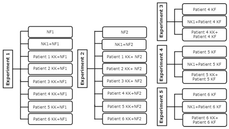
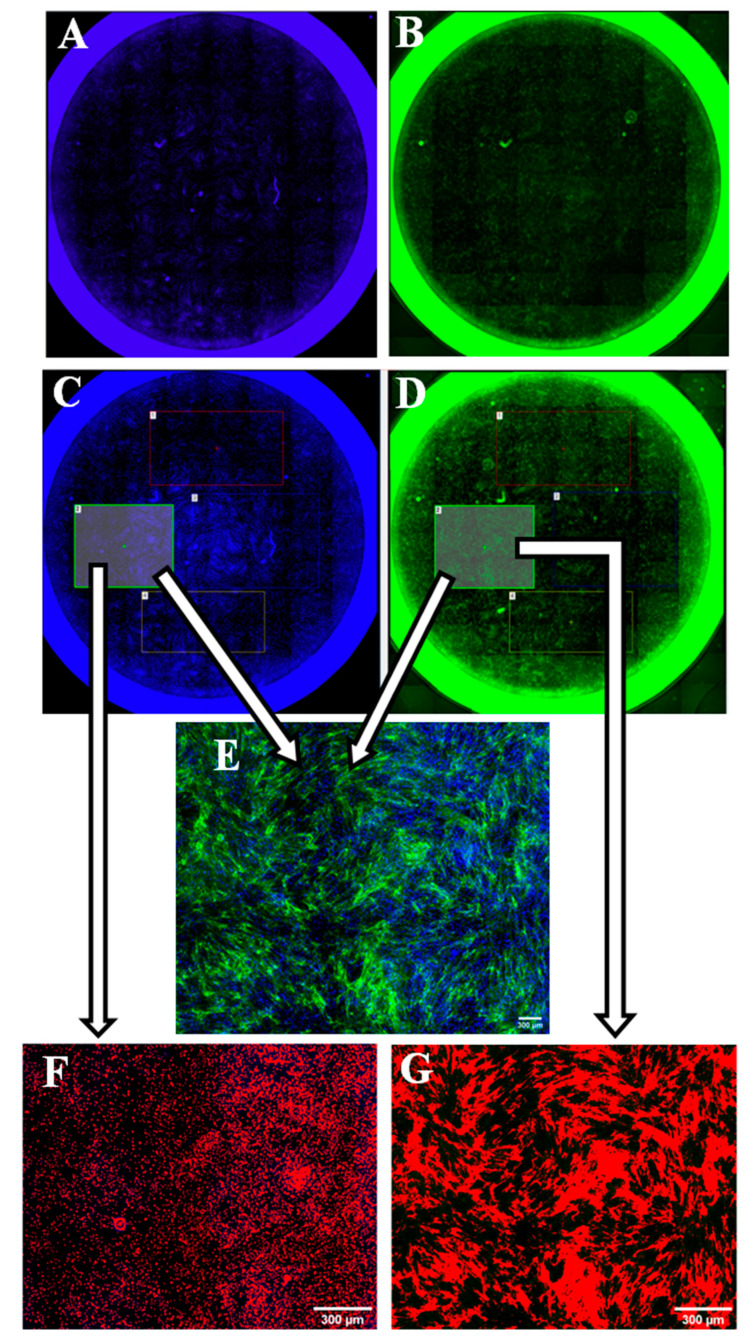
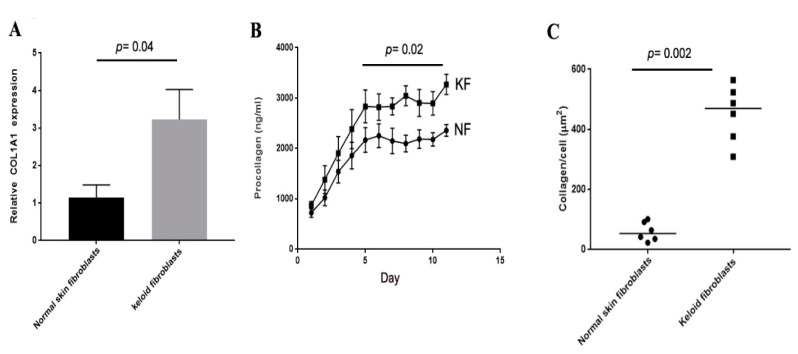
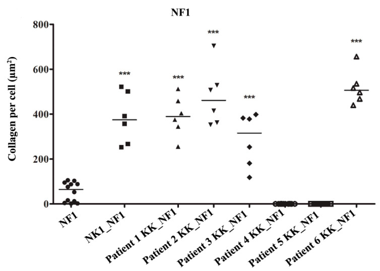
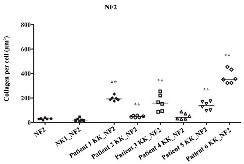
Interactions between keratinocytes and fibroblasts in the skin layers are crucial in normal tissue development, wound healing, and scarring. This study has investigated the role of keloid keratinocytes in regulating collagen production by primary fibroblasts in vitro. Keloid cells were obtained from removed patients' tissue whereas normal skin cells were discarded tissue obtained from elective surgery procedures. Fibroblasts and keratinocytes were isolated, cultured, and a transwell co-culture system were used to investigate the effect of keratinocytes on collagen production using a 'scar-in-a-jar' model. Keloid fibroblasts produced significantly more collagen than normal skin fibroblasts in monoculture at the RNA, secreted protein, and stable fibrillar protein level. When keloid keratinocytes were added to normal skin fibroblasts, expression of collagen was significantly upregulated in most samples, but when added to keloid fibroblasts, collagen I production was significantly reduced. Interestingly, keloid keratinocytes appear to decrease collagen production by keloid fibroblasts. This suggests that signaling in both keratinocytes and fibroblasts is disrupted in keloid pathology.

Keywords: coculture techniques; collagen type I; fibroblasts; keloids; keratinocytes; wound healing.


Conflict of interest statement

The authors declare no conflict of interest.

Figures

Figure 1
Figure 1
Flowchart of the experimental design. Normal fibroblast (NF), normal keratinocyte (NK), keloid fibroblast (KF), keloid keratinocyte (KK).
Figure 2
Figure 2
Whole well imaging and collagen quantification process. The left column shows the process of counting cell nuclei (whole well imaging (A), regions of interest (ROI) selection (C), and nuclei count (F)). The right column shows the process of measuring the collagen binary area (whole well imaging (B), ROI selection (D), and collagen threshold (G)). (E): collagens and nuclei merged.
Figure 3
Figure 3
Collagen production of keloid fibroblasts is significantly higher than normal skin fibroblasts in monoculture. qPCR for COIA1 shows higher levels in keloid fibroblasts compared to normal skin fibroblasts at 24 h after plating in culture (A). Fluorescent Resonance Energy Transfer (FRET) assay shows increased levels of procollagen peptide in cell media over a period of 11 d from keloid fibroblasts (KF) compared to normal skin fibroblasts (NF) (B). Immunohistochemistry for COLIA1 using the ‘scar-in-a-jar’ model shows significantly higher collagen I deposited by keloid fibroblasts compared to normal skin fibroblasts (C).
Figure 4
Figure 4
Keloid keratinocytes significantly increase collagen production by normal skin fibroblasts. Keloid keratinocytes and normal skin keratinocytes were co-cultured with normal fibroblasts from patient 1 (NF1). Both normal keratinocytes and 4/6 keloid keratinocyte samples significantly increased COLI produced by NF1. *** = p-value < 0.001.
Figure 5
Figure 5
Comparison of the COL1A production evaluated by immunohistochemistry staining of normal fibroblasts culture from patient 1 (NF1) (A), when co-cultured with normal keratinocytes (NK1) (B), or keloid keratinocytes (Patient 1 keloid keratinocytes (KK)) (C).
Figure 6
Figure 6
Keloid keratinocytes significantly increase collagen production by normal skin fibroblasts. Keloid keratinocytes and normal skin keratinocytes were co-cultured with normal fibroblasts from patient 2 (NF2). 5/6 keloid keratinocyte samples increased NF2 COLI production. No effect was seen with normal keratinocytes or with 1 keloid keratinocyte sample. ** = p-value < 0.01.
Figure 7
Figure 7
Comparison of the COL1A production evaluated by immunohistochemistry staining of normal fibroblasts culture from patient 2 (NF2) (A), when co-cultured with normal keratinocytes (NK1) (B), or keloid keratinocytes (Patient 1 keloid keratinocytes (KK)) (C).
Figure 8
Figure 8
Keloid keratinocytes significantly decrease collagen production by keloid fibroblasts. Keloid fibroblasts from patients 4, 5 and 6 were cultured with matched keloid keratinocytes or normal keratinocytes (NK1). Keloid keratinocytes significantly reduced COLI produced by the matched keloid fibroblasts (AC). * = p-value < 0.05, *** = p-value < 0.001.
Figure 9
Figure 9
Comparison of the COL1A production evaluated by immunohistochemistry staining of keloid fibroblasts culture from patient 4 (KF) (A), when co-cultured with normal keratinocytes (NK1) (B), or keloid keratinocytes (Patient 4 keloid keratinocytes (KK)) (C).

References

    1. Slemp A.E., Kirschner R.E. Keloids and scars: A review of keloids and scars, their pathogenesis, risk factors, and management. Curr. Opin. Pediatr. 2006;18:396–402. doi: 10.1097/01.mop.0000236389.41462.ef. - DOI - PubMed
    1. Gauglitz G.G., Korting H.C., Pavicic T., Ruzicka T., Jeschke M.G. Hypertrophic scarring and keloids: Pathomechanisms and current and emerging treatment strategies. Mol. Med. 2011;17:113–125. doi: 10.2119/molmed.2009.00153. - DOI - PMC - PubMed
    1. Podobed O.V., Prozorovskiĭ N.N., Kozlov E.A., Tsvetkova T.A., Vozdvidzhensky C.I., Delvig A.A. Comparative study of collagen in hypertrophic and keloid cicatrix. Vopr. Med. Khim. 1996;42:240–245. - PubMed
    1. Uitto J., Perejda A.J., Abergel R.P., Chu M.L., Ramirez F. Altered steady-state ratio of type I/III procollagen mRNAs correlates with selectively increased type I procollagen biosynthesis in cultured keloid fibroblasts. Proc. Natl. Acad. Sci. USA. 1985;82:5935–5939. doi: 10.1073/pnas.82.17.5935. - DOI - PMC - PubMed
    1. Syed F., Ahmadi E., Iqbal S.A., Singh S., McGrouther D.A., Bayat A. Fibroblasts from the growing margin of keloid scars produce higher levels of collagen I and III compared with intralesional and extralesional sites: Clinical implications for lesional site-directed therapy. Br. J. Dermatol. 2011;164:83–96. doi: 10.1111/j.1365-2133.2010.10048.x. - DOI - PubMed

LinkOut - more resources