Review
. 2020 Jul 8;8(7):1017.
doi: 10.3390/microorganisms8071017.

Enteroviruses and T1D: Is It the Virus, the Genes or Both which Cause T1D

Affiliations
Review

Enteroviruses and T1D: Is It the Virus, the Genes or Both which Cause T1D

Shirin Geravandi et al. Microorganisms. .

Abstract

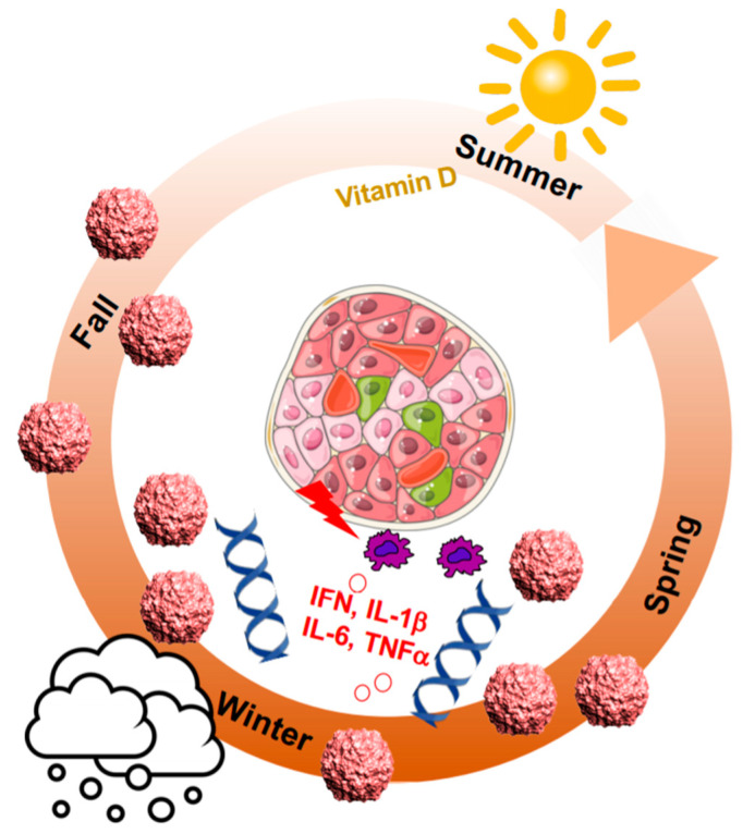
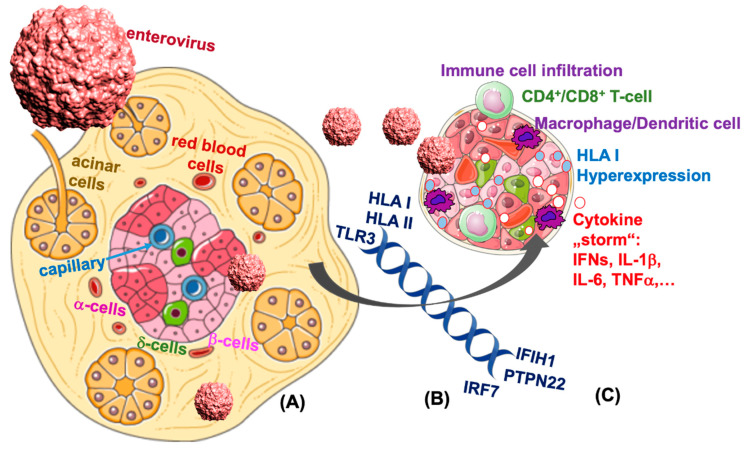
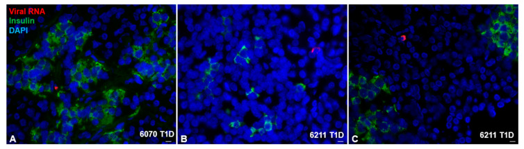
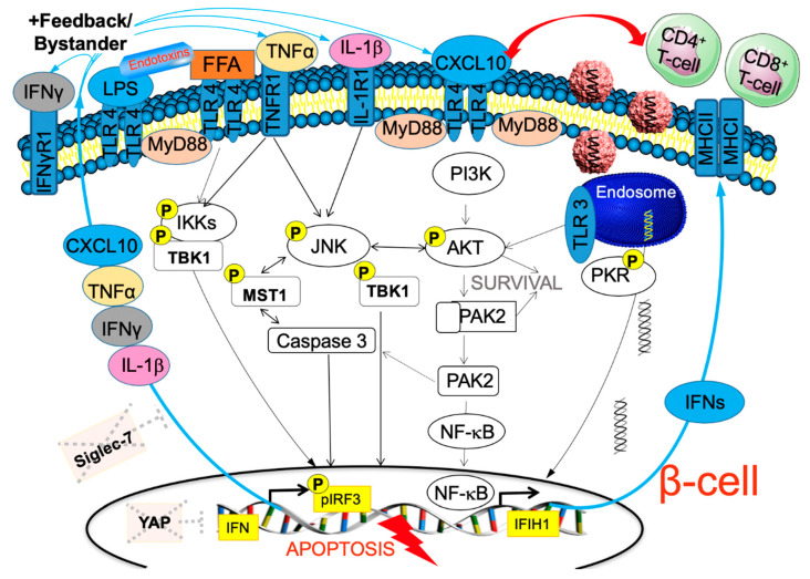
Type 1 diabetes (T1D) is a chronic autoimmune disorder that results from the selective destruction of insulin-producing β-cells in the pancreas. Up to now, the mechanisms triggering the initiation and progression of the disease are, in their complexity, not fully understood and imply the disruption of several tolerance networks. Viral infection is one of the environmental factors triggering diabetes, which is initially based on the observation that the disease's incidence follows a periodic pattern within the population. Moreover, the strong correlation of genetic susceptibility is a prerequisite for enteroviral infection associated islet autoimmunity. Epidemiological data and clinical findings indicate enteroviral infections, mainly of the coxsackie B virus family, as potential pathogenic mechanisms to trigger the autoimmune reaction towards β-cells, resulting in the boost of inflammation following β-cell destruction and the onset of T1D. This review discusses previously identified virus-associated genetics and pathways of β-cell destruction. Is it the virus itself which leads to β-cell destruction and T1D progression? Or is it genetic, so that the virus may activate auto-immunity and β-cell destruction only in genetically predisposed individuals?

Keywords: HLA; Hippo; IF1H1; IFIH1; TLR3; YAP; beta-cell; coxsackievirus; enterovirus; type 1 diabetes.


Conflict of interest statement

The authors declare no conflict of interest.

Figures

Figure 1
Figure 1
Not only environmental factors but also gene regulation show seasonal patterns. T1D (type 1 diabetes) diagnosis peaks in the colder months of late autumn to early spring, where viral infections come together with less sunlight exposure, less exercise outside, a change in diet together with an increase in pro-inflammatory cytokines and a change towards pro-inflammatory gene networks.
Figure 2
Figure 2
β-cell destruction in T1D is associated with viral response pathways. β-cells are highly vulnerable to enteroviral infection. (A) Several genetic mutations in the viral response pathway in T1D may lead to the potentiation in viral response. (B) A consequent “storm” of pro-inflammatory cytokines and chemokines lead to HLA I hyperexpression and attract cytotoxic T-cells and macrophages and subsequently to the loss of β-cells (C) and manifestation of T1D.
Figure 3
Figure 3
Coxsackieviral RNA in the T1D pancreas. Representative images of T1D donors 6070 and 6211 from the nPOD cohort. Viral RNA was found within the endocrine area (A) and outside the islets (B,C) shown by co-staining of viral RNA probes (red), insulin (green) and DAPI (nuclei; blue). Tissues were first probed for viral RNA, and then stained for insulin after a previously established protocol ([77]; Busse et al.). Scale bar depicts 10 µm.
Figure 4
Figure 4
β-cell in the storm. Our hypothetical model on how chronic potentiation of proinflammatory pathways leads to β-cell destruction. Coxsackieviruses enter the β-cell through the Coxsackie–adenovirus receptor (CAR) and bind to endosomal TLR3. While the virus promotes the AKT-JNK axis for initial host cell survival, parallel activation of viral response pathways through PKR-TBK-IRF3 leads to the transcriptional activation of the IFN response and production of interferons, which increase surface MHCs, recognized by cytotoxic CD8- and CD4-T-cells causing “bystander damage”, and β-cell apoptosis through a “storm” of cytokines and chemokines, which all find their receptors on the surface of the β-cell, and a vicious cycle is initiated with the full activation of the apoptotic machinery including JNK-MST1-Caspase 3-NFκB. Bacterial toxins as well as chronically elevated free fatty acids (FFA) are also associated with β-cell damage and act through TLR4 activation and similar downstream pro-inflammatory pathways. While many cells can counteract such damage cycles with a potent survival machinery, the β-cell is deficient of the Hippo terminator YAP, which would balance the viral IRF3 response. Furthermore, Siglec-7, which balances immune activation, is diminished in a chronic diabetogenic pro-inflammatory milieu in the β-cell.

References

    1. Jacobsen L.M., Newby B.N., Perry D.J., Posgai A.L., Haller M.J., Brusko T.M. Immune mechanisms and pathways targeted in type 1 diabetes. Curr. Diabetes Rep. 2018;18:90. doi: 10.1007/s11892-018-1066-5. - DOI - PMC - PubMed
    1. Wallberg M., Cooke A. Immune mechanisms in type 1 diabetes. Trends Immunol. 2013;34:583–591. doi: 10.1016/j.it.2013.08.005. - DOI - PubMed
    1. Pociot F., Lernmark A. Genetic risk factors for type 1 diabetes. Lancet. 2016;387:2331–2339. doi: 10.1016/S0140-6736(16)30582-7. - DOI - PubMed
    1. Herold K.C., Vignali D.A., Cooke A., Bluestone J.A. Type 1 diabetes: Translating mechanistic observations into effective clinical outcomes. Nat. Rev. Immunol. 2013;13:243–256. doi: 10.1038/nri3422. - DOI - PMC - PubMed
    1. Hyoty H., Taylor K.W. The role of viruses in human diabetes. Diabetologia. 2002;45:1353–1361. doi: 10.1007/s00125-002-0852-3. - DOI - PubMed