. 2020 Jul 10;11(1):276.
doi: 10.1186/s13287-020-01781-w.

Bone marrow mesenchymal stem cell-derived exosomes protect cartilage damage and relieve knee osteoarthritis pain in a rat model of osteoarthritis

Affiliations

Bone marrow mesenchymal stem cell-derived exosomes protect cartilage damage and relieve knee osteoarthritis pain in a rat model of osteoarthritis

Lei He et al. Stem Cell Res Ther. .

Abstract

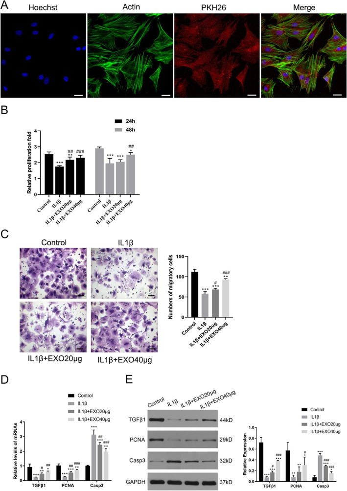
Background: This study aimed to investigate the effect of bone marrow mesenchymal stem cell (BMSC)-derived exosome injection on cartilage damage and pain relief in both in vitro and in vivo models of osteoarthritis (OA).

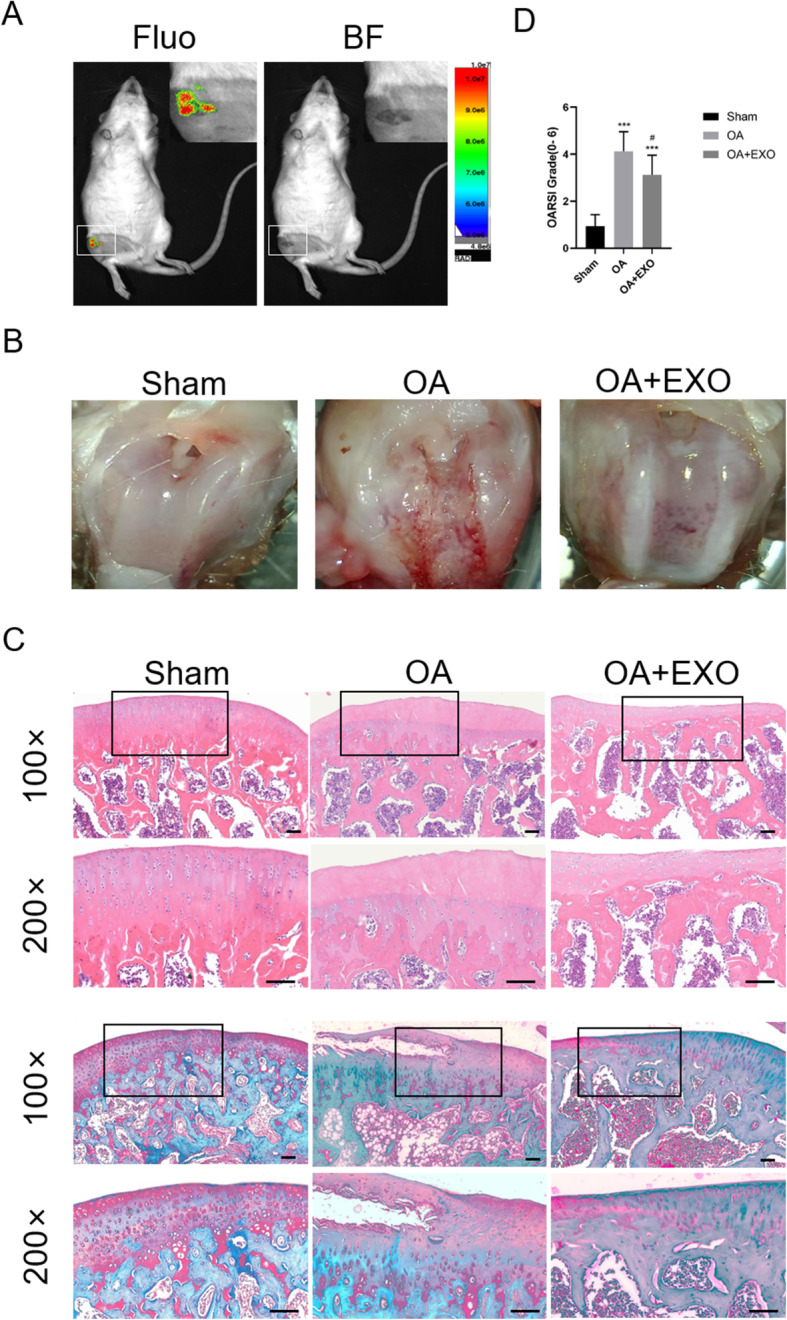
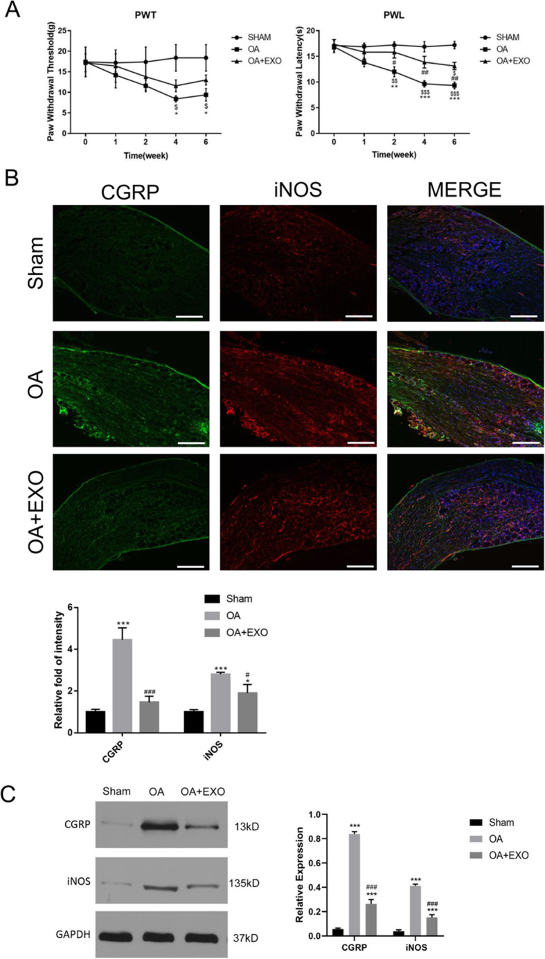
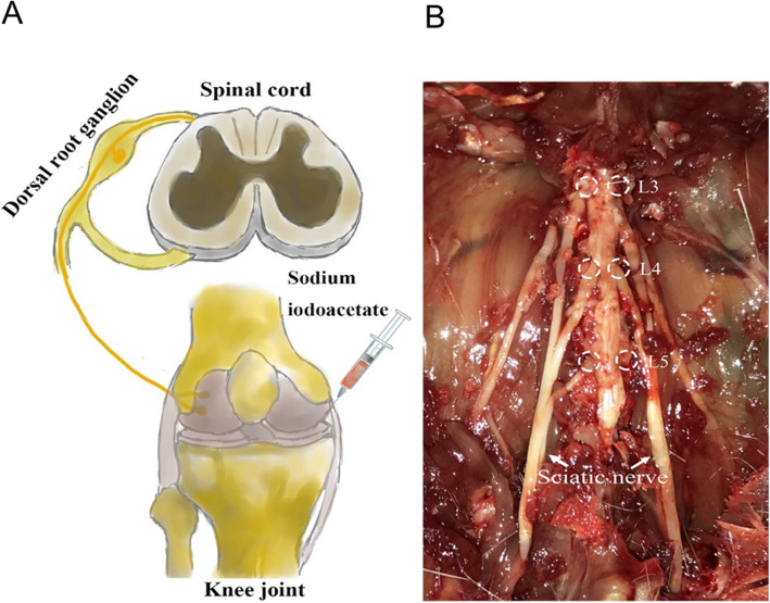
Methods: The BMSCs were extracted from rat bone marrow of the femur and tibia. Chondrocytes were treated with IL-1β to establish the in vitro model of OA. Chondrocyte proliferation and migration were assessed by CCK-8 and transwell assay, respectively. A rat model of OA was established by injection of sodium iodoacetate. At 6 weeks after the model was established, the knee joint specimens and dorsal root ganglion (DRG) of rats were collected for histologic analyses. For pain assessment, paw withdrawal threshold (PWT) and paw withdrawal latency (PWL) were evaluated before model establishment and at 1, 2, 4, and 6 weeks after model establishment.

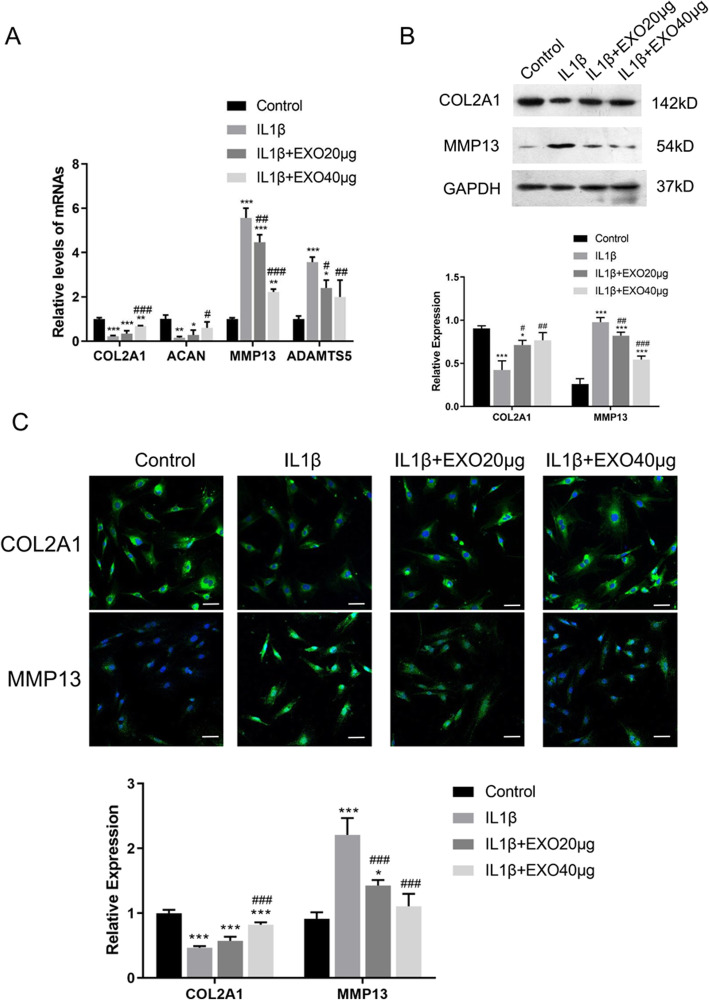
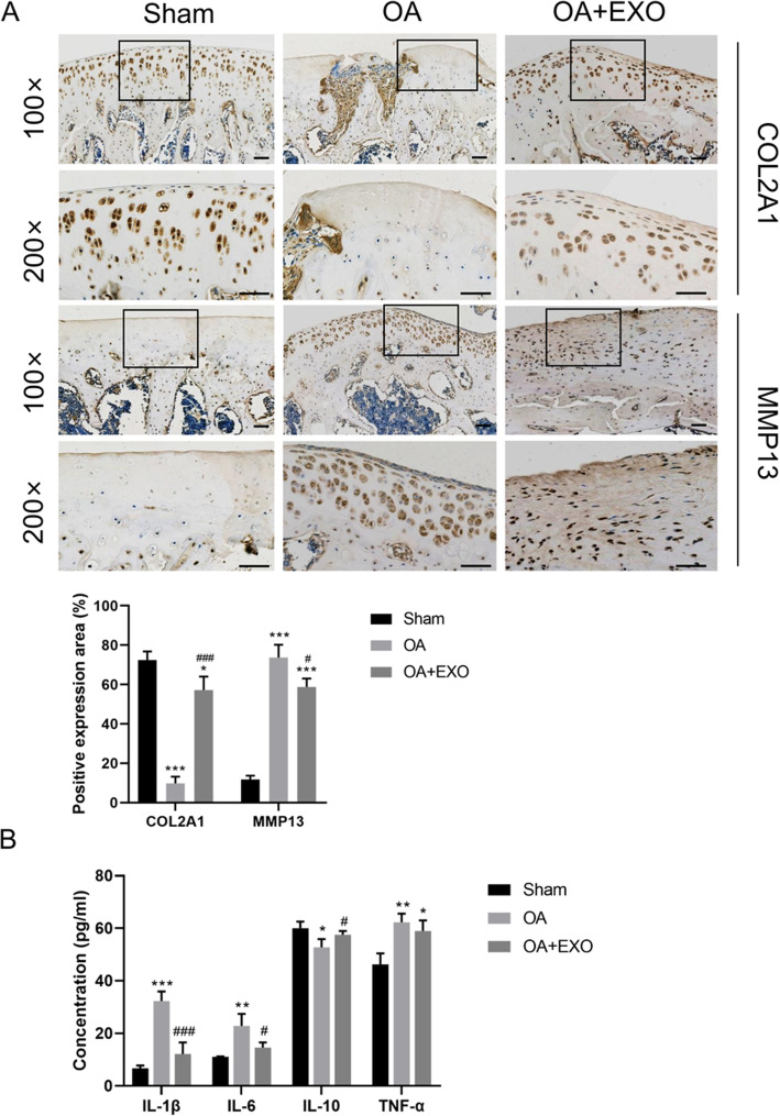
Results: Exosomes can be endocytosed with the chondrocytes in vitro. Exosome treatment significantly attenuated the inhibitory effect of IL-1β on the proliferation and migration of chondrocytes. Exosome pre-treatment significantly attenuated IL-1β-induced downregulation of COL2A1 and ACAN and upregulation of MMP13 and ADAMTS5. In the animal study, exosome treatment significantly upregulated COL2A1 protein and downregulated MMP13 protein in the cartilage tissue of the OA rat. At weeks 2, 4, and 6, the PWL value was significantly improved in the exosome-treated OA rats as compared with the untreated OA animals. Moreover, exosome treatment significantly alleviated the upregulation of CGRP and iNOS in the DRG tissue of OA rats.

Conclusion: BMSC-derived exosomes can effectively promote cartilage repair and extracellular matrix synthesis, as well as alleviate knee pain in the OA rats.

Keywords: BMSC-derived exosomes; Chondrocytes; Osteoarthritis; Pain relief.


Conflict of interest statement

The authors declare that there are no conflicts of interest.

Figures

Fig. 1
Fig. 1
Exosomes attenuated IL-1β-induced inhibitions on the proliferation and migration of chondrocytes. a Immunofluorescence staining of chondrocytes and BMSC-derived exosomes (PKH26) showed that exosomes gathered inside the chondrocytes. b The proliferation of chondrocytes was evaluated by CCK-8 assay. c Chondrocyte migration was determined by the transwell migration assay. Chondrocytes (1 × 106 cells, in 100 μl serum-free medium) were added to the upper chamber in a 24-well plate, in which the lower chamber contained 600 μl of complete medium. Five fields were randomly selected from each sample for quantification. All experiments were repeated independently at least three biological replicates. In the in vitro model of chondrocyte degeneration, PCR (d) and western blot (e) assays were performed to determine the mRNA and protein levels of growth factor (TGFβ1), proliferation marker (PCNA), and apoptosis marker (Casp3). Scale bar = 50 μm. *< 0.05, **< 0.01, ***< 0.001, compared with the control group. #< 0.05, ##< 0.01, ###< 0.001, compared with the IL-1β group
Fig. 2
Fig. 2
Exosomes attenuated IL-1β-induced downregulation of anabolic markers and upregulation of catabolic markers in cartilage degradation. a mRNA levels of MMP13, ADAMTS5, COL2A1, and ACAN were determined by RT-PCR. b Western blot analysis of protein levels of COL2A1 and MMP13. c Immunofluorescence staining of COL2A1 and MMP13. Rat chondrocytes were seeded at a density of 1 × 104 cells/well in the glass bottom culture dishes. Five fields were randomly selected from each sample for quantification. All experiments were repeated independently at least three biological replicates. Scale bar = 50 μm. *< 0.05, **< 0.01, ***< 0.001, compared with the control group. #< 0.05, ##< 0.01, ###< 0.001, compared with the IL-1β group
Fig. 3
Fig. 3
Exosomes alleviate cartilage damage in OA rats. a In vivo imaging of the rat after injection of the exosomes in the joint cavity. Fluo, fluorescence, BF, brightfield. b Gross morphological images of rat’s knee. c Images of H&E and Safranin O/Fast Green staining of knee joint specimens. Scale bar = 50 μm. d Osteoarthritis Research Society International (OARSI) score for the cartilage among different groups. ***< 0.001, compared with the sham group. #< 0.05, compared with the OA group. n = 8 for each group
Fig. 4
Fig. 4
a Immunohistochemical staining of COL2A1 and MMP13 proteins in the cartilage tissue. Scale bar = 50 μm. b Detection of serum inflammatory factors in OA rats by ELISA. *< 0.05, **< 0.001, ***< 0.001, compared with the sham group. #< 0.05, ###< 0.001, compared with the OA group. n = 8 for each group
Fig. 5
Fig. 5
Exosomes relieved pain in OA rats. a PWT and PWL were used to evaluate mechanical pain sensitivity and hyperalgesia. $< 0.05, $$< 0.01, $$$< 0.001, compared with the baseline level before model establishment (week 0). *< 0.05, **< 0.01, ***< 0.001, compared with the sham group. #< 0.05, ##< 0.01, compared with the OA group. n = 8 for each group. b Immunofluorescence staining of CGRP and iNOS protein in the dorsal root ganglion (DRG) tissue. Scale bar = 200 μm. *< 0.05, ***< 0.001, compared with the sham group. #< 0.05, ###< 0.001, compared with the OA group. n = 4 for each group. c Western blot analysis of protein levels of CGRP and iNOS in the DRG tissue. ***< 0.001, compared with the sham group. ###< 0.001, compared with the OA group. n = 4 for each group
Fig. 6
Fig. 6
Schematic diagram of knee joint pain caused by intraarticular injection of sodium iodoacetate to establish the rat model of knee joint OA (a). Neuronal damage and excitability increase in L3–5 DRG are important causes of pain and peripheral sensitization, which participate in the pathogenesis of OA pain (b)

References

    1. Chen D, Wang T, Shen J, Han L, Hamilton JL, Zhao W, et al. Osteoarthritis: toward a comprehensive understanding of pathological mechanism. Bone Res. 2017;5:16044. doi: 10.1038/boneres.2016.44. - DOI - PMC - PubMed
    1. Loeser RF, Goldring SR, Scanzello CR, Goldring MB. Osteoarthritis: a disease of the joint as an organ. Arthritis Rheum. 2012;64:1697–707. - PMC - PubMed
    1. Felson DT. Clinical practice. Osteoarthritis of the knee. N Engl J Med. 2006;354:841–848. doi: 10.1056/NEJMcp051726. - DOI - PubMed
    1. Knee osteoarthritis. Am Fam Physician. 2011;83:1294. - PubMed
    1. Loeser RF. Aging and osteoarthritis: the role of chondrocyte senescence and aging changes in the cartilage matrix. Osteoarthr Cartil. 2009;17:971–9. - PMC - PubMed

Publication types