Clinical Trial
. 2021 Mar;24(1):140-149.
doi: 10.1038/s41391-020-0254-y. Epub 2020 Jul 10.

Proof-of-principle Phase I results of combining nivolumab with brachytherapy and external beam radiation therapy for Grade Group 5 prostate cancer: safety, feasibility, and exploratory analysis

Affiliations
Clinical Trial

Proof-of-principle Phase I results of combining nivolumab with brachytherapy and external beam radiation therapy for Grade Group 5 prostate cancer: safety, feasibility, and exploratory analysis

Zhigang Yuan et al. Prostate Cancer Prostatic Dis. 2021 Mar.

Abstract

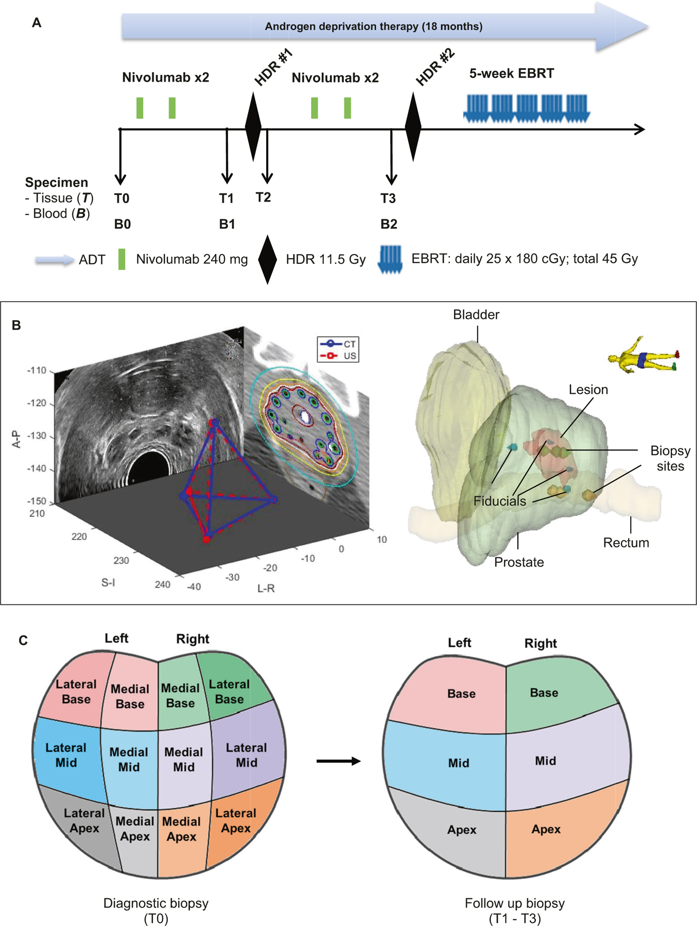
Background: To determine whether combining brachytherapy with immunotherapy is safe in prostate cancer (PCa) and provides synergistic effects, we performed a Phase I/II trial on the feasibility, safety, and benefit of concurrent delivery of anti-PD-1 (nivolumab) with high-dose-rate (HDR) brachytherapy and androgen deprivation therapy (ADT) in patients with Grade Group 5 (GG5) PCa.

Methods: Eligible patients were aged 18 years or older with diagnosis of GG5 PCa. Patients received ADT, nivolumab every two weeks for four cycles, with two cycles prior to first HDR, and two more cycles prior to second HDR, followed by external beam radiotherapy. The primary endpoint was to determine safety and feasibility. This Phase I/II trial is registered with ClinicalTrials.gov (NCT03543189).

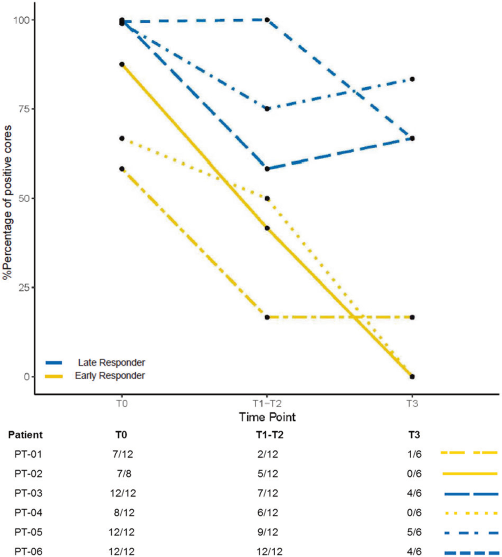
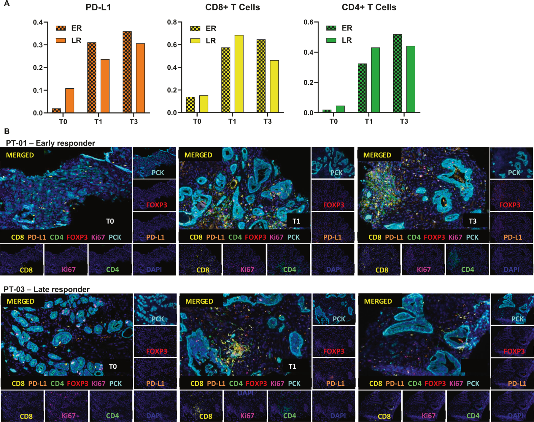
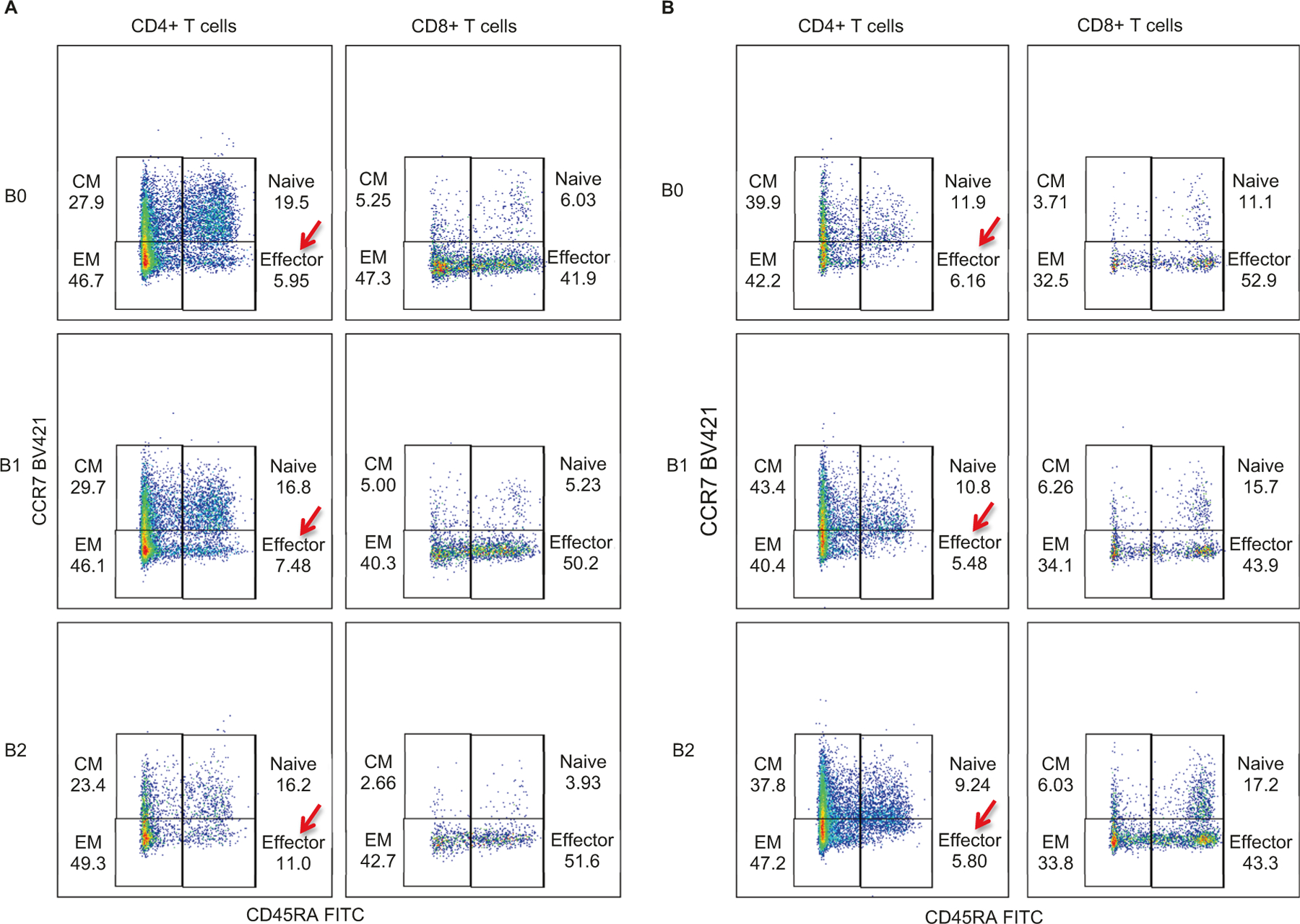
Results: Between September 2018 and June 2019, six patients were enrolled for the Phase I safety lead-in with a minimum observation period of 3 months after nivolumab administration. Overall, nivolumab was well tolerated in combination with ADT and HDR treatment. One patient experienced a grade 3 dose-limiting toxicity (elevated Alanine aminotransferase and Aspartate aminotransferase) after the second cycle of nivolumab. Three patients (50%) demonstrated early response with no residual tumor detected in ≥4 of 6 cores on biopsy post-nivolumab (4 cycles) and 1-month post-HDR. Increase in CD8+ and FOXP3+/CD4+ T cells in tissues, and CD4+ effector T cells in peripheral blood were observed in early responders.

Conclusion: Combination of nivolumab with ADT and HDR is well tolerated and associated with evidence of increased immune infiltration and antitumor activity.


Conflict of interest statement

Conflict of interest J.Z. reports honorarium from AstraZeneca, Merck, Sanofi and Bayer for speaker programs and advisory boards. R.J. serves as an advisor for Pfizer and a speaker for Dava Oncology. R.L. is on Clinical trial protocol committee for Cold Genesys and BMS, and serves as a scientific advisor/consultant for BMS and Ferring. S.K. reports research funds from BMS and Astra Zeneca. The remaining authors declare that they have no conflict of interest.

Figures

Fig. 1
Fig. 1. Study design and systematically coordinated tissue biopsy system.
a Study schema. ADT: Androgen deprivation therapy. EBRT: External beam radiation therapy. HDR: High dose brachytherapy. Nivo: nivolumab. B0/T0: Baseline blood and diagnostic biopsy. B1/T1: Specimen collected after two cycles of nivolumab, and prior to HDR#1 brachytherapy. T2: Specimen collection within 2 h post HDR#1. B2/T3: Specimen collection after 4 cycles of nivolumab and four weeks after HDR#1, but prior to HDR#2. b Tissue collection schema. A carefully designed coordinates system was used to determine the location of the 6-core biopsy sites (left apex, left mid, left base, right apex, right mid, and right base). Four fiducial markers were placed into the apex, base, left mid and right mid gland. Biopsies were performed under ultrasound guidance by combining the prostate template grid, the ultrasound transducer stepper, and the location of the fiducial marker. Mirada’s multimodality registration was used to rigidly register the MRI image to the planning CT using local registration. The biopsy sites were documented for follow-up biopsy reference. c Prostate biopsy locations. Left panel: T0 is diagnostic biopsy prior to treatment; Right panel: T1 is post-nivolumab (2 Cycles) and Pre high dose radiation (HDR-Brachytherapy) biopsy; T2 is within 2 h post-HDR brachytherapy, T3 is post-nivolumab (4 cycles) and 1 month post-HDR brachytherapy.
Fig. 2
Fig. 2. Timing of pathologic response to treatment.
Pathological responses were assessed through analyzing serial 6-core biopsies, which were obtained at three different time points: pre-HDR#1 (T1), post-HDR#1 (T2), and pre-HDR#2 (T3). Percentage of positive cores were averaged for the samples from the time points T1 and T2 since the interval of biopsy between T1 and T2 was approximately 2 h. Number of positive cores over number of core biopsies for each patient at different time point was listed.
Fig. 3
Fig. 3. Detection of human CD8 (yellow), CD4 (green), PCK (cian), FOXP3 (red), Ki67 (magenta) and PD-L1 (orange) in FFPE prostate cancer biopsies by IHC-IF.
a Quantification of PD-L1 (left), CD8+ (middle) and CD4+ (right) in multiplex images of early responders (ER) and late responders (LR) patients; Three patients per group. b Representative Multiplex IHC images of patients 1 and 3 (PT-01 and PT-03) at T0 (Diagnostic biopsy), T1 (Specimen collected after two cycles of nivolumab, and prior to HDR#1) and T3 (Specimen collected after 4 cycles of nivolumab and four weeks after HDR#1, but prior to HDR#2).
Fig. 4
Fig. 4. Quantification of CD4+ and CD8+ T cells in patients peripheral blood samples.
a Quantification of CD4+ and CD8+ T cells in blood samples of early responder patient PT-01 at different stages: B0 (Baseline blood), B1 (Blood collected after two cycles of nivolumab, and prior to HDR#1) and B2 (Specimen collected after 4 cycles of nivolumab and four weeks after HDR#1, but prior to HDR#2). b Quantification of CD4+ and CD8+ T cells in blood samples of late responder patient PT-03.

References

    1. Wolchok JD, Chiarion-Sileni V, Gonzalez R, Rutkowski P, Grob JJ, Cowey CL, et al. Overall survival with combined nivolumab and ipilimumab in advanced melanoma. N. Engl J Med 2017;377:1345–56. - PMC - PubMed
    1. Garon EB, Rizvi NA, Hui R, Leighl N, Balmanoukian AS, Eder JP, et al. Pembrolizumab for the treatment of non-small-cell lung cancer. N. Engl J Med 2015;372:2018–28. - PubMed
    1. Motzer RJ, Tannir NM, McDermott DF, Aren Frontera O, Melichar B, Choueiri TK, et al. Nivolumab plus ipilimumab versus sunitinib in advanced renal-cell carcinoma. N. Engl J Med 2018;378:1277–90. - PMC - PubMed
    1. Bellmunt J, de Wit R, Vaughn DJ, Fradet Y, Lee JL, Fong L, et al. Pembrolizumab as second-line therapy for advanced urothelial carcinoma. N. Engl J Med 2017;376:1015–26. - PMC - PubMed
    1. Topalian SL, Hodi FS, Brahmer JR, Gettinger SN, Smith DC, McDermott DF, et al. Safety, activity, and immune correlates of anti-PD-1 antibody in cancer. N. Engl J Med 2012;366:2443–54. - PMC - PubMed

Publication types

Substances

Associated data