Review
. 2020:2173:21-51.
doi: 10.1007/978-1-0716-0755-8_2.

Optogenetic Techniques for Manipulating and Sensing G Protein-Coupled Receptor Signaling

Affiliations
Review

Optogenetic Techniques for Manipulating and Sensing G Protein-Coupled Receptor Signaling

Nohely Abreu et al. Methods Mol Biol. 2020.

Abstract

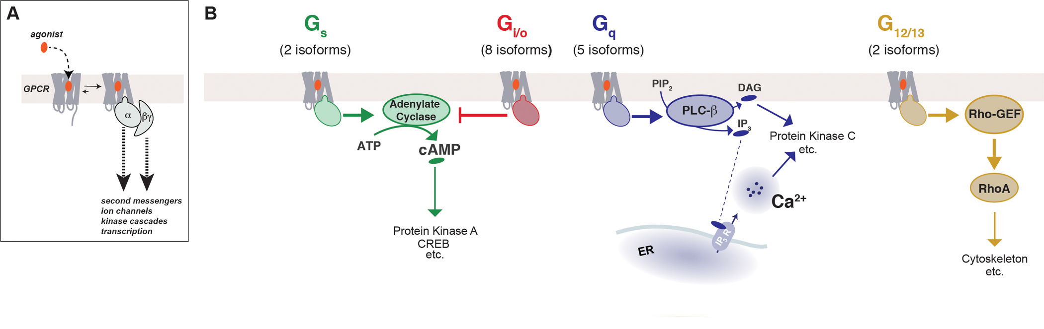
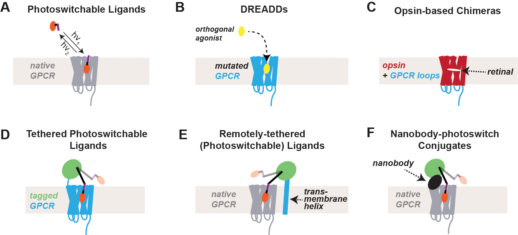
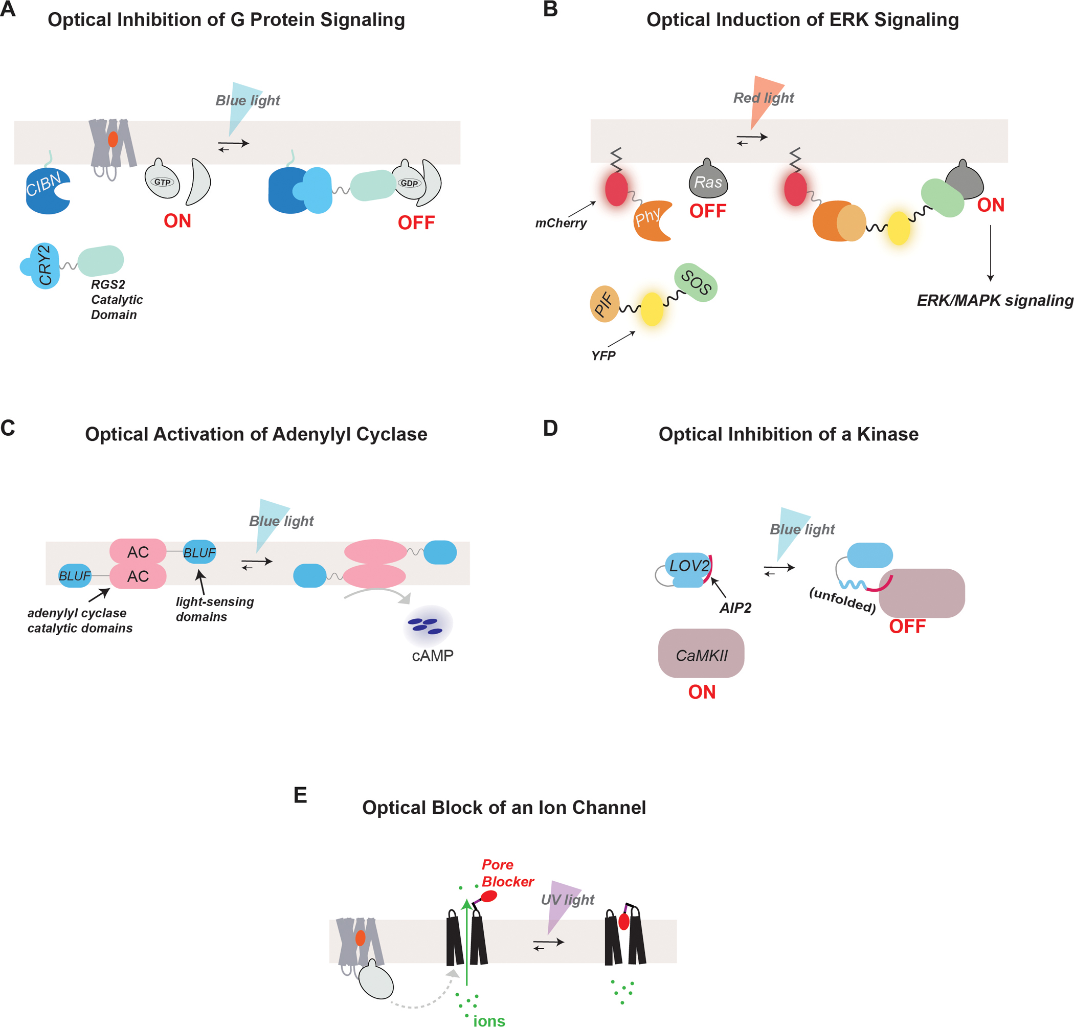
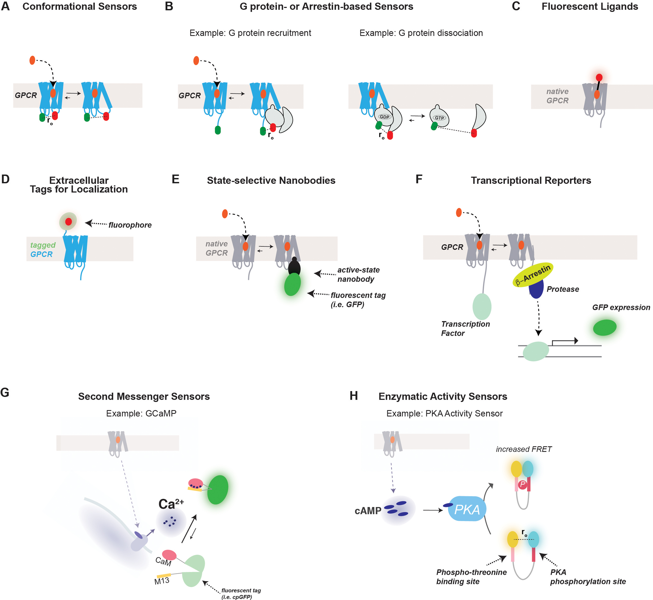
G protein-coupled receptors (GPCRs) form the largest class of membrane receptors in the mammalian genome with nearly 800 human genes encoding for unique subtypes. Accordingly, GPCR signaling is implicated in nearly all physiological processes. However, GPCRs have been difficult to study due in part to the complexity of their function which can lead to a plethora of converging or diverging downstream effects over different time and length scales. Classic techniques such as pharmacological control, genetic knockout and biochemical assays often lack the precision required to probe the functions of specific GPCR subtypes. Here we describe the rapidly growing set of optogenetic tools, ranging from methods for optical control of the receptor itself to optical sensing and manipulation of downstream effectors. These tools permit the quantitative measurements of GPCRs and their downstream signaling with high specificity and spatiotemporal precision.

Keywords: Calcium; G protein; G protein-coupled receptors (GPCR); LOV domain; Optogenetics; Photopharmacology.


Figures

Figure 1.
Figure 1.
Basic mechanisms of GPCR signaling (A) GPCRs bind ligands which initiate a conformational change in the receptor that allows the receptor to couple to and signal through heterotrimeric G proteins, which activates various second messengers and effectors. (B) Canonical signaling pathways mediated by the four families of Gα protein subtypes.
Figure 2.
Figure 2.
Chemical and optical methods for the targeted control of GPCRs (A) Photoswitchable ligands are typically composed of a well-characterized ligand (orange) and a light-sensitive moiety (purple) that either isomerizes or releases the ligand in the presence of light. This system allows the control of native GPCRs by controlling ligand availability with high time resolution. The temporal resolution of reversal is limited by ligand unbinding and spatial targeting is limited by diffusion. (B) DREADDs are mutated GPCRs that are engineered to respond to a synthetic agonist but to no longer respond to a native ligand. By targeting DREADD expression, one can achieve genetically-targeted chemical (i.e. “chemogenetic”) control of GPCR signaling. (C) Opsins are naturally-occurring light-activatable GPCRs that have been taken advantage of for the optical control of GPCR activation. To study and manipulate signaling pathways associated with specific GPCRs, chimeras may be designed that incorporate the intracellular loops and/or the C-terminal tail of the GPCR of interest. (D) Photoactivation of specific GPCRs can be achieved by tethering a photoswitchable ligand to a self-labeling tag, such as SNAP (green). This system requires heterologous expression of a tagged GPCR which allows the incorporation of receptor mutants or variants but can lead to overexpression. Tethered photoswitchable ligands allow the highest temporal resolution of both ligand binding and un-binding. (E) Photoactivation of native GPCRs with genetic targeting can be accomplished by the expression of a tagged transmembrane domain that is tethered to a photoswitchable ligand. (F) Native GPCRs may also be optically-controlled via photoswitchable ligands tethered via self-labeling tags (green) to nanobodies that recognizes an extracellular site on the receptor. Nanobody-tethered photoswitchable ligands can either be delivered directly or can be genetically-encoded for heterologous expression and cell-type targeting.
Figure 3.
Figure 3.
Optical methods for the control of GPCR effectors (A) The CRY-CIB system can be used for blue light-induced association of two proteins. For example, optical inhibition of G protein-mediated signaling has been achieved by utilizing the CRY-CIB system to induce the translocation of RGS2 to the plasma membrane with blue light. This recruitment brings RGS2 in close proximity to membrane-bound Gα-GTP which allows it to effectively inhibit G protein signaling. (B) The Phy-PIF system can be used for red light-induced association of two proteins. For example, photocontrol of ERK signaling can be achieved through the induction of the surface translocation of SOS using the Phy-PIF system. Phy was fused to a CAAX motif for membrane incorporation, while SOS was fused to PIF. Red light induces dimerization of Phy and PIF, translocating SOS to the membrane to promote interaction with and activation of Ras GTPase and initiate ERK signaling. Fluorescent proteins were incorporated into the constructs to observe their expression and dynamic localization. (C) Optical control of cAMP production can be achieved by heterologously expressing bPAC, a photoactivatable adenylyl cyclase from Beggiatoa, which has increased catalytic activity in the presence of blue light. (D) The LOV domain has been utilized to gain photocontrol of the activity of various signaling proteins. Following blue light illumination, the Jalpha helix of the LOV domain unfolds allowing it to release functional peptides or proteins for light-gated activity. In the illustrated case, LOV2 was fused to a CaMKII inhibitory peptide, AIP2, which becomes released in the presence of blue light. (E) Photoswitchable tethered ligands can be employed for the optical control of ion channels. For example, a photoswitchable pore blocker may be attached to an ion channel using cysteine chemistry. The pore blocker is designed such that it fits into the opening of the channel only in the presence of light to effectively block ion conductance.
Figure 4.
Figure 4.
Optical sensing of GPCRs and their downstream effectors. (A) FRET-based sensors may be used to detect the conformational changes associated with GPCR activation following ligand binding. Typically, donor and acceptor fluorophores (i.e. fluorescent proteins) are introduced into the intracellular loops of the receptor which move relative to each other during activation. Often these fluorophores disrupt coupling to G proteins making them more typically used for understanding receptor pharmacology or structure/function relationships rather than signaling properties. (B) FRET-based sensors have also been developed to sense G protein recruitment to the receptor (left) or dissociation of G protein heterotrimers (right). For G protein recruitment, a fluorophore is typically fused to the C-terminal tail of the receptor while the Gα subunit is tagged with a fluorophore in an internal site to avoid disrupting function and receptor specificity. To sense G protein dissociation, the Gα subunit is tagged as described above, while another fluorophore is typically placed on the N terminus of either Gβ or Gγ. Similar sensors have also been developed to sense the recruitment or conformational changes of β-arrestins. (C) Ligands conjugated to fluorophores have been developed to aid in their visualization both in vitro and in vivo. (D) GPCRs can be tagged with a fluorophore, either a fluorescent protein or dye conjugation to a tag (i.e. SNAP, CLIP, or Halo) to permit the observation of their surface and subcellular localizations in living cells. (E) Fluorophore-tagged nanobodies have been developed which can be genetically encoded and used to detect the conformational state of a receptor, G protein or β-arrestin. Accumulation of fluorescence in a given location (i.e. plasma membrane or endosomes) indicates a population of receptor or effectors in a state recognized by the NB. (F) GPCR activation can also be sensed using a transcriptional reporter, such as the Tango assay. In this assay a transcription factor and a protease site are incorporated into the C-terminal tail of the receptor, while β-arrestin is tagged with a protease. Receptor activation leads to β-arrestin-protease recruitment, leading to the cleavage and release of the transcription factor which will permit expression of a reporter gene (i.e. GFP). (G) A classic sensor for the detection of the second messenger, Ca2+, is GCaMP. Ca2+ binding to CaM permits its association with the M13 peptide which induce the closure and increased fluorescence of cpGFP. Many permutations of GCaMP and other related calcium sensors exist with variable kinetics, sensitivity, subcellular targeting and spectral properties to allow the ideal construct to be used for the relevant application. (H) FRET-based enzymatic activity sensors are widely used to detect kinase activity. In the case of the PKA sensor, AKAR, the sensor contains N- and C-terminal donor and acceptor fluorophores, a PKA phosphorylation site, and a domain that binds to the phosphorylated residue. Following phosphorylation by active PKA, association of the two sites leads to increased FRET between the fluorophores.

Similar articles

Cited by

References

    1. Agnetta L, Kauk M, Canizal MCA, Messerer R, Holzgrabe U, Hoffmann C, and Decker M (2017). A Photoswitchable Dualsteric Ligand Controlling Receptor Efficacy. Angew Chem Int Ed Engl 56, 7282–7287. - PubMed
    1. Airan RD, Thompson KR, Fenno LE, Bernstein H, and Deisseroth K (2009). Temporally precise in vivo control of intracellular signalling. Nature 458, 1025–1029. - PubMed
    1. Albizu L, Cottet M, Kralikova M, Stoev S, Seyer R, Brabet I, Roux T, Bazin H, Bourrier E, Lamarque L, et al. (2010). Time-resolved FRET between GPCR ligands reveals oligomers in native tissues. Nat Chem Biol 6, 587–594. - PMC - PubMed
    1. Allen MD, and Zhang J (2006). Subcellular dynamics of protein kinase A activity visualized by FRET-based reporters. Biochem Biophys Res Commun 348, 716–721. - PubMed
    1. Alon S, Huynh GH, and Boyden ES (2019). Expansion microscopy: enabling single cell analysis in intact biological systems. FEBS J 286, 1482–1494. - PMC - PubMed

Publication types

MeSH terms

LinkOut - more resources