. 2020 Jul 1;143(7):2009-2026.
doi: 10.1093/brain/awaa163.

Molecular and cellular correlates of human nerve regeneration: ADCYAP1/PACAP enhance nerve outgrowth

Affiliations

Molecular and cellular correlates of human nerve regeneration: ADCYAP1/PACAP enhance nerve outgrowth

Georgios Baskozos et al. Brain. .

Erratum in

Abstract

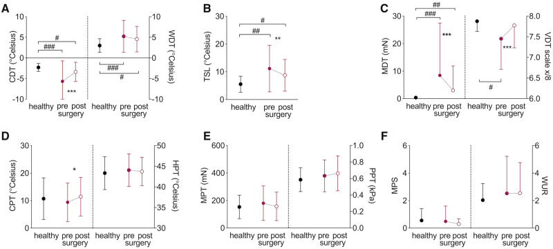
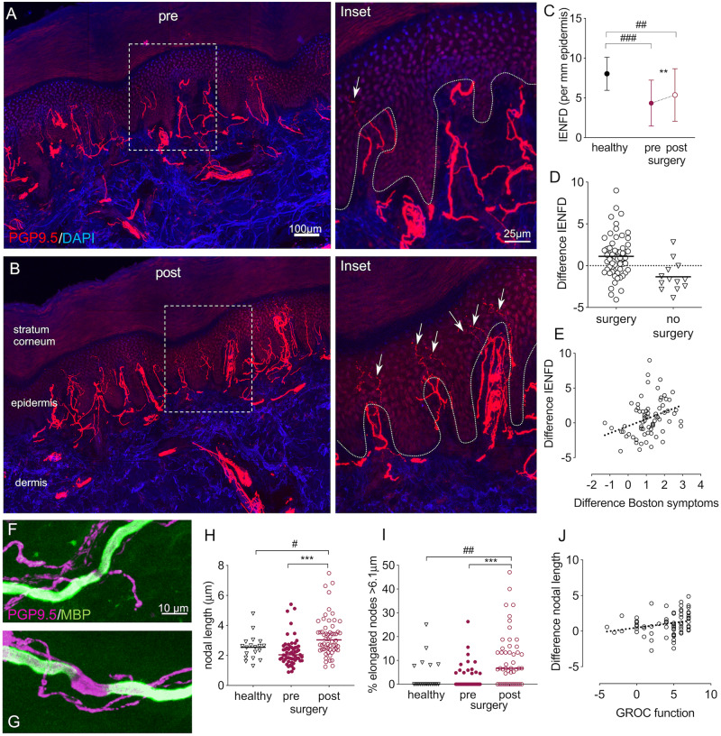
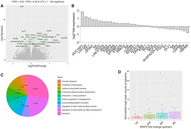
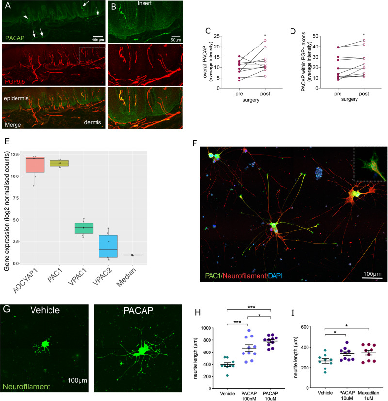
We only have a rudimentary understanding of the molecular and cellular determinants of nerve regeneration and neuropathic pain in humans. This cohort study uses the most common entrapment neuropathy (carpal tunnel syndrome) as a human model system to prospectively evaluate the cellular and molecular correlates of neural regeneration and its relationship with clinical recovery. In 60 patients undergoing carpal tunnel surgery [36 female, mean age 62.5 (standard deviation 12.2) years], we used quantitative sensory testing and nerve conduction studies to evaluate the function of large and small fibres before and 6 months after surgery. Clinical recovery was assessed with the global rating of change scale and Boston Carpal Tunnel Questionnaire. Twenty healthy participants provided normative data [14 female, mean age 58.0 (standard deviation 12.9) years]. At 6 months post-surgery, we noted significant recovery of median nerve neurophysiological parameters (P < 0.0001) and improvements in quantitative sensory testing measures of both small and large nerve fibre function (P < 0.002). Serial biopsies revealed a partial recovery of intraepidermal nerve fibre density [fibres/mm epidermis pre: 4.20 (2.83), post: 5.35 (3.34), P = 0.001], whose extent correlated with symptom improvement (r = 0.389, P = 0.001). In myelinated afferents, nodal length increased postoperatively [pre: 2.03 (0.82), post: 3.03 (1.23), P < 0.0001] suggesting that this is an adaptive phenomenon. Transcriptional profiling of the skin revealed 31 differentially expressed genes following decompression, with ADCYAP1 (encoding pituitary adenylate cyclase activating peptide, PACAP) being the most strongly upregulated (log2 fold-change 1.87, P = 0.0001) and its expression was associated with recovery of intraepidermal nerve fibres. We found that human induced pluripotent stem cell-derived sensory neurons expressed the receptor for PACAP and that this peptide could significantly enhance axon outgrowth in a dose-dependent manner in vitro [neurite length PACAP 1065.0 µm (285.5), vehicle 570.9 μm (181.8), P = 0.003]. In conclusion, carpal tunnel release is associated with significant cutaneous reinnervation, which correlates with the degree of functional improvement and is associated with a transcriptional programme relating to morphogenesis and inflammatory processes. The most highly dysregulated gene ADCYAP1 (encoding PACAP) was associated with reinnervation and, given that this peptide signals through G-protein coupled receptors, this signalling pathway provides an interesting therapeutic target for human sensory nerve regeneration.

Keywords: ADCYAP1; carpal tunnel syndrome; nerve regeneration; peripheral nerve injury; pituitary adenylate cyclase-activating peptide.


Figures

Figure 1
Figure 1
Recovery of somatosensory function. Raw quantitative sensory testing data are presented as mean and standard deviations for healthy participants (n =20, black circles) and patients with CTS before (closed red circle) and after surgery (n =60, open red circle). (A) Cold (CDT) and warm detection thresholds (WDT); (B) thermal sensory limen (TSL); (C) mechanical (MDT) and vibration detection thresholds (VDT); (D) cold (CPT) and heat pain thresholds (HPT); (E) mechanical (MPT) and pressure pain thresholds (PPT); (F) mechanical pain sensitivity (MPS) and wind-up ratio (WUR). Significant differences for paired/independent t-tests after Benjamini Hochberg correction are indicated between groups with #P <0.05, ##P <0.01, ###P <0.0001 and within groups with *P <0.05, **P <0.01, ***P <0.0001.
Figure 2
Figure 2
Histological evidence nerve regeneration in target tissue. Immunohistochemically stained sections of index finger skin of a patient with CTS before (A) and 6 months after surgery (B). Cell nuclei are apparent in blue (DAPI) and axons in red (PGP9.5). After surgery, an increased number of small fibres (arrows on ×4 magnified regions of interest on the right) penetrate the dermal-epidermal border (dotted line). (C) Quantification revealed a reduced IENFD in patients with CTS compared to healthy controls, which increased after surgery but failed to reach normal levels. (D) The difference in IENFD post- compared to pre-surgery demonstrated a high interindividual variability, with a continuing small fibre degeneration in patients who were not operated on. (E) A more pronounced intraepidermal nerve fibre regeneration (difference IENFD) correlates with improved symptoms (r = 0.389, P =0.001, difference Boston symptom scale data multiplied with −1). (FJ) Changes in nodal architecture. Example of normal (F) and elongated node (G) in skin sections stained immunohistochemically with PGP9.5 and myelin basic protein (MBP). (H) Median nodal length and (I) the median percentage of elongated nodes >6.1 μm increase following surgery and remain higher than healthy controls. (J) The correlation between difference in mean nodal length and the global rating of change scale (GROC) for hand function marginally fails to reach significance after Benjamini Hochberg correction (r = 0.316, P =0.008). Significant differences for paired/independent t-tests after Benjamini Hochberg correction are indicated between groups with #P <0.05, ##P <0.01, ###P <0.0001 and within groups with **P <0.01, ***P <0.0001. Group differences based on n =59 patients with CTS and n =20 healthy control subjects.
Figure 3
Figure 3
Molecular signature for nerve regeneration in skin. (A) Volcano plot showing the range of transcriptional changes following carpal tunnel surgery compared to before surgery; x-axis log2 (fold change), y-axis −log10 (P-value). Significant DEGs according to FDR adjusted P-value are highlighted and labelled. ADCYAP1, i.e. the gene encoding PACAP, was the most upregulated gene. (B) Gene expression changes (log2 fold change) for DEGs. (C) Top 20 biological process gene ontology (GO) terms from GO enrichment analysis with number of DEGs of total genes marked for each term on the pie chart. (D) ADCYAP1 is upregulated at baseline in all IENFD fold change quarters compared to the lowest quarter of patients with negative IENFD fold change, who continue to degenerate after surgery. Graph shows mean normalized log2 read counts at baseline, standard errors and single datapoints.
Figure 4
Figure 4
PACAP is expressed in human skin and induces neurite outgrowth in hiPSCd sensory neurons. (A) Immunohistochemical staining in human finger skin demonstrating presence of PACAP (green) within sensory nerve fibres (PGP9.5, arrows) and in the deeper epidermis (arrowhead). (B) Enlarged images of PACAP and PGP9.5 co-localization within sensory afferents. Quantification of PACAP staining intensity reveals an increase in intensity post-surgery compared to baseline within (C) an area including epidermal and subepidermal plexus and (D) PGP9.5+ sensory neurons. *P <0.047. (E) Gene expression strength of ADCYAP1, its main receptor PAC1 as well as secondary receptors VPAC1 and VPAC2 and the median expression of all genes in hiPSCd sensory neurons. The y-axis shows RNA-seq log2 normalized gene counts. Box plots show the median and interquartile range with single data-points depicting each experiment (n =3 cell lines, two differentiations each). (F) Immunohistochemical staining of hiPSCd sensory neurons demonstrating expression of the PACAP receptor (PAC1, green) in the soma as well as at the growth cones (×5 magnified inset). (G and H) PACAP given to dissociated mature (27 ± 3 weeks old) and replated hiPSCd sensory neurons enhances neurite outgrowth compared to vehicle in a dose-dependent manner (NHDF line). (I) The selective PAC1 agonist maxadilan induces a similar regenerative effect on hiPSCd sensory neurons as PACAP (AD2 line, 27 ± 3 weeks old). Data are presented as mean, standard error of the mean (SEM) and single data-points, *P <0.05, **P <0.01, ***P <0.0001.

References

    1. Alexa A, Rahnenfuhrer J. topGO: Enrichment analysis for gene ontology. R package version 2.34.0. ed; 2018. Available at: https://bioconductor.org/packages/release/bioc/html/topGO.html.
    1. Anand P, Terenghi G, Warner G, Kopelman P, WilliamsChestnut RE, Sinicropi DV.. The role of endogenous nerve growth factor in human diabetic neuropathy. Nat Med 1996; 2: 703–7. - PubMed
    1. Aruga J. The role of Zic genes in neural development. Mol Cell Neurosci 2004; 26: 205–21. - PubMed
    1. Baskozos G Dawes JM Austin JS Antunes-Martins A Mcdermott L Clark AJ, et al. . . Comprehensive analysis of long noncoding RNA expression in dorsal root ganglion reveals cell-type specificity and dysregulation after nerve injury. Pain 2019; 160: 463–85. - PMC - PubMed
    1. Benjamini Y, Hochberg Y.. Controlling the false discovery rate: a practical and powerful approach to multiple testing. J R Stat Soc 1994; 57: 289–300.

Publication types

Substances