Review
. 2020 Aug 1;12(8):593-606.
doi: 10.1093/jmcb/mjaa035.

Tumor organoids to study gastroesophageal cancer: a primer

Affiliations
Review

Tumor organoids to study gastroesophageal cancer: a primer

Ramon U Jin et al. J Mol Cell Biol. .

Abstract

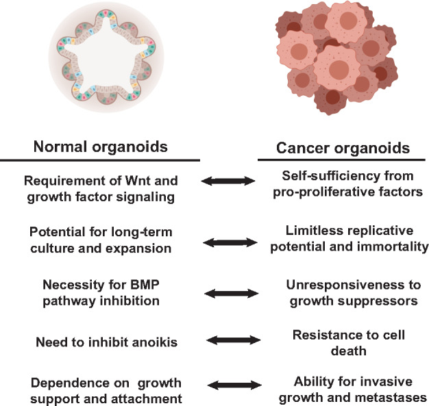
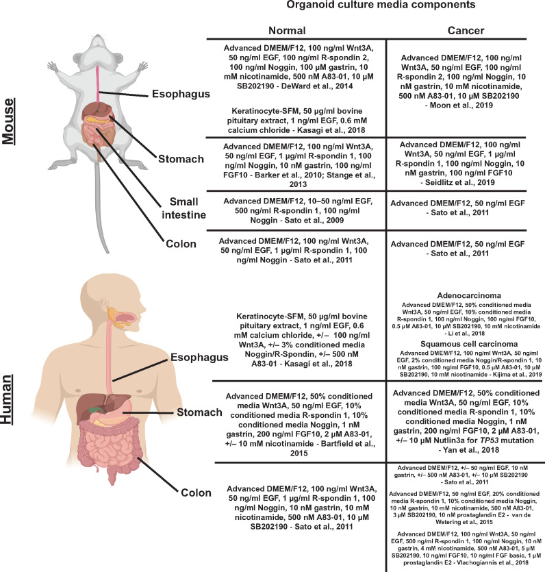
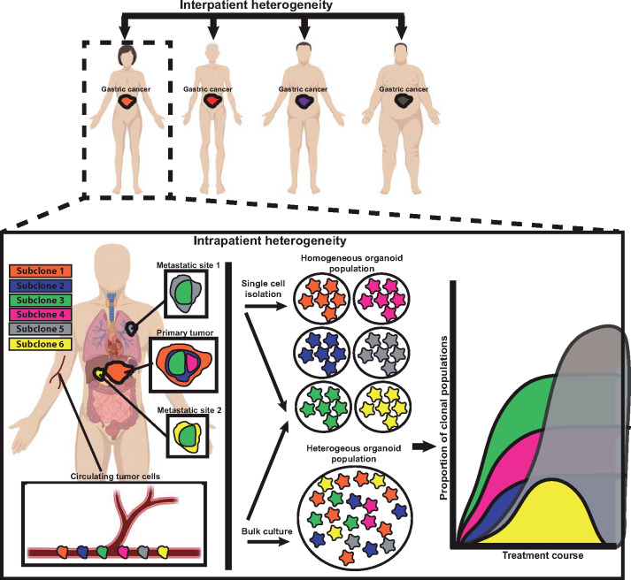
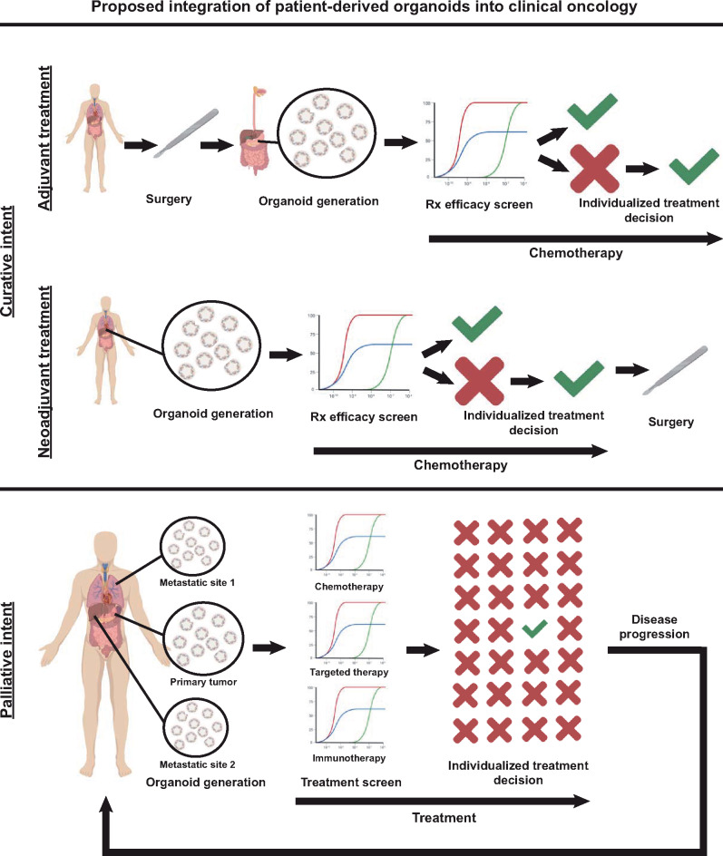
Gastroesophageal cancers are leading causes of cancer death. Our attempts at adopting molecularly based treatment approaches have been slow and ineffective even though we begin to identify specific targetable gene mutations and pathways. It is clear that we should no longer treat all gastroesophageal cancers as a homogeneous disease, which is what we do when we use non-specific chemotherapy. However, we currently cannot monitor successful gene/pathway targeting, nor understand how/when tumors develop resistance, nor predict which patients will derive maximal benefit. To improve outcomes, we must precisely detail the heterogeneity of these tumors to then individualize cancer therapy as well as develop novel avenues to study and predict treatment effects in individual patients. To this end, patient-derived organoids, in which tumor cells from individual patients are grown in a Petri dish, are a new versatile system that allows for timely expandability, detailed molecular characterization, and genetic manipulation with the promise of enabling predictive assessment of treatment response. In this review, we will explore the development and basic techniques for organoid generation, and discuss the current and potential future applications of this exciting technology to study the basic science of carcinogenesis and to predict/guide cancer patient care in the clinics.

Keywords: cancer evolution; cancer model; personalized medicine; precision oncology; targeted therapy; tumor heterogeneity.


Figures

Figure 1
Figure 1
Comparison between growth parameters and culture requirements of normal vs. cancer organoid cultures. Normal organoid culture conditions are designed to allow indefinite expansion, mimicking certain aspects of carcinogenesis. Cancers by definition are prone to indefinite expansion in the absence of external queues, so when cancers are cultured in organoid conditions, they may variably not require certain culture media components. All figures are created with BioRender.com.
Figure 2
Figure 2
Major components of culture media for normal and cancer GI organoids derived from humans and mice. The culture media composition is essential for the establishment and maintenance of normal and cancer organoids. We detail here the important differences in culturing techniques used to culture normal and cancer GI organoids from humans and mice including different approaches to generate organoids from the same starting tissues.
Figure 3
Figure 3
Organoids can help study cancer heterogeneity and evolution. Cancer heterogeneity exists patient to patient (‘interpatient’) and within the same patient between different metastatic sites, as a function of time during treatment course, and even within regions of the same tumor site (‘intrapatient’). Such heterogeneity can be modeled by generating organoids from single cells that can each be grown as subclones that model a tumor cell population within the patient. On the other hand, growing organoids in bulk from tumors may reflect the overall behavior and response of a tumor as a whole and preserve key subclone‒subclone interactions and overall tumor clonal architecture. Circulating tumor cells often travel as single cells that can be grown in bulk conditions or grown as subclones without the need for cell separation. In either case, organoids can provide valuable and clinically applicable information regarding therapy response at the clonal level, though this tendency of tumor cells (and thus organoids derived from them) to be heterogeneous must be kept in mind when interpreting results. For example, in the hypothetical plot, the yellow subclone is responsive to therapy, whereas the orange subclone continues to grow.
Figure 4
Figure 4
Proposed integration of PDOs into clinical oncology. Organoids hold promise to impact clinical oncology decision-making in both localized and advanced disease. Curative treatment of localized disease often involves multi-modality treatment including systemic treatment after (‘adjuvant’) or before (‘neoadjuvant’) surgical resection. Generating patient organoids in the adjuvant setting from the surgical resection specimen or in the neoadjuvant setting from initial diagnostic biopsy samples will allow screening of systemic treatments to predict regimens with maximal curative efficacy and to avoid ineffective treatments. For patients with advanced disease, organoids can be generated from multiple sites (primary tumor and accessible metastatic sites). These patients can then begin standard of care first-line treatment. During this time, PDOs can be used to screen established and novel therapies including chemotherapeutics, targeted agents, and immunotherapy, to individualize the choice of treatment prior to disease progression. This iterative approach can be adopted for each subsequent line of therapy.

Similar articles

Cited by

References

    1. Anderson W.F., Camargo M.C., Fraumeni J.F. Jr, et al. (2010). Age-specific trends in incidence of noncardia gastric cancer in US adults. JAMA 303, 1723–1728. - PMC - PubMed
    1. Bang Y.J., Van Cutsem E., Feyereislova A., et al. (2010). Trastuzumab in combination with chemotherapy versus chemotherapy alone for treatment of HER2-positive advanced gastric or gastro-oesophageal junction cancer (ToGA): a phase 3, open-label, randomised controlled trial. Lancet 376, 687–697. - PubMed
    1. Barker N., Huch M., Kujala P., et al. (2010). Lgr5+ve stem cells drive self-renewal in the stomach and build long-lived gastric units in vitro. Cell Stem Cell 6, 25–36. - PubMed
    1. Barker N., Ridgway R.A., van Es J.H., et al. (2009). Crypt stem cells as the cells-of-origin of intestinal cancer. Nature 457, 608–611. - PubMed
    1. Barker N., van Es J.H., Kuipers J., et al. (2007). Identification of stem cells in small intestine and colon by marker gene Lgr5. Nature 449, 1003–1007. - PubMed

Publication types