Review
. 2020 Dec 1:450:168-183.
doi: 10.1016/j.neuroscience.2020.06.046. Epub 2020 Jul 9.

The Cerebellar Nuclei and Dexterous Limb Movements

Affiliations
Review

The Cerebellar Nuclei and Dexterous Limb Movements

Ayesha R Thanawalla et al. Neuroscience. .

Abstract

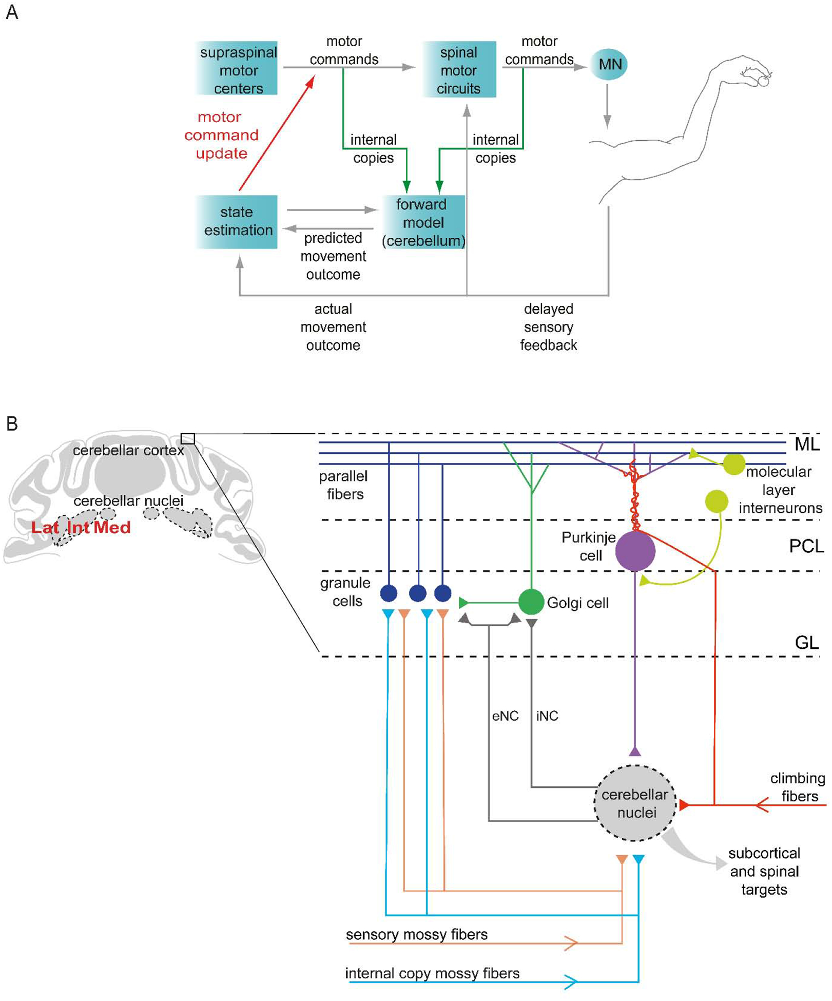
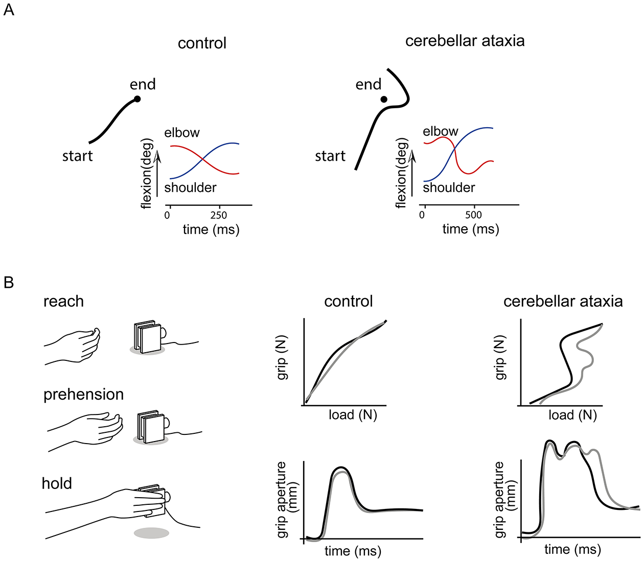
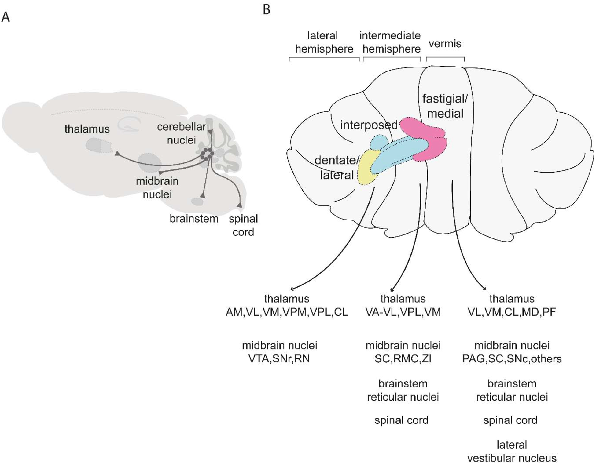
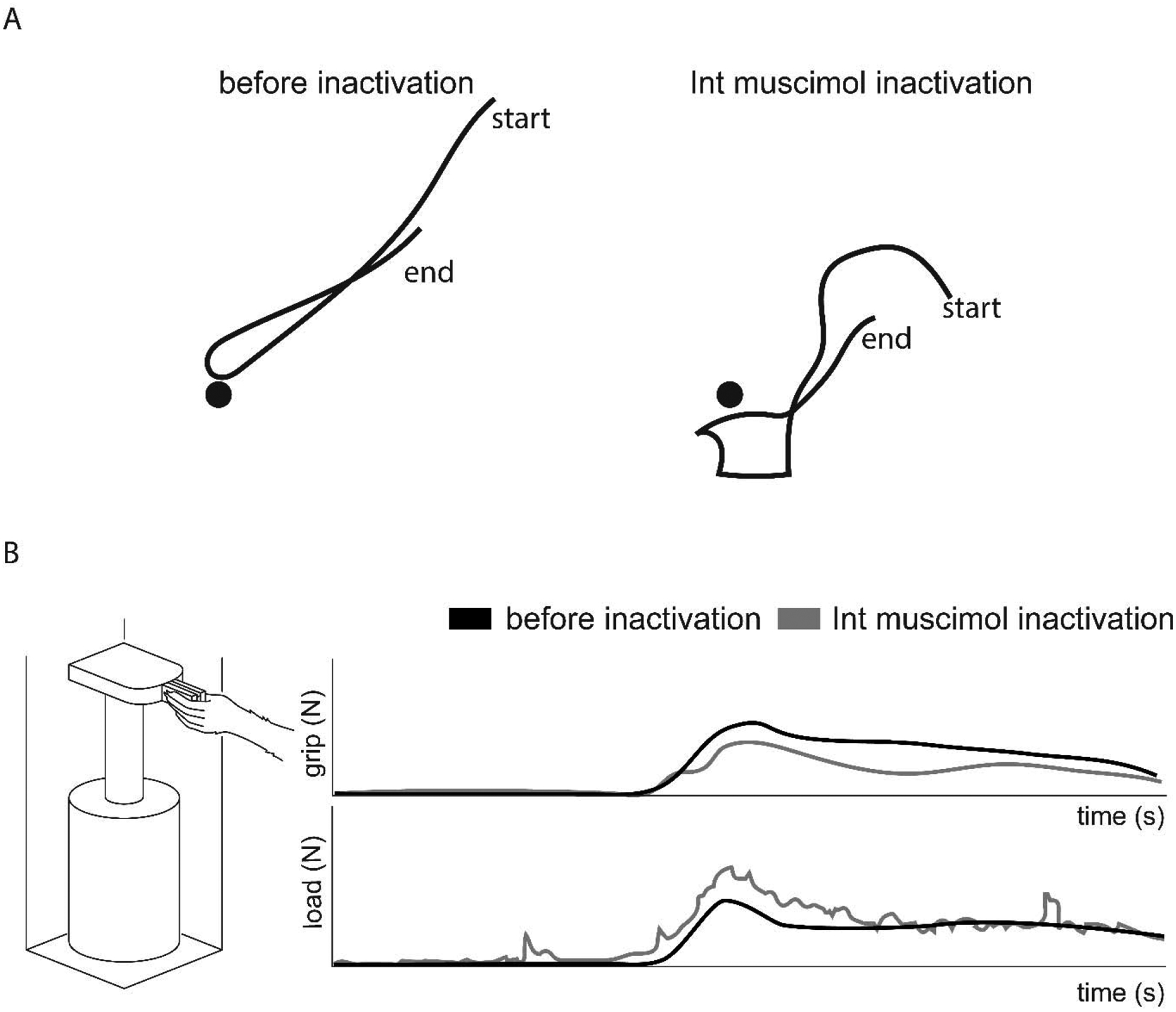
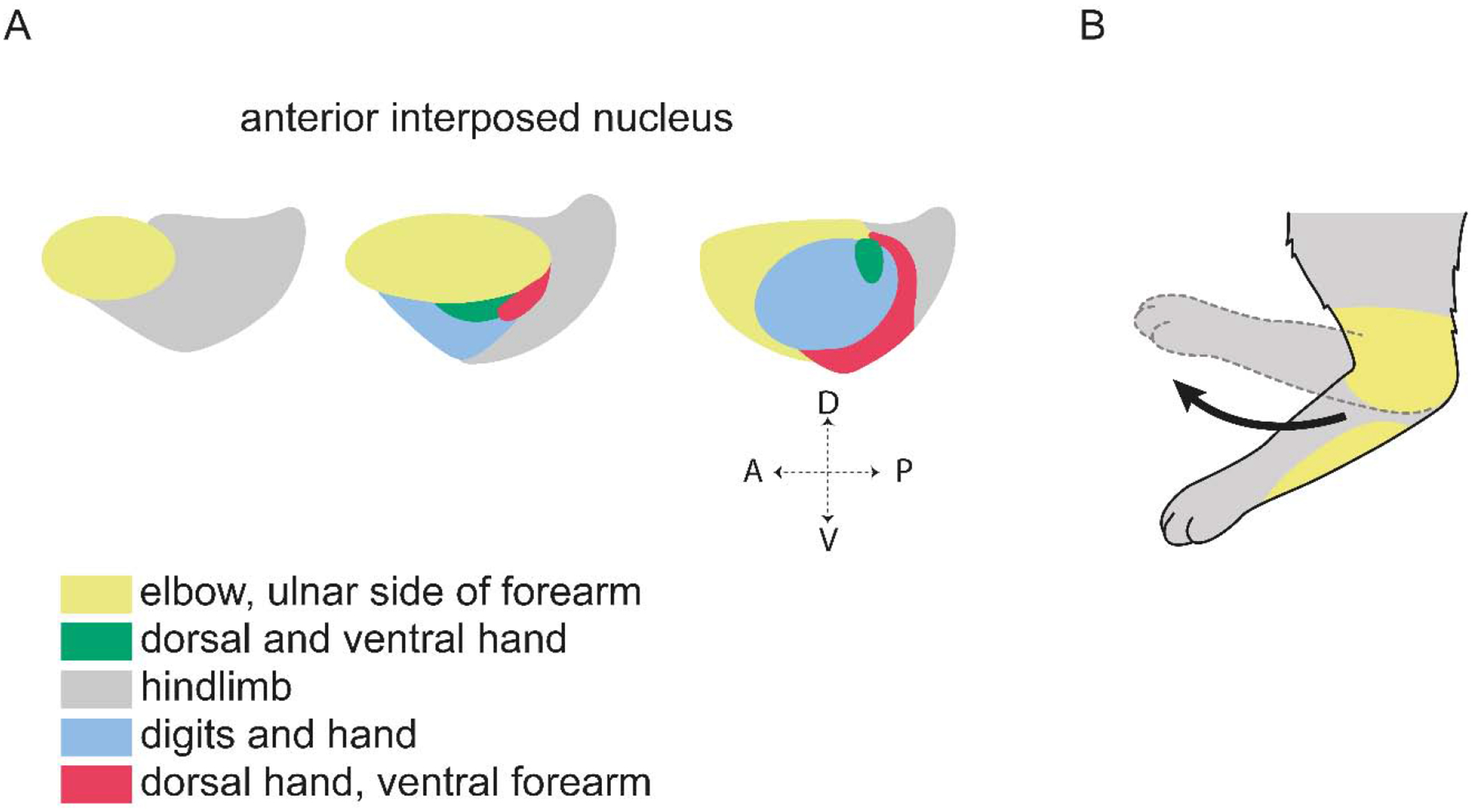
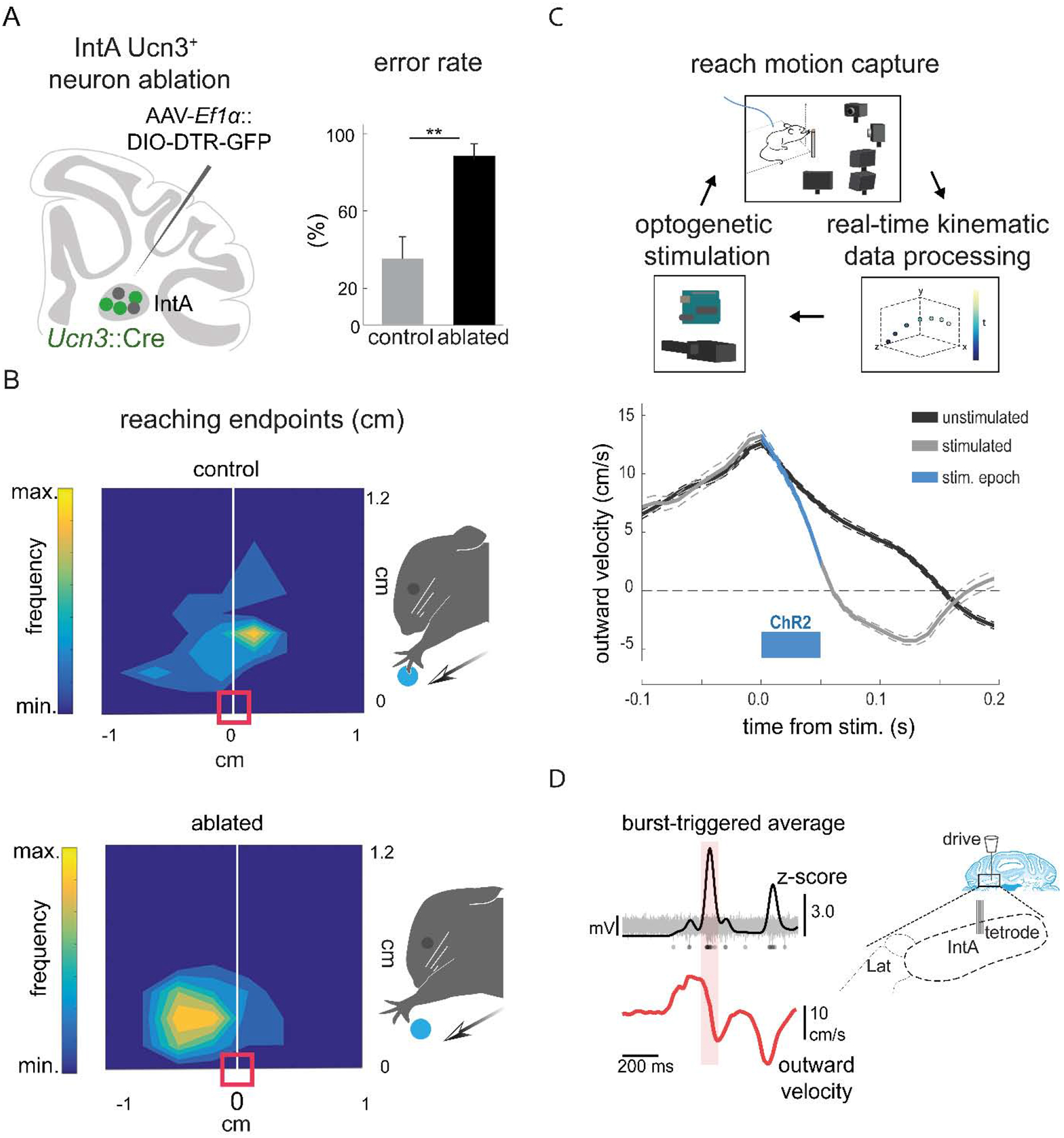
Dexterous forelimb movements like reaching, grasping, and manipulating objects are fundamental building blocks of the mammalian motor repertoire. These behaviors are essential to everyday activities, and their elaboration underlies incredible accomplishments by human beings in art and sport. Moreover, the susceptibility of these behaviors to damage and disease of the nervous system can lead to debilitating deficits, highlighting a need for a better understanding of function and dysfunction in sensorimotor control. The cerebellum is central to coordinating limb movements, as defined in large part by Joseph Babinski and Gordon Holmes describing motor impairment in patients with cerebellar lesions over 100 years ago (Babinski, 1902; Holmes, 1917), and supported by many important human and animal studies that have been conducted since. Here, with a focus on output pathways of the cerebellar nuclei across mammalian species, we describe forelimb movement deficits observed when cerebellar circuits are perturbed, the mechanisms through which these circuits influence motor output, and key challenges in defining how the cerebellum refines limb movement.

Keywords: Ataxia; Cerebellar nuclei; Dysmetria; Grasp; Internal copy; Reach.


Figures

Figure 1.
Figure 1.. Cerebellar circuits for forelimb movement.
(A) During limb movement, motor commands elicit muscle contraction, generating sensory feedback that is used to update the estimate of limb state and adjust motor output from supraspinal and spinal targets. Yet sensory feedback delays imply a need for a more rapid internal feedback mechanism. Copies of motor commands (internal copies) are thought to be conveyed to the cerebellum, where a forward model generates a prediction of movement outcome, enabling more rapid online refinement. Reducing mismatch between predictions and sensory-reported outcome can also be used to adapt subsequent movements. Cerebellar output is thus tasked with recruiting the necessary motor structures to update motor commands and adjust limb movement (Azim and Alstermark, 2015). (B) The cerebellar cortex receives inputs that deliver sensory and internal copy signals from the spinal cord and brainstem (mossy fibers), and teaching-related signals from the inferior olive (climbing fibers) (Huang et al., 2013; Ishikawa et al., 2015; Miall, 2016; Lang et al., 2017). Neurons in the cerebellar nuclei, the primary output of the cerebellum, also receive input from mossy fiber and climbing fiber collaterals. The activity of neurons in the cerebellar nuclei is shaped by inhibition from Purkinje cells located in the Purkinje cell layer (PCL). Granule cells located in the granular layer (GL) receive mossy fiber signals and provide excitatory input to Purkinje cells via parallel fibers that extend through the molecular layer (ML). Purkinje cells also receive strong excitatory input from climbing fibers, as well as inhibitory input from molecular layer interneurons. Mossy fiber inputs to granule cells are regulated by inhibitory Golgi cells. Neurons in the cerebellar nuclei send inhibitory nucleo-cortical fibers (iNC) to Golgi cells, and excitatory nucleo-cortical fibers (eNC) to granule cells and Golgi cells. Cerebellar nuclear neurons project to many subcortical and spinal targets (Miall, 2016).
Figure 2.
Figure 2.. Reaching and grasping deficits in patients with cerebellar ataxia.
(A) When asked to perform fast, accurate reaches toward a target (filled black circles), control subjects exhibit straight trajectories, while patients with cerebellar ataxia display dysmetria, in this case showing a tendency to overshoot the target (traces show the trajectory of the index finger). Patients also exhibit curved trajectories, increased shoulder flexion (blue traces), and inappropriate flexion and extension of the elbow (red traces), as compared to controls (Bastian et al., 1996). (B) During reach-to-grasp movements (left), patients with cerebellar ataxia show abnormal grip force relative to load force during the grasp (top), and exaggerated grip aperture (bottom) compared to control subjects (traces show two example trials) (Brandauer et al., 2008).
Figure 3.
Figure 3.. Major targets of the cerebellar nuclei.
(A) Schematic of pathways originating from the cerebellar nuclei. (B) Targets of the fastigial/medial nucleus include the thalamus (VL: ventrolateral, VM: ventromedial, CL: centrolateral, MD: mediodorsal, PF: parafascicular), midbrain nuclei (PAG: periaqueductal gray, SC: superior colliculus, SNc: substantia nigra pars compacta), brainstem reticular nuclei, spinal cord, lateral vestibular nucleus, and other regions described in (Fujita et al., 2020). Targets of the interposed nuclei include the thalamus (VA-VL: ventral anterior-ventrolateral, VPL: ventral posterolateral, VM: ventromedial), midbrain nuclei (SC: superior colliculus, RMC: magnocellular red nucleus, ZI: zona incerta), brainstem reticular nuclei, and spinal cord (Houck and Person, 2015; Low et al., 2018; Sathyamurthy et al., 2020). Targets of the dentate/lateral nucleus include the thalamus (AM: anteromedial, VL: ventrolateral, VM: ventromedial, VPM: ventral posteromedial: VPL ventral posterolateral, CL: centrolateral) and midbrain nuclei (VTA: ventral tegmental area, SNr: substantia nigra pars reticulata, RN: red nucleus) (Carta et al., 2019; Dacre et al., 2019; Sakayori et al., 2019). For simplicity, schematics are restricted to tracing studies performed in mice.
Figure 4.
Figure 4.. Reversible inactivation of the Int nucleus results in abnormal forelimb movement.
(A) Cats exhibit dysmetric trajectories when reaching toward a target (filled black circles) and during limb retraction after muscimol inactivation of the Int nucleus (Bracha et al., 1999). (B) During a grasp, lift, and hold task (left), muscimol inactivation of the Int nucleus in monkeys results in reduced grip force during the hold phase (top) and ataxic movement with dynamic tremor visible in the load force (bottom) (Monzee et al., 2004).
Figure 5.
Figure 5.. Organization of forelimb areas in the IntA nucleus.
(A) The IntA nucleus is topographically organized based on Purkinje cell termination from microzones with specific climbing fiber receptive fields. Shown here are forelimb and hindlimb cutaneous and nociceptive receptive fields (Garwicz and Ekerot, 1994; Ekerot et al., 1997). (B) Microstimulation of the nuclear zone receiving indirect cutaneous input from the ulnar side of the forelimb results in flexion of the elbow, moving the limb region corresponding to the receptive field away from the putative stimulus (Ekerot et al., 1995).
Figure 6.
Figure 6.. The IntA nucleus ensures endpoint precision during skilled reaching.
(A) Schematic summarizing strategy for selective ablation of Ucn3+ IntA neurons using a viral diphtheria toxin receptor-mediated strategy (left). Ablation of Ucn3+ IntA neurons results in increased pellet reach-to-grasp errors and reduces pellet retrieval success (right) (Low et al., 2018). (B) Contour density plots depict the endpoint position of the wrist with respect to the pellet. Arrows depict the direction of reach and the red box indicates the position of the pellet. Ablation of Ucn3+ IntA neurons results in hypermetric reaching and overshooting the target (Low et al., 2018). (C) Schematic of the behavioral setup for real-time kinematic analysis and closed-loop optogenetic manipulation of the IntA nucleus (top). Averaged velocity traces from a single animal illustrate that optogenetic activation of IntA neurons during reaching reduces outward velocity of the limb (bottom) (Becker and Person, 2019). (D) Tetrode recordings in IntA during reaching reveal that the amplitude of IntA neuronal activity (top) peaks during abrupt reductions in limb outward velocity (Becker and Person, 2019).

References

    1. Adrian MH, and John WK (2012). Theoretical Models of Motor Control and Motor Learning In Routledge Handbook of Motor Control and Motor Learning (Routledge; ).
    1. Aizenman CD, Huang EJ, and Linden DJ (2003). Morphological correlates of intrinsic electrical excitability in neurons of the deep cerebellar nuclei. J Neurophysiol 89, 1738–1747 doi: 10.1152/jn.01043.2002. - DOI - PubMed
    1. Alstermark B, and Ekerot CF (2013). The lateral reticular nucleus: a precerebellar centre providing the cerebellum with overview and integration of motor functions at systems level. A new hypothesis. J Physiol 591, 5453–5458 doi: 10.1113/jphysiol.2013.256669. - DOI - PMC - PubMed
    1. Alstermark B, and Isa T (2012). Circuits for skilled reaching and grasping. Annual review of neuroscience 35, 559–578 doi: 10.1146/annurev-neuro-062111-150527. - DOI - PubMed
    1. Ankri L, Husson Z, Pietrajtis K, Proville R, Lena C, Yarom Y, Dieudonne S, and Uusisaari MY (2015). A novel inhibitory nucleo-cortical circuit controls cerebellar Golgi cell activity. eLife 4, e06262 doi: 10.7554/eLife.06262. - DOI - PMC - PubMed

Publication types