. 2020 Aug;24(15):8452-8465.
doi: 10.1111/jcmm.15404. Epub 2020 Jul 11.

Sotrastaurin, a PKC inhibitor, attenuates RANKL-induced bone resorption and attenuates osteochondral pathologies associated with the development of OA

Affiliations

Sotrastaurin, a PKC inhibitor, attenuates RANKL-induced bone resorption and attenuates osteochondral pathologies associated with the development of OA

Cong Pang et al. J Cell Mol Med. 2020 Aug.

Abstract

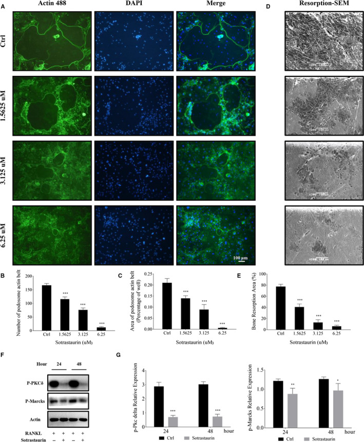
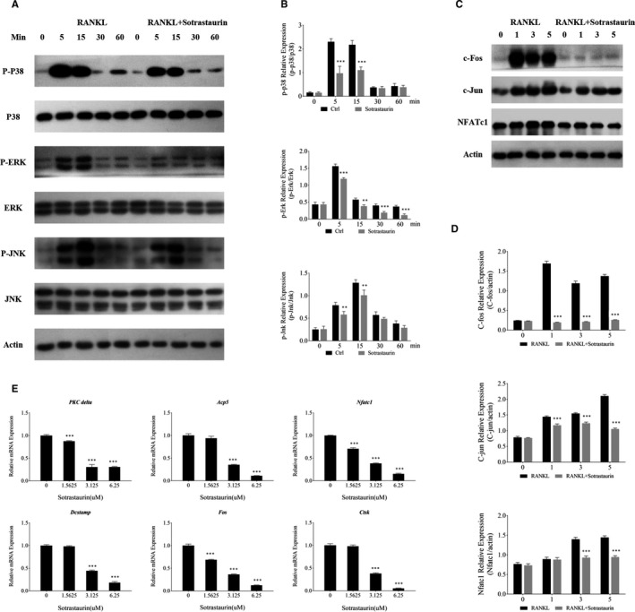
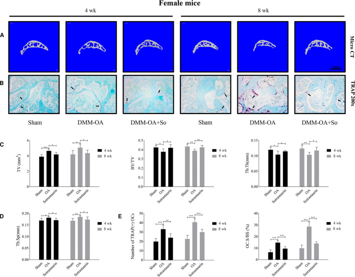
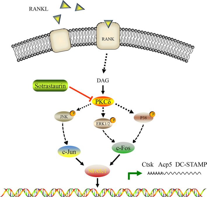
Osteoarthritis (OA) is a common degenerative disease that affects the musculoskeletal structure of the whole joint, which is characterized by progressive destruction of both articular cartilage and subchondral bone. Treatment of the bone pathologies, particularly osteoclast-mediated subchondral bone loss in the early stages of OA, could prevent subsequent cartilage degeneration and progression of OA. In the present study, the PKC inhibitor, Sotrastaurin, was found to inhibit RANKL-induced osteoclast formation in vitro in a dose- and time-dependent manner. In particular, SO exerted its anti-osteoclastic effect predominantly at the early stages of RANKL stimulation, suggesting inhibitory effects on precursor cell fusion. Using mature osteoclasts cultured on bovine bone discs, we showed that SO also exerts anti-resorptive effects on mature osteoclasts bone resorptive function. Mechanistically, SO attenuates the early activation of the p38, ERK and JNK signalling pathways, leeding to impaired induction of crucial osteoclast transcription factors c-Jun, c-Fos and NFATc1. We also showed that SO treatment significantly inhibited the phosphorylation of PKCδ and MARCKS, an upstream regulator of cathepsin K secretion. Finally, in animal studies, SO significantly alleviates the osteochondral pathologies of subchondral bone destruction as well as articular cartilage degeneration following DMM-induced OA, markedly improving OARSI scores. The reduced subchondral bone loss was associated with marked reductions in TRAP(+) osteoclasts in the subchondral bone tissue. Collectively, our data provide evidence for the protective effects of SO against OA by preventing aberrant subchondral bone and articular cartilage changes. Thus, SO demonstrates potential for further development as an alternative therapeutic option against OA.

Keywords: PKCδ; osteoclast; sotrastaurin; subchondral bone; therapeutics.


Conflict of interest statement

The authors have no conflicts of interests to declare.

Figures

FIGURE 1
FIGURE 1
Sotrastaurin inhibited osteoclast formation induced by RANKL in vitro. A, The chemical structure of sotrastaurin. B, The effect of sotrastaurin on the activity of BMMs (bone marrow macrophages) in 144 h was assessed by CCK‐8 cell cytotoxicity/ proliferation test. C, Stimulation of M‐CSF (macrophage colony‐stimulating factor) dependent BMMS was performed with RANKL (receptor activator of nuclear factor‐κB ligand) for 6 d in the absence or presence of sotrastaurin in specified concentration, and subsequently fixated and stained for activity of TRAP. D‐F, Quantifiy the number and acreage of TRAP‐positive polynucleated osteoclasts (nuclei > 3). The amount and dimension of osteoclasts in each sotrastaurin concentration group were compared with the untreated controls. G and H, Sotrastaurin affected the osteoclastogenesis in a time‐dependent manner. BMMs (M‐CSF dependence) were stimulated with RANKL and treated with 6.25 μmol/L sotrastaurin within specified days, then fixated and stained to detect the activity of TRAP. G, I, J, Quantifiy the number and acreage of TRAP‐positive polynucleated osteoclasts (nuclei > 3). The amount and dimension of osteoclasts in each sotrastaurin concentration group were compared with the untreated controls. Data are expressed as means ± SD (*P < .05, **P < .01 and ***P < .001); SD, standard deviation. Scale bar, 100 μm. All experiments were performed at least three times
FIGURE 2
FIGURE 2
Sotrastaurin inhibits podosome actin belt formation and impairs osteoclasts mediated bone resorption in vitro. A, Effect of sotrastaurin on the formation of podosome actin belt. M‐CSF‐dependent BMMS were seeded onto 96 well plates and stimulated with RANKL without or with the specified concentration of sotrastaurin (0, 1.625, 3.125 and 6.25 μmol/L) for 6 d. Cells were fixed and stained for immunofluorescence. Typical fluorescence images of osteoclasts’ podosome actin belts (green) and nuclei (blue, DAPI). B and C, The number and area of podosome actin belts were quantified. Scale bar, 100 μm. D, Effect of sotrastaurin on osteoclast bone resorption activity. BMM‐derived pre‐osteoclasts were cultured on bone slices and stimulated with RANKL (100 ng/mL) in the absence or presence of increasing concentrations of sotrastaurin for 6 d. The cells were removed by ultrasound and the resorption pits were evaluated under a scanning electron microscope. Scale bar, 100 μm. E, Relative to the untreated controls, area of bone resorption pits under each treatment concentration was quantified. F, BMMs were treated with 6.25 μmol/L sotrastaurin, cells were incubated for 24 and 48 h and then harvested. The expression levels of phosphorylated PCKδ and MARCKS in protein lysates were analysed by Western blot. Expression of β‐Actin was analysed as an internal loading control. G, Densitometric analyses of the expression of the phosphorylation‐PKCδ and phosphorylation‐MARCKS were reported as p‐PKCδ/ β‐Actin and p‐MARCKS/ β‐Actin. Data are presented as the mean ± SD (*P < .05, **P < .01 and ***P < .001); SD: standard deviation. DAPI: 6‐diamidino‐2‐phenylindole
FIGURE 3
FIGURE 3
Sotrastaurin exerts its anti‐osteoclast effect by the inhibition of MAPKs signalling cascade. A, Sotrastaurin inhibited the activation phosphorylation of p38, JNK and ERK induced by RANKL. After pre‐treatment with 6.25 μmol/L sotrastaurin for 1 h, and then stimulated with RANKL for a specified time, Western blot analysis was performed with specific antibodies to mitogen‐activated protein kinases (p38, ERK and JNK) signalling cascade. β‐actin was used to act as an internal loading control. B, Relative change in the phosphorylation status of p38, JNK and ERK were determined by optical density analysis of each phosphorylated band, and each one was expressed as the ratio of its total protein counterpart. C, Sotrastaurin attenuated the expression of c‐Fos, c‐Jun and NFATc1 induced by RANKL. The total protein extracts of BMMs cultured in the medium containing RANKL and 6.25 μmol/L sotrastaurin for 0, 1, 3 and 5 d were analysed by Western blotting and specific antibodies to c‐Fos, c‐Jun and NFATc1. β‐actin was used to act as an internal loading control. D, The changes in protein expression level after sotrastaurin treatment were quantified by relative β‐actin density analysis and expressed as the ratio compared with the untreated group stimulated by RANKL alone. E, Sotrastaurin inhibited osteoclast marker genes induced by RANKL in a dose‐dependent manner. Real‐time quantitative PCR assessed the expression of PKCδ, ACP5, NFATc1, DC‐STAMP, c‐Fos and CTSK in osteoclasts treated with specified concentrations of sotrastaurin. The expression of the target genes were standardized with reference to the housekeeping gene GADPH and then displayed a decreased change compared with the untreated control groups. Data are presented as the mean ± SD (*P < .05, **P < .01 and ***P < .001); SD, standard deviation. MARCKs, myristoylated alanine‐rich C‐kinase substrate
FIGURE 4
FIGURE 4
Sotrastaurin protects against subchondral bone loss in vivo after destabilization of the medial meniscus. A, Representative three‐dimensional micro‐CT images of the sagittal views of the medial compartment of tibial subchondral bone from Sham (Sham + PBS), OA (DMM + PBS) or Sotrastaurin (DMM + Sotrastaurin) groups at 4 and 8 weeks after operation. Scale bar, 1 mm. B, Representative images of knee joint subchondral bone were stained by fast green and osteoclasts in subchondral bone were stained by tartrate‐resistant acid phosphatase (TRAP) at 4 and 8 weeks after surgery (magnification, 200×); Black arrow indicates TRAP‐positive cells. C and D, Histogram of three‐dimensional structural parameters of tibial subchondral bone: (TV) tissue volume, (BV/TV) trabecular bone volume/tissue volume, (Tb.Th) trabecular thickness, and (Tb.Sp) trabecular separation. E, The number and area percentage of osteoclasts stained by TRAP in sections were measured. n = 6 per group. Data are expressed as means ± SD (*P < .05, **P < .01 and ***P < .001); SD, standard deviation
FIGURE 5
FIGURE 5
Histological and histomorphometric evaluation of the sotrastaurin effect on cartilage degradation of DMM model in sections. A, Representative images of cartilage stained by Safranin O/Fast green (magnification, 100 × or 200×) from tissue sections of Sham, OA (DMM + PBS) or Sotrastaurin (DMM + Sotrastaurin) groups; The thickness of hyaline cartilage (HC) and calcified cartilage (CC) were marked by dotted line; n = 6 per group. B, HC and CC thickness of articular cartilage. C, Osteoarthritis Research Society International (OARSI)‐modified Mankin scores to cartilage structure damage of knee joint on mice. Data are presented as the mean ± SD (*P < .05, **P < .01 and ***P < .001); SD, standard deviation
FIGURE 6
FIGURE 6
Schematic diagram and potential mechanisms of sotrastaurin inhibition effect on osteoclast resorption. Sotrastaurin inhibits osteoclast resorption activity by inhibiting PKCδ/ MAPKs/ (c‐Fos/c‐Jun)/ NFATc1 signalling pathway

Similar articles

Cited by

References

    1. Gu Y‐T, Chen J, Meng Z‐L, et al. Research progress on osteoarthritis treatment mechanisms. Biomed Pharmacother. 2017;93:1246‐1252. - PubMed
    1. Cross M, Smith E, Hoy D, et al. The global burden of hip and knee osteoarthritis: estimates from the global burden of disease 2010 study. Ann Rheum Dis. 2014;73(7):1323‐1330. - PubMed
    1. Iijima H, Aoyama T, Ito A, et al. Effects of short‐term gentle treadmill walking on subchondral bone in a rat model of instability‐induced osteoarthritis. Osteoarthritis Cartilage. 2015;23(9):1563‐1574. - PubMed
    1. Rahmati M, Mobasheri A, Mozafari M. Inflammatory mediators in osteoarthritis: a critical review of the state‐of‐the‐art, current prospects, and future challenges. Bone. 2016;85:81‐90. - PubMed
    1. Iijima H, Aoyama T, Ito A, et al. Destabilization of the medial meniscus leads to subchondral bone defects and site‐specific cartilage degeneration in an experimental rat model. Osteoarthritis Cartilage. 2014;22(7):1036‐1043. - PubMed

Publication types

MeSH terms