

Endomyocardial Biopsy Characterization of Heart Failure With Preserved Ejection Fraction and Prevalence of Cardiac Amyloidosis
- PMID: 32653448
- PMCID: PMC7604801
- DOI: 10.1016/j.jchf.2020.04.007
Endomyocardial Biopsy Characterization of Heart Failure With Preserved Ejection Fraction and Prevalence of Cardiac Amyloidosis
Abstract
Objectives: This study prospectively evaluated endomyocardial biopsies in patients with heart failure with preserved ejection fraction (HFpEF) to identify histopathologic phenotypes and their association with clinical characteristics.
Background: Myocardial tissue analysis from a prospectively defined HFpEF cohort reflecting contemporary comorbidities is lacking.
Methods: Patients with HFpEF (EF ≥50%) referred to the Johns Hopkins HFpEF Clinic between August 2014 and September 2018 were enrolled for right heart catheterization and endomyocardial biopsy. Clinical features, echocardiography, hemodynamics, and tissue histology were determined and compared with controls (unused donor hearts) and HF with reduced EF (HFrEF).
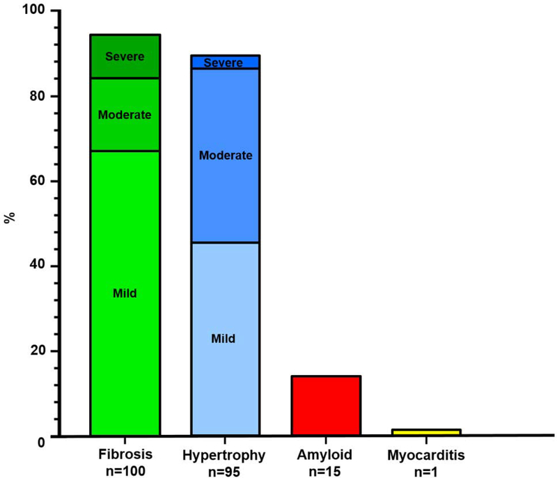
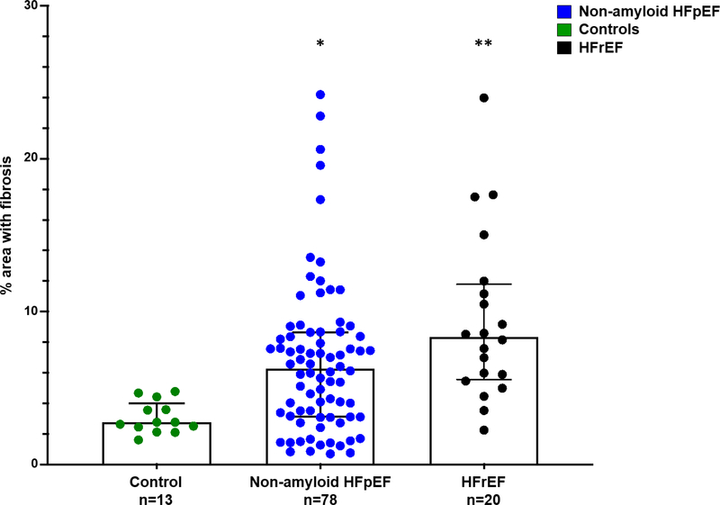
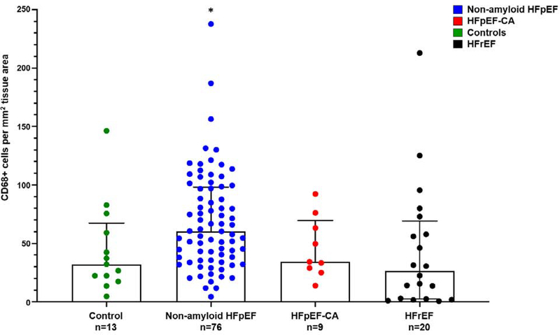
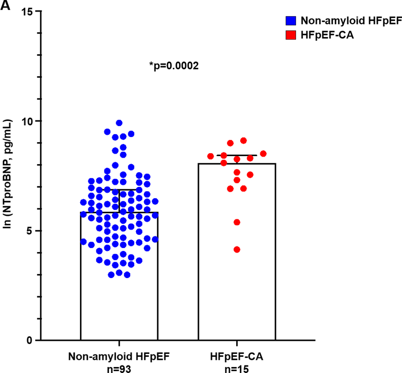
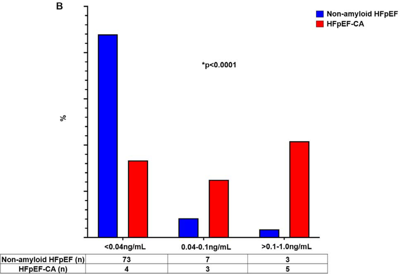
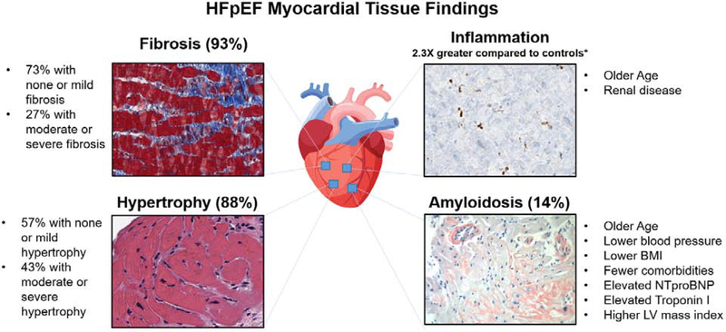
Results: Of the 108 patients enrolled, median age was 66 years (25th to 75th percentile: 57 to 74 years), 61% were women, 57% were African American, 62% had a previous HF hospitalization, median systolic blood pressure was 141 mm Hg (25th to 75th percentile: 125 to 162 mm Hg), body mass index (BMI) was 37 kg/m2 (25th to 75th percentile: 32 to 45 kg/m2), and 97% were on a loop diuretic. Myocardial fibrosis and myocyte hypertrophy were often present (93% and 88%, respectively); however, mild in 71% with fibrosis and in 52% with hypertrophy. Monocyte infiltration (CD68+ cells/mm2) was greater in patients with HFpEF versus controls (60.4 cells/mm2 [25th to 75th percentile: 36.8 to 97.8] vs. 32.1 cells/mm2 [25th to 75th percentile: 22.3 to 59.2]; p = 0.02) and correlated with age and renal disease. Cardiac amyloidosis (CA) was diagnosed in 15 (14%) patients (HFpEF-CA: 7 patients with wild-type transthyretin amyloidosis [ATTR], 4 patients with hereditary ATTR, 3 patients with light-chain amyloidosis, and 1 patient with AA (secondary) amyloidosis), of which 7 cases were unsuspected. Patients with HFpEF-CA were older, with lower BMI, higher left ventricular mass index, and higher N-terminal pro-B-type natriuretic peptide and troponin I levels.
Conclusions: In this large, prospective myocardial tissue analysis of HFpEF, myocardial fibrosis and hypertrophy were common, CD68+ inflammation was increased, and CA prevalence was 14%. Tissue analysis in HFpEF might improve precision therapies by identifying relevant myocardial mechanisms.
Keywords: HFpEF; amyloidosis; biopsy; fibrosis; hypertrophy; inflammation.
Copyright © 2020. Published by Elsevier Inc.
Figures
References
-
- Benjamin EJ, Virani SS, Callaway CW et al. Heart Disease and Stroke Statistics-2018 Update: A Report From the American Heart Association. Circulation 2018;137:e67–e492. - PubMed
-
- Owan TE, Hodge DO, Herges RM, Jacobsen SJ, Roger VL, Redfield MM. Trends in prevalence and outcome of heart failure with preserved ejection fraction. N Engl J Med 2006;355:251–9. - PubMed
-
- Steinberg BA, Zhao X, Heidenreich PA et al. Trends in patients hospitalized with heart failure and preserved left ventricular ejection fraction: prevalence, therapies, and outcomes. Circulation 2012;126:65–75. - PubMed
-
- Sharma K, Hill T, Grams M et al. Outcomes and Worsening Renal Function in Patients Hospitalized With Heart Failure With Preserved Ejection Fraction. The American journal of cardiology 2015;116:1534–40. - PubMed
-
- Sharma K, Vaishnav J, Kalathiya R et al. Randomized Evaluation of Heart Failure With Preserved Ejection Fraction Patients With Acute Heart Failure and Dopamine: The ROPA-DOP Trial. JACC Heart failure 2018;6:859–870. - PubMed
Publication types
MeSH terms
Substances
Grants and funding
LinkOut - more resources
Full Text Sources
Medical
Research Materials
Miscellaneous
