Review
. 2020 Sep:198:108136.
doi: 10.1016/j.exer.2020.108136. Epub 2020 Jul 10.

Biology of keratorefractive surgery- PRK, PTK, LASIK, SMILE, inlays and other refractive procedures

Affiliations
Review

Biology of keratorefractive surgery- PRK, PTK, LASIK, SMILE, inlays and other refractive procedures

Steven E Wilson. Exp Eye Res. 2020 Sep.

Abstract

The outcomes of refractive surgical procedures to improve uncorrected vision in patients-including photorefractive keratectomy (PRK), laser in-situ keratomileusis (LASIK), Small Incision Lenticule Extraction (SMILE) and corneal inlay procedures-is in large part determined by the corneal wound healing response after surgery. The wound healing response varies depending on the type of surgery, the level of intended correction of refractive error, the post-operative inflammatory response, generation of opacity producing myofibroblasts and likely poorly understood genetic factors. This article details what is known about these specific wound healing responses that include apoptosis of keratocytes and myofibroblasts, mitosis of corneal fibroblasts and myofibroblast precursors, the development of myofibroblasts from keratocyte-derived corneal fibroblasts and bone marrow-derived fibrocytes, deposition of disordered extracellular matrix by corneal fibroblasts and myofibroblasts, healing of the epithelial injury, and regeneration of the epithelial basement membrane. Problems with epithelial and stromal cellular viability and function that are altered by corneal inlays are also discussed. A better understanding of the wound healing response in refractive surgical procedures is likely to lead to better treatments to improve outcomes, limit complications of keratorefractive surgical procedures, and improve the safety and efficiency of refractive surgical procedures.

Keywords: Apoptosis; Cornea; Corneal fibroblasts; Corneal haze; Corneal inlays; Corneal scar; LASIK; Laser thermal keratoplasty (LTK); Mitomycin C; Myofibroblasts; PRK; PTK; Radial keratotomy; Small incision lenticule extraction (SMILE); Thermal conductive keratoplasty (CK); Wound healing.


Figures

Fig. 1.
Fig. 1.
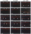
TUNEL assay to detect apoptosis in rabbit corneas at different time points after −4.5D PRK, −9D PRK and −9D LASIK. Note that corneas that have −9D PRK have greater keratocyte apoptosis (arrows), especially at the 4 hour time point. The apoptosis response persists for at least one week and includes bone marrow-derived cells that infiltrate the cornea after surgery. After −9D LASIK, the keratocyte apoptosis response is less than −9D PRK and is localized deeper in the corneal stroma along the interface and away from the overlying epithelium. e indicates epithelium stained with DAPI. Mag. 200X. Reprinted with permission from Mohan et al. Exp. Eye Res. 2003;76:71-87.
Fig. 2.
Fig. 2.
Quantitative analysis of TUNEL+ apoptotic stromal cells at each time point for −4.5D PRK, −9D PRK and −9D LASIK, with 8 corneas in each group at each time point. Note that from 4 to 72 hours stromal cell apoptosis was significantly greater in the −9D PRK group. The −9D LASIK group had lower stromal cell apoptosis from 4 to 72 hours than the −9D PRK group and was also lower than the −4.5D PRK group at the 4 hour and 24 hour time points. Modified with permission from Mohan et al. Exp. Eye Res. 2003;76:71-87.
Fig. 3.
Fig. 3.
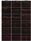
Stromal cell mitosis detected with immunohistochemistry for the mitosis marker Ki-67 at different time points after −4.5D PRK, −9D PRK and −9D LASIK. Note there is much greater stromal cell mitosis (arrows) in both PRK groups compared to the LASIK group. Epithelial cells normally undergo mitosis as a part of their life cycle. Mag. 200X. Reprinted with permission from Mohan et al. Exp. Eye Res. 2003;76:71-87.
Fig. 4.
Fig. 4.
Quantitative analysis of Ki-67+ mitotic stromal cells at each time point for −4.5D PRK, −9D PRK and −9D LASIK, with 8 corneas in each group at each time point. Note that stromal cell mitosis is much greater in the −9D PRK group, especially at 24 hours after surgery. Stromal cell mitosis remained significantly higher in the PRK groups compared to the LASIK group at one week after surgery. Modified with permission from Mohan et al. Exp. Eye Res. 2003;76:71-87.
Fig. 5.
Fig. 5.
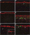
Subepithelial myofibroblasts detected with immunohistochemistry for alpha smooth muscle actin (SMA, green, arrows) after −4.5D, −9D PRK or −9D LASIK in rabbits. A. Very few, if any, SMA+ cells are detected in an unwounded rabbit cornea. B. Few SMA+ cells are detected in a cornea that had −4.5D PRK at one month after surgery. C. Similarly few SMA+ cells are detected at one week after −9D LASIK (insert shows the entry point of the microkeratome blade (arrowhead). At one month after −9D PRK (not shown) SMA+ myofibroblasts are only found at the peripheral edge of the LASIK flap where the epithelial basement membrane was damaged by the LASIK microkeratome. D. At one week after −9D PRK, only a single SMA+ myofibroblasts in detected in the subepithelial stroma. E. By one month after −9D PRK, numerous SMA+ myofibroblasts are present in the anterior stroma. F. At three months after −9D PRK, the SMA+ myofibroblasts have decreased. By 6 months after −9D PRK (not shown) few if any SMA+ myofibroblasts can be detected in the stroma. red is propidium iodide staining of nuclei. Mag. 200x Reprinted with permission from Mohan et al. Exp. Eye Res. 2003;76:71-87.
Fig. 6.
Fig. 6.
Quantitative analysis of alpha-smooth muscle actin+ myofibroblasts at each time point for −4.5D PRK, −9D PRK and −9D LASIK, with 8 corneas in each group at each time point. Note myofibroblasts were much greater in the −9D PRK group, with only a few myofibroblasts detected in corneas that had −4.5D PRK and none detected in the central cornea of eyes that had −9D LASIK. Modified with permission from Mohan et al. Exp. Eye Res. 2003;76:71-87.
Fig. 7.
Fig. 7.
Immunohistochemistry for the mitosis marker Ki-67 (green) with DAPI straining (blue) of all cell nuclei in rabbit corneas that had −9.0 D PRK for myopia with mitomycin C or vehicle balanced salt solution (BSS) application at the time of surgery and studied at 24 hours or 4 weeks later. Note there was marked cell proliferation (arrowheads) in the anterior stroma in the BSS group (C) at 24 hours after PRK, with some stromal cell mitosis continuing at 4 weeks after surgery. Markedly reduced proliferation of anterior stromal cells was found at 24 hours and at four weeks after PRK surgery in corneas treated with 0.02% mitomycin C for two minutes. If rabbit corneas were treated with 0.002% mitomycin C for 12 seconds, there were more anterior stromal cells undergoing mitosis at 24 hours and four weeks after PRK than with 0.02% mitomycin C, but less than in the BSS group. Mag. 200X. Reprinted with permission from Netto, et al. J. Ref. Surgery, 2006;22:562-574.
Fig. 8.
Fig. 8.
Flap edge fibrosis (arrows) that is commonly noted after LASIK at the slit lamp. Mag. 40X. Reprinted with permission from Medeiros CS et al. Invest. Ophthalmol. Vis. Sci. 2018;59:4044–4053.
Fig. 9.
Fig. 9.
Microkeratome compared to femtosecond laser flap inflammation. At 24 hours after flap formation in rabbit corneas, corneas were processed for immunohistochemistry for the monocyte marker CD11b (green). Note that with the microkeratome monocytes were only detected at the edges of the flap (arrows). Arrowheads indicate the interface. The 15 kHz Intralase laser required the 2.5 μJ setting to cut the flap and resulted in much greater inflammation at the flap edge (arrows) and along the interface (arrowheads). This resulted in much more flap edge DLK, and DLK in the central cornea, and necessitated the use of more frequent topical corticosteroids during the early postoperative period.36 Both flap edge and interface inflammation were decreased with the 30 kHz Intralase using the 0.9 μJ energy setting. The 60 kHz Intralase resulted in flap edge inflammation (arrows) similar to the microkeratome and little inflammation along the interface (arrowheads). Mag 200x. Reprinted with permission from Netto et al J. Ref. Surg. 2007;23:667-676.
Fig. 10.
Fig. 10.
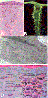
The role of myofibroblasts in corneal wound healing following radial keratotomy (RK), (A, owl monkey and B, cat) and penetrating corneal injury (C) in the rabbit. In histologic sections of the cornea, 14 days after RK, the gaping wound becomes filled with proliferating fibroblastic-like cells that have migrated in from the adjacent corneal stroma (A). These cells when probed for the presence of filamentous actin using the molecular probe, Phalloidin, show intense staining indicating the presence of extensive actin filament arrays within cells immediately adjacent to and within the wound (B). Transmission electron microscopy of these fibroblastic cells shows the presence of microfilaments, actin bundles with electron dense plaques that contain myosin (arrow) indicating the presence of ‘stress fibers’—the electron microscopic marker for myofibroblasts (C). A model for the role of myofibroblasts in corneal wound healing is presented in D, where actinomyosin filament contraction exerts force on newly synthesized collagen and the adjacent wound margin through fibronectin interactions to contract and close the gaping wound. Graciously provided by James V. Jester, PhD, U.C. Irvine.
Fig. 11.
Fig. 11.
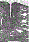
Histopathological evaluation of a human cornea several years after RK. An epithelial plug extends deep into the corneal stroma. Surrounding the epithelial plug there is an intact EBM and acellular areas that appear to represent a Bowman’s-like layer (arrows). e is epithelium. s is stroma. Mag. 25X
Fig. 12.
Fig. 12.
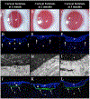
Rabbit corneas at one month, two months and three months after deep non-perforating linear incisions across the central cornea (Marino et al., 2017). At one month (A) the incisions are seen as dense linear opacities, but became progressively fainter at two months (B) and three months (C) after surgery. Surprisingly, no SMA+ myofibroblasts were detected anywhere in these corneas at one month (D), two months (E) or three months (F) after surgery. A single EBM (arrows) surrounded the epithelial plugs in all eyes at one month (G), two months (H) and three months (I) 23,000 mag. Disorganized collagen type 3, that is not normally detected in the corneal stroma, was present beneath the epithelial plugs of all corneal incisions at one month (J), and intensified at 2 months (K) and three months (L) after surgery, adjacent to high densities of cells that were likely corneal fibroblasts since they were SMA- cells. 25X mag. Blue is DAPI staining of all cell nuclei. e is epithelium; s is stroma, and * indicates artifactual separation of the epithelium and stroma that occurs during sectioning of some specimens. Reprinted with permission from Marino GK et al. J. Ref Surg. 2017;33:337-346.

Similar articles

Cited by

References

    1. Alió JL, Mulet ME, Zapata LF, Vidal MT, De Rojas V, Javaloy J, 2004. Intracorneal inlay complicated by intrastromal epithelial opacification. Arch. Ophthalmol 122, 1441–6. - PubMed
    1. Antonios R, Abdul Fattah M, Arba Mosquera S, Abiad BH, Sleiman K, Awwad ST, 2017. Single-step transepithelial versus alcohol-assisted photorefractive keratectomy in the treatment of high myopia: a comparative evaluation over 12 months. Br. J. Ophthalmol 101, 1106–1112. - PubMed
    1. Ayala Espinosa MJ, Alió Y Sanz J, Ismail MM, Sánchez Castro P, 2000. [ Experimental corneal histological study after thermokeratoplasty with holmium laser]. Arch. Soc. Esp. Oftalmol 75, 619–25. - PubMed
    1. Beer SMC, Werner L, Nakano EM, Santos RT, Hirai F, Nitschke EJ, Francesconi Benicio C, Campos MSQ, A 3-year follow-up study of a new corneal inlay: clinical results and outcomes B.r J. Ophthalmol Published Online First: 24 August 2019. doi: 10.1136/bjophthalmol-2019-314314 - DOI - PubMed
    1. Carones F, Fiore T, Brancato R, 1999. Mechanical vs. alcohol epithelial removal during photorefractive keratectomy. J. Ref. Surg 15, 556–562. - PubMed

Publication types

MeSH terms