

Case Reports
doi: 10.1007/s00415-020-10057-5.
Epub 2020 Jul 11.
Frontal encephalopathy related to hyperinflammation in COVID-19
Affiliations
- PMID: 32654063
- PMCID: PMC7353824
- DOI: 10.1007/s00415-020-10057-5
Item in Clipboard
Case Reports
Frontal encephalopathy related to hyperinflammation in COVID-19
J Neurol.
2021 Jan.
No abstract available
Conflict of interest statement
The authors declare that they have no conflict of interest.
Figures

Clinical symptoms, diagnostic work-up, treatment, and inflammatory profile during the disease course. CSF cerebrospinal fluid, ARDS acute respiratory distress syndrome, EEG electroencephalography, CT computed tomography, LP lumbar puncture, MRI magnetic resonance imaging, FDG-PET fluorodeoxyglucose-positron emission tomography, CRP C-reactive protein, IL-6 interleukin-6, IL-8 interleukin-8, TNFα tumor necrosis factor-alpha, IL-12 interleukin-12, IL-1β interleukin-1Beta, IL-10 interleukin-10; < , under limit of detection. Reference range: CRP < 0,50 mg/dl; ferritin 11–306 ng/ml; IL-6 < 5,9 pg/ml; IL-8 < 70 pg/ml; TNFα < 8,1 pg/ml, IL-10 < 9 pg/ml
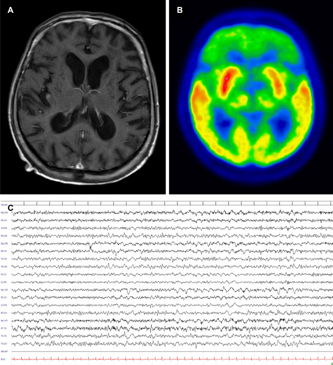
Magnetic resonance imaging (MRI), 18F-FDG-PET/CT, and electroencephalography (EEG) findings. Normal MRI findings in MRI on T1 with gadolinium (a). 18F-FDG-PET/CT showing severe frontal lobe hypometabolism (b). EEG alterations observed at time of first neurologic evaluation with prominent and bilateral frontal slowing (c). Electroencephalographic acquisition settings: 10–20 system, longitudinal montage. Recording speed: 30 s/page; sensitivity: 7 μV/mm; time constant: 0.1 s; high-frequency filter: 70 Hz
References
-
- Santomasso BD, Park JH, Salloum D, et al. Clinical and biological correlates of neurotoxicity associated with car t-cell therapy in patients with B-cell acute lymphoblastic leukemia. Cancer Discov. 2018;8:958–971. doi: 10.1158/2159-8290.CD-17-1319. - DOI - PMC - PubMed
Publication types
MeSH terms
Substances
LinkOut - more resources
Full Text Sources
Medical
