. 2020 Oct;57(10):4060-4068.
doi: 10.1007/s12035-020-02020-2. Epub 2020 Jul 11.

Optogenetic Activation of Dopamine Receptor D1 and D2 Neurons in Anterior Cingulate Cortex Differentially Modulates Trigeminal Neuropathic Pain

Affiliations

Optogenetic Activation of Dopamine Receptor D1 and D2 Neurons in Anterior Cingulate Cortex Differentially Modulates Trigeminal Neuropathic Pain

Sufang Liu et al. Mol Neurobiol. 2020 Oct.

Abstract

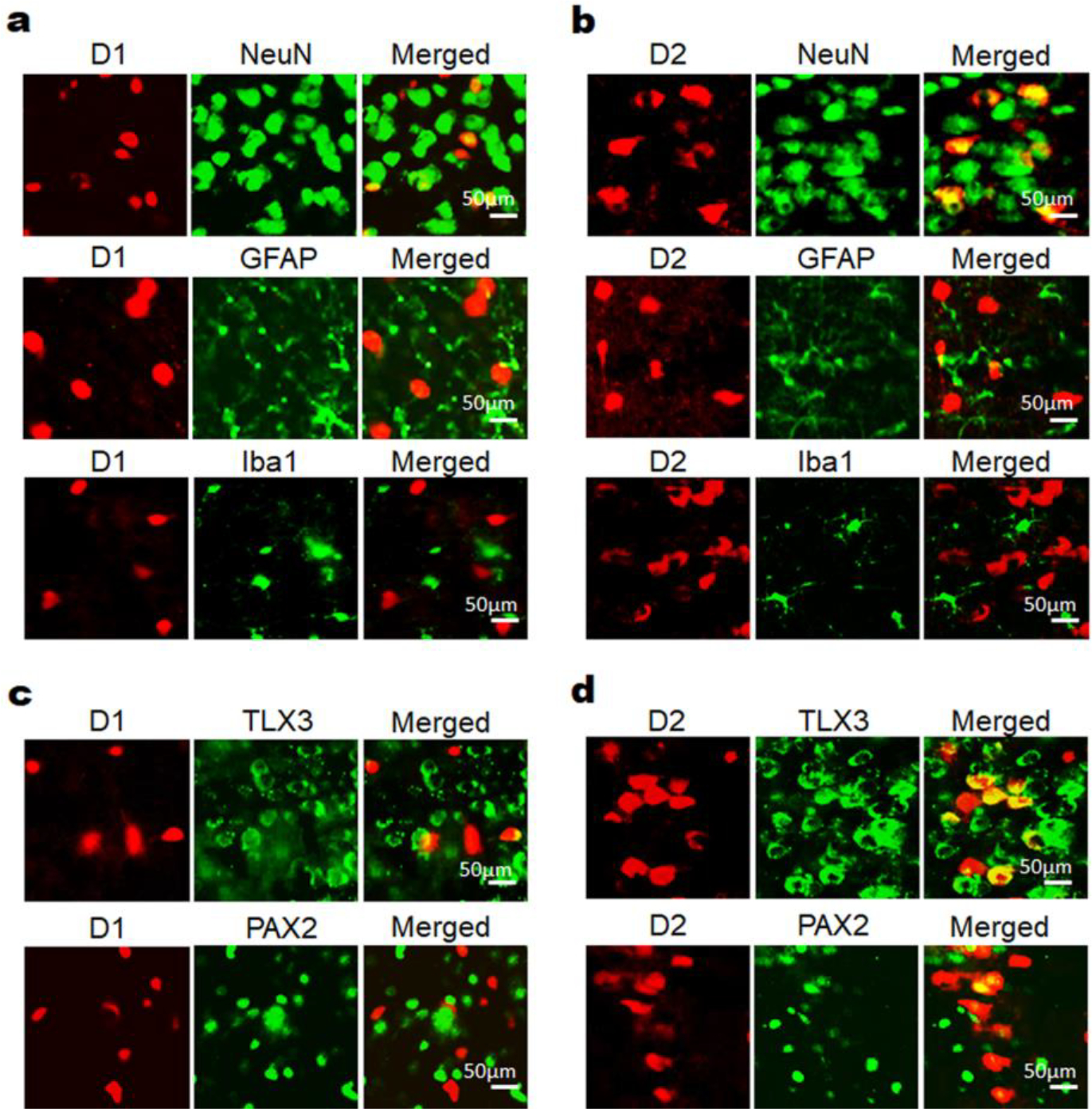
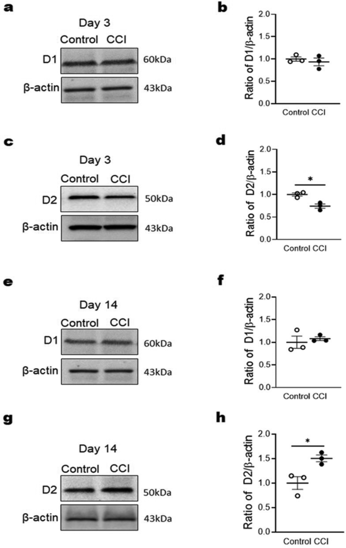
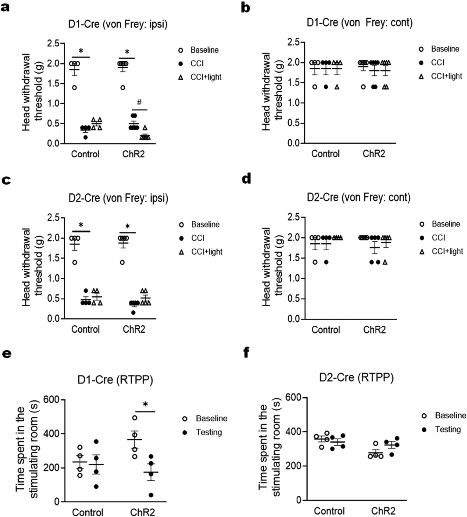
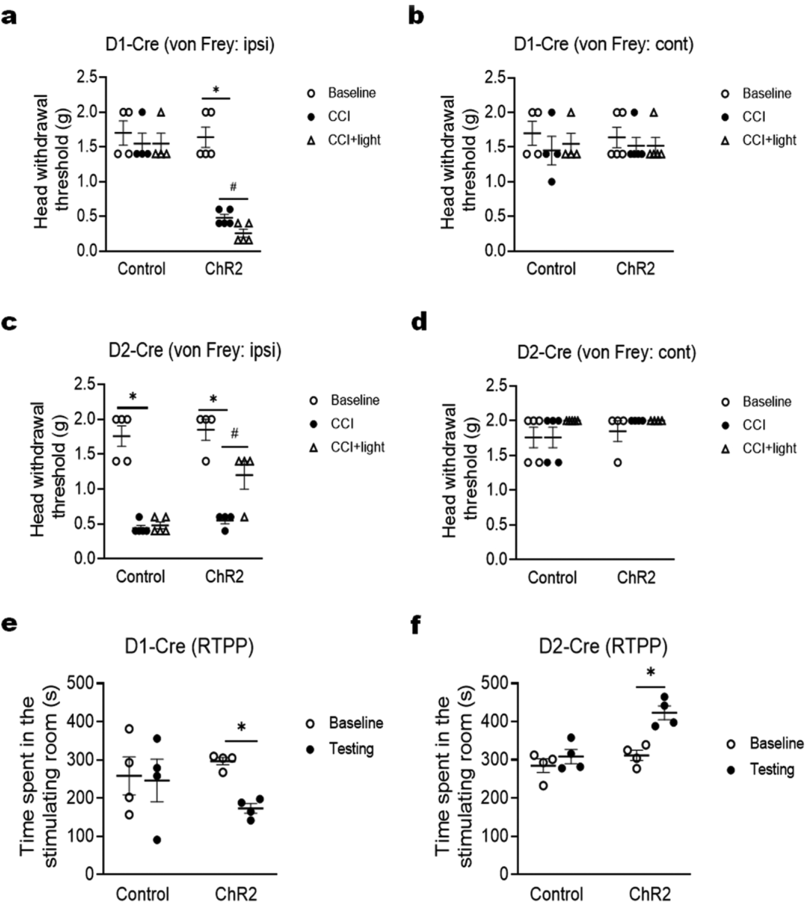
Anterior cingulate cortex (ACC) is a critical brain center for chronic pain processing. Dopamine signaling in the brain has been demonstrated to contribute to descending pain modulation. However, the role of ACC dopamine receptors in chronic neuropathic pain remains unclear. In this study, we investigated the effect of optogenetic activation of ACC dopamine receptors D1- and D2-expressing neurons on trigeminal neuropathic pain. Chronic constriction injury of infraorbital nerve (CCI-ION) was carried out to induce trigeminal neuropathic pain in mice. We conducted optogenetic stimulation to specifically activate D1- and D2-expressing neurons in the ACC. Western blotting and immunofluorescence staining were used to examine ACC D1 and D2 expression and localization. The von Frey and real-time place preference tests were performed to measure evoked mechanical pain and nonreflexive emotional pain behaviors, respectively. We observed that dopamine receptors D1 and D2 in the ACC are primarily expressed in excitatory neurons and that the D2 receptor is differentially regulated in the early and late phases of trigeminal neuropathic pain. Optogenetic activation of D1-expressing neurons in the ACC markedly exacerbates CCI-ION-induced trigeminal neuropathic pain in both early and late phases, but optogenetic activation of D2-expressing neurons in the ACC robustly ameliorates such pain in its late phase. Our results suggest that dopamine receptors D1 and D2 in the ACC play different roles in the modulation of trigeminal neuropathic pain.

Keywords: Anterior cingulate cortex; Dopamine receptors; Optogenetic stimulation; Trigeminal neuropathic pain.


Conflict of interest statement

CONFLICT OF INTEREST

The authors have no conflict of interest to declare.

Figures

Fig. 1
Fig. 1
Dopamine receptors D1 and D2 are primarily expressed in excitatory neurons of the ACC. D1-Cre:Ai14 and D2-Cre:Ai14 mouse lines were generated to label D1 and D2, respectively, with red fluorescent protein tdTomato. The offspring mice were used for immunofluorescence staining with different cell markers. (a) Immunofluorescence staining with NeuN, GFAP andIba1 using ACC sections of D1-Cre:Ai14 mice showed that tdTomato-labeled D1 in the ACC was exclusively expressed in NeuN-positive neurons, but not GFAP-positive or Iba1-positive glial cells. (b) Immunofluorescence staining with NeuN, GFAP and Iba1 using ACC sections of D2-Cre:Ai14 mice showed that tdTomato-labeled D2 in the ACC was also exclusively expressed in NeuN-positive neurons, but not GFAP-positive or Iba1-positive glial cells. (c) Immunofluorescence staining with TLX3 and PAX2 using ACC sections of D1-Cre:Ai14 mice showed that tdTomato-labeled D1 in the ACC was mostly expressed in TLX3-positive excitatory neurons, but not in PAX2-positive inhibitory neurons. (d) Immunofluorescence staining with TLX3 and PAX2 using ACC sections of D2-Cre:Ai14 mice showed that tdTomato-labeled D2 in the ACC was expressed in both TLX3-positive excitatory neurons and PAX2-positive inhibitory neurons, but the majority of D2 was co-labeled with TLX3. The immunofluorescence staining experiments were repeated three times to confirm the data shown in this figure. Scale bar, 50 μm.
Fig. 2
Fig. 2
Dopamine receptors D1 and D2 in the ACC are differentially regulated by CCI-ION. Western blotting was performed to examine change in the expression of these receptors in the ACC on day 3 and day 14 after CCI-ION. (a and b) ACC D1 expression had no significant alteration on day 3 after CCI-ION. (c and d) ACC D2 expression was significant decreased on day 3 after CCI-ION. (e and f) ACC D1 expression had no significant alteration on day 14 after CCI-ION. (g and h) ACC D2 expression was significant increased on day 14 after CCI-ION. n = 3 per group. *P < 0.05 vs. the sham control group.
Fig. 3
Fig. 3
Optogenetic activation of ACC dopamine receptor D1-expressing neurons, but not D2-expressing neurons, increases trigeminal neuropathic pain on day 3 after CCI-ION. (a and c) In both D1-Cre (a) and D2-Cre (c) mice, the CCI-ION significantly decreased head withdrawal threshold in the ipsilateral side; however, optogenetic activation of ACC D1 neurons (a), but not D2 neurons (c), further decreased the head withdrawal threshold. (b and d) In both D1-Cre (b) and D2-Cre (d) mice, the CCI-ION and optogenetic stimulation had no effect on head withdrawal threshold in the contralateral side. (e and f) In the real-time place preference (RTPP) test, optogenetic activation of ACC D1 neurons (e), but not D2 neurons (f), significantly decreased the time spent in stimulating room. n = 6 per group. *P < 0.05 vs. the corresponding Baseline values; #P < 0.05 vs. the corresponding CCI group.
Fig. 4
Fig. 4
Optogenetic activation of ACC dopamine receptor D1-expressing neurons and D2-expressing neurons oppositely modulates trigeminal neuropathic pain on day 14 after CCI-ION. (a and c) In D1-Cre mice (a), optogenetic activation of ACC D1 neurons further decreased head withdrawal threshold in the ipsilateral side; in D2-Cre mice (c), optogenetic activation of ACC D2 neurons reversely increased head withdrawal threshold in the ipsilateral side. (b and d) In both D1-Cre (b) and D2-Cre (d) mice, the CCI-ION and optogenetic stimulation had no effect on head withdrawal threshold in the contralateral side. (e and f) In the real-time place preference (RTPP) test, optogenetic activation of ACC D1 neurons (e) significantly decreased the time spent in stimulating room, but optogenetic activation of ACC D1 neurons (f) significantly increased the time spent in stimulating room. n = 6 per group. *P < 0.05 vs. the corresponding Baseline values; #P < 0.05 vs. the corresponding CCI group.

Similar articles

Cited by

References

    1. Lechin F, van der Dijs B, Lechin ME, Amat J, Lechin AE, Cabrera A, Gomez F, Acosta E, Arocha L, Villa S, et al. (1989) Pimozide therapy for trigeminal neuralgia. Arch Neurol 46 (9):960–963. doi:10.1001/archneur.1989.00520450030015 - DOI - PubMed
    1. Zhang J, Yang M, Zhou M, He L, Chen N, Zakrzewska JM (2013) Non-antiepileptic drugs for trigeminal neuralgia. Cochrane Database Syst Rev (12):CD004029. doi:10.1002/14651858.CD004029.pub4 - DOI - PMC - PubMed
    1. Dosenovic S, Kadic AJ, Miljanovic M, Biocic M, Boric K, Cavar M, Markovina N, Vucic K, Puljak L (2017) Interventions for Neuropathic Pain: An Overview of Systematic Reviews. Anesthesia and Analgesia 125 (2):643–652. doi:10.1213/Ane.0000000000001998 - DOI - PubMed
    1. Al-Quliti KW (2015) Update on neuropathic pain treatment for trigeminal neuralgia. The pharmacological and surgical options. Neurosciences (Riyadh) 20 (2):107–114. doi:10.17712/nsj.2015.2.20140501 - DOI - PMC - PubMed
    1. Jones MR, Urits I, Ehrhardt KP, Cefalu JN, Kendrick JB, Park DJ, Cornett EM, Kaye AD, Viswanath O (2019) A Comprehensive Review of Trigeminal Neuralgia. Curr Pain Headache Rep 23 (10):74. doi:10.1007/s11916-019-0810-0 - DOI - PubMed

Substances

LinkOut - more resources