. 2020 Sep;34(9):11641-11657.
doi: 10.1096/fj.202000544R. Epub 2020 Jul 12.

LPAR2 receptor activation attenuates radiation-induced disruption of apical junctional complexes and mucosal barrier dysfunction in mouse colon

Affiliations

LPAR2 receptor activation attenuates radiation-induced disruption of apical junctional complexes and mucosal barrier dysfunction in mouse colon

Pradeep K Shukla et al. FASEB J. 2020 Sep.

Abstract

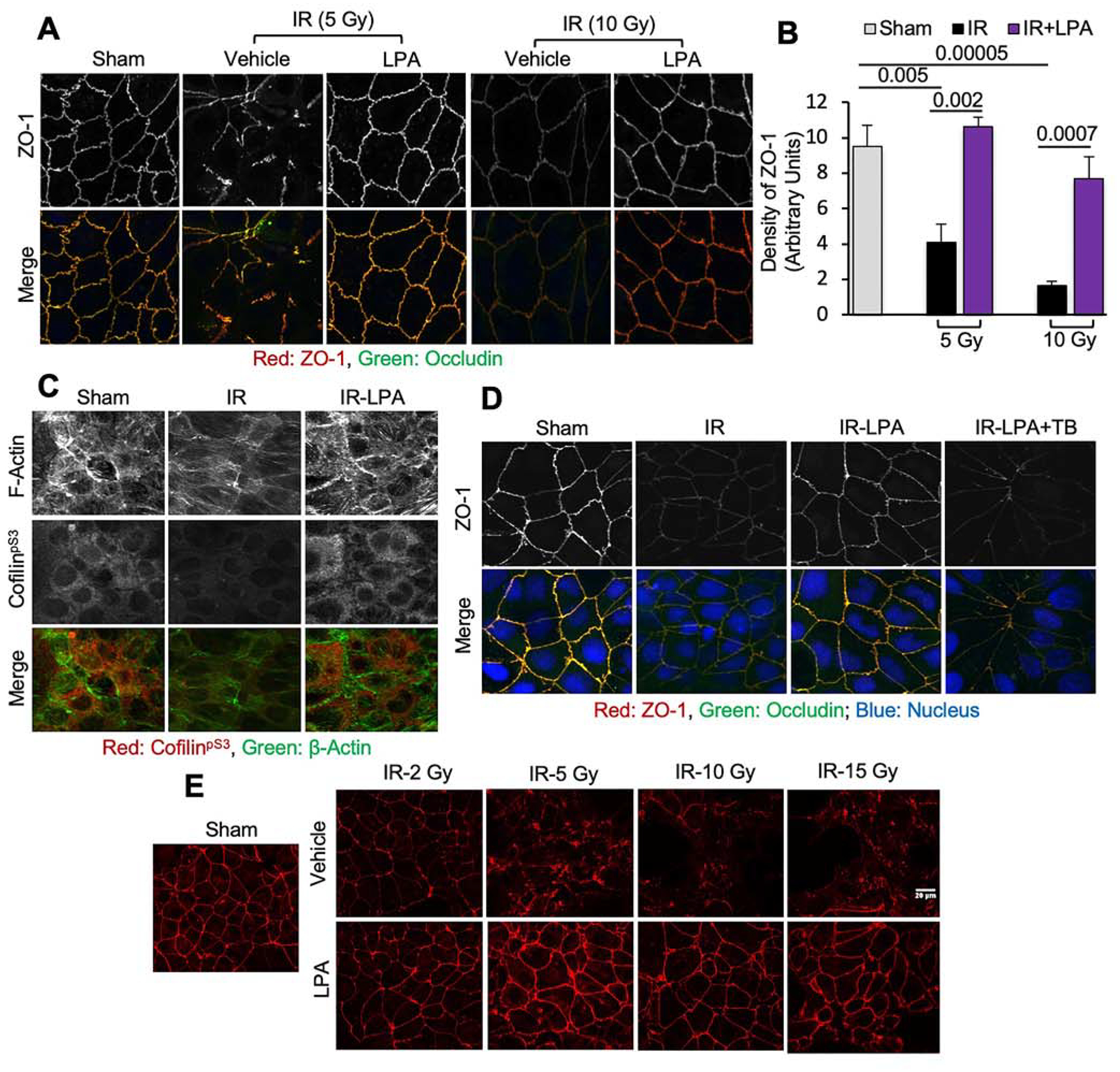
The tight junction (TJ) and barrier function of colonic epithelium is highly sensitive to ionizing radiation. We evaluated the effect of lysophosphatidic acid (LPA) and its analog, Radioprotein-1, on γ-radiation-induced colonic epithelial barrier dysfunction using Caco-2 and m-ICC12 cell monolayers in vitro and mice in vivo. Mice were subjected to either total body irradiation (TBI) or partial body irradiation (PBI-BM5). Intestinal barrier function was assessed by analyzing immunofluorescence localization of TJ proteins, mucosal inulin permeability, and plasma lipopolysaccharide (LPS) levels. Oxidative stress was analyzed by measuring protein thiol oxidation and antioxidant mRNA. In Caco-2 and m-ICC12 cell monolayers, LPA attenuated radiation-induced redistribution of TJ proteins, which was blocked by a Rho-kinase inhibitor. In mice, TBI and PBI-BM5 disrupted colonic epithelial tight junction and adherens junction, increased mucosal permeability, and elevated plasma LPS; TJ disruption by TBI was more severe in Lpar2-/- mice compared to wild-type mice. RP1, administered before or after irradiation, alleviated TBI and PBI-BM5-induced TJ disruption, barrier dysfunction, and endotoxemia accompanied by protein thiol oxidation and downregulation of antioxidant gene expression, cofilin activation, and remodeling of the actin cytoskeleton. These data demonstrate that LPAR2 receptor activation prevents and mitigates γ-irradiation-induced colonic mucosal barrier dysfunction and endotoxemia.

Keywords: endotoxemia; intestine; irradiation; lysophosphatidic acid; tight junction.


Conflict of interest statement

Conflict of interest

GT is a founder and stockholder in RxBio Inc. that has licensed the intellectual property for RP-1.

Figures

Figure 1:
Figure 1:. LPA attenuates radiation-induced disruption of tight junctions in the intestinal epithelial monolayers.
Caco-2 and m-ICC12 cell monolayers on Transwell inserts were irradiated (2–15 Gy) with or without LPA (10 μM) administered 30 min prior to irradiation. One hour after irradiation, tight junction integrity was examined. A & B: Caco-2 cell monolayers exposed to radiation with or without LPA were fixed and stained for occludin (green) and ZO-1 (red). Confocal images (A) and fluorescence density at the junctions (B) are presented. Values are Mean ± SEM (n = 4). The corresponding p-values (above bars) indicate significant differences between groups. NS indicates that the p-value is greater than 0.05. C: Caco-2 cell monolayers exposed to radiation (10 Gy) with or without LPA were stained for F-actin (green) and cofilinpS3 (red). D: Caco-2 cell monolayers were pretreated with toxin-B (TB) 30 min before LPA treatment. Monolayers were irradiated (10 Gy) 30 min after LPA treatment and stained for occludin (green), ZO-1 (red), and nucleus (blue). E: Confocal images for ZO-1 in m-ICC12 cell monolayers that were treated with LPA or vehicle before irradiation.
Figure 2:
Figure 2:. Effect of LPA2 receptor deficiency on the radiation-induced disruption of tight junction and adherens junction.
Wild type (WT) and LPA2 receptor knockout (Lpar2−/−) mice were subjected to TBI (9.5 Gy). Two hours after irradiation, colonic sections were stained for tight junction and adherens junction proteins. A: Merged fluorescence images for occludin (green), ZO-1 (red), and nucleus (blue). B: Fluorescence images for E-Cadherin (green), β-Catenin (red), and nucleus (blue). Representative images from 4 mice per each irradiated group and 2 mice for control groups are presented.
Figure 3:
Figure 3:. RP1 blocks TBI-induced disruption of tight junction and adherens junction, barrier dysfunction, and endotoxemia.
A-D: Wild type mice were injected with RP1 (0.5 mg/kg) or vehicle (Veh) 30 min prior to TBI (9.5 Gy) (IR and IR+RP1); the control group was sham-treated. At 2 hours post-irradiation, colonic sections were co-stained for occludin, ZO-1, and nucleus (A) or E-cadherin, β-catenin, and nucleus (B). Confocal images were captured, and fluorescence densities at the junctions were measured. Fluorescence densities for ZO-1 (C) and E-cadherin (D) are presented. Values are Mean ± SEM (n = 4; each value is an average of fluorescence values from 10 regions within the individual colonic section). E-H: At 4 hours post-irradiation, colonic mucosal extracts were immunoblotted (E), and the band densities for occludin (OCLN; F), claudin-3 (CLDN; G), and β-catenin (H). Values are Mean ± SEM (n = 4). I-K: At 2 and 4 hours post-irradiation, mucosal permeability in vivo in the colon (E) and ileum (F) and the plasma LPS levels (G) were measured. Values are Mean ± SEM (n = 4). In all graphs, the numbers above the bars are p-values for significant differences between groups indicated by the horizontal bars. “NS” indicates no significant difference between groups with the p-values greater than 0.05. The experiment was repeated with similar results.
Figure 4:
Figure 4:. RP1 mitigates TBI-induced disruption of AJC, mucosal barrier dysfunction, and endotoxemia.
Adult wild type mice were subjected to TBI (9.5 Gy). At 24 hours after irradiation, mice were injected with RP1 (0.5 mg/kg daily) or vehicle (Veh); the control group was sham-treated. At 24 and 48 hours after RP1 treatment, junctional integrity (A-F), gut permeability in vivo (G & H), and endotoxemia (I) were evaluated. Sections of the colon were co-stained for occludin, ZO-1, and nucleus (A), E-cadherin, β-catenin, and nucleus (B) or claudin-3, Claudin-2, and nucleus (C). Confocal fluorescence images were captured, and fluorescence densities at the junctions were measured. Fluorescence densities for ZO-1 (D) and β-catenin (E) and Claudin-3 (F) are presented. Mucosal permeability in vivo in the colon (G) and ileum (H) and plasma LPS levels (I) were measured. Values are Mean ± SEM (n = 4). The corresponding p-values (above bars) indicate significant differences between groups. NS indicates that the p-value is greater than 0.05. The experiment was repeated with similar results. Similar results were also produced in a similar experiment analyzed at seven days post-irradiation.
Figure 5:
Figure 5:. RP1 alleviates TBI-induced downregulation of antioxidant gene expression.
Wild type (WT) mice were subjected to TBI (9.5 Gy). At 24 hours after irradiation, mice were injected with RP1 (0.5 mg/kg daily) or vehicle (Veh); the control group was sham-treated. At 24 hours after RP1 treatment, RNA extracted from colonic mucosa was analyzed for mRNA for Gpx1 (A), SOD1 (B), CAT (C), Prdx1 (D), and Nrf2 (E) by RT-qPCR. Values are Mean ± SEM (n = 4). The corresponding p-values (above bars) indicate significant differences between groups. NS indicates that the p-value is greater than 0.05.
Figure 6:
Figure 6:. RP1 blocks PBI-induced disruption of AJC, barrier dysfunction, and endotoxemia.
Adult wild type mice were injected with RP1 (3 mg/kg) or vehicle (Veh) daily starting one day after partial body irradiation (PBI; 15.6 Gy); the control group was sham-treated. At varying times after irradiation, colonic sections were co-stained for occludin, ZO-1, and nucleus (A) or E-cadherin, β-catenin, and nucleus (B). Confocal fluorescence images were captured, and the fluorescence densities of ZO-1 (C) and E-cadherin (D) at the junctions were measured. Values are Mean ± SEM (n = 4). The corresponding p-values (above bars) indicate significant differences between groups. NS indicates that the p-value is greater than 0.05. Similar results were produced in a similar experiment when examined at 72 and 96 hours post-irradiation.
Figure 7:
Figure 7:. RP1 mitigates PBI-induced oxidative stress.
Adult mice were injected with RP1 (3 mg/kg) or vehicle (Veh) daily starting one day after partial body irradiation (PBI-BM5; 15.6 Gy); the control group was sham-treated. At 48 hours after irradiation (or 24 hours after RP1), colonic sections were stained for reduced-protein thiols, oxidized-protein thiols, and NRF2. Antioxidant gene expression was analyzed by RT-qPCR. A & B: Confocal images for protein thiols were captured (A), and the fluorescence densities were measured (B). C & D: Colonic sections were co-stained for F-actin (green), NRF2 (red) and nucleus (blue) (C). The protein extracts were immunoblotted for NRF2, and the band densities were measured. E-J: RNA isolated from colonic mucosa was analyzed for mRNA for Nrf2 (E), SOD1 (F), Gpx1 (G), CAT (H), Trx1 (I), and Prdx1 (J) by RT-qPCR. Values are Mean ± SEM (n = 4). The corresponding p-values (above bars) indicate significant differences between groups. NS indicates that the p-value is greater than 0.05. Similar results were produced in a similar experiment when examined at 72 and 96 hours post-irradiation.
Figure 8:
Figure 8:. RP1 mitigates PBI-induced F-actin remodeling and its association with apical junctional proteins.
Adult mice were injected with RP1 (3 mg/kg) or vehicle (Veh) daily starting one day after partial body irradiation (PBI-BM5; 15.6 Gy); the control group was sham-treated. A-E: At 48 hours after irradiation (or 24 hours after RP1), detergent-insoluble fractions of colonic mucosa were immunoblotted for tight junction and adherens junction proteins as well as NRF2 and β-actin (A). Band densities for E-cadherin (B), β-catenin (C), CLDN-3 (D), ZO-1 (E), and β-actin (F) were measured. G: Cryosections of the colon were fixed and stained for F-actin (red) and nucleus (blue). H & I: Colonic sections were co-stained for cofilinpS3 (red), F-actin (green) and nucleus (blue) (H). Mucosal extracts were immunoblotted for cofilinpS3 and β-actin (I). Band densities were measured, and the cofilinpS3 band densities presented by values normalized to corresponding actin band densities (I). In all graphs, values are Mean ± SEM (n = 3). The corresponding p-values (above bars) indicate significant differences between groups. NS indicates that the p-value is greater than 0.05.
Figure 9:
Figure 9:. Schematic outlining working model of the potential mechanisms associated with radiation-induced gut barrier dysfunction and its prevention by LPAR2 agonists.
Radiation induces oxidative stress by the production of reactive oxygen species (ROS) and down-regulation of antioxidant gene expression. Oxidative stress disrupts tight junctions by either signaling a cascade that targets tight junction directly or induces remodeling of the actin cytoskeleton via cofilin activation that leads to tight junction disruption. LPA and analogs that activate the LPAR2 receptor prevent and mitigate radiation-induced tight junction disruption, barrier dysfunction, and endotoxemia by blocking radiation-induced oxidative stress.

References

    1. Anderson JM, and Van Itallie CM (2009) Physiology and function of the tight junction. Cold Spring Harb Perspect Biol 1, a002584. - PMC - PubMed
    1. Rao R (2008) Oxidative stress-induced disruption of epithelial and endothelial tight junctions. Front Biosci 13, 7210–7226 - PMC - PubMed
    1. Van Itallie CM, and Anderson JM (2006) Claudins and epithelial paracellular transport. Annu Rev Physiol 68, 403–429 - PubMed
    1. Basuroy S, Seth A, Elias B, Naren AP, and Rao R (2006) MAPK interacts with occludin and mediates EGF-induced prevention of tight junction disruption by hydrogen peroxide. Biochem J 393, 69–77 - PMC - PubMed
    1. Basuroy S, Sheth P, Kuppuswamy D, Balasubramanian S, Ray RM, and Rao RK (2003) Expression of kinase-inactive c-Src delays oxidative stress-induced disassembly and accelerates calcium-mediated reassembly of tight junctions in the Caco-2 cell monolayer. J Biol Chem 278, 11916–11924 - PubMed

Publication types

MeSH terms

LinkOut - more resources