. 2020 Sep;40(9):2110-2116.
doi: 10.1111/liv.14601. Epub 2020 Jul 20.

Liver histopathology in severe COVID 19 respiratory failure is suggestive of vascular alterations

Affiliations

Liver histopathology in severe COVID 19 respiratory failure is suggestive of vascular alterations

Aurelio Sonzogni et al. Liver Int. 2020 Sep.

Abstract

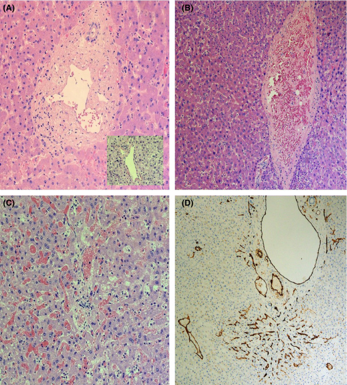
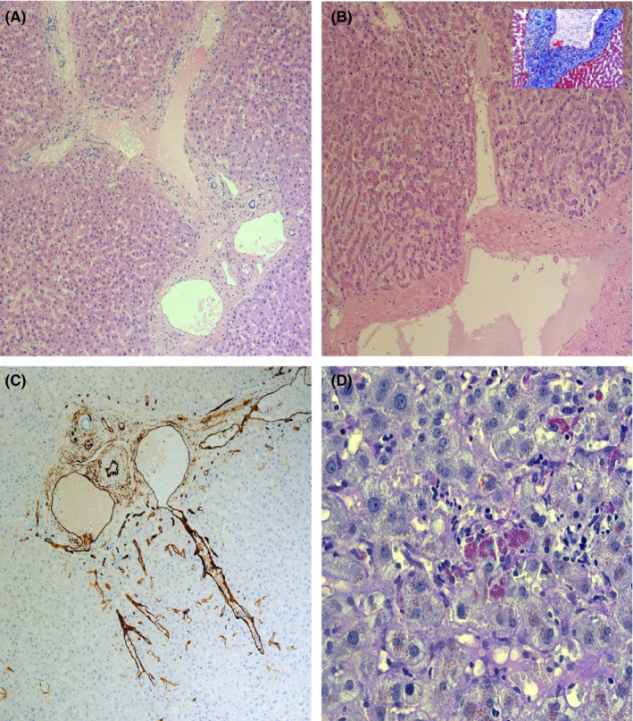
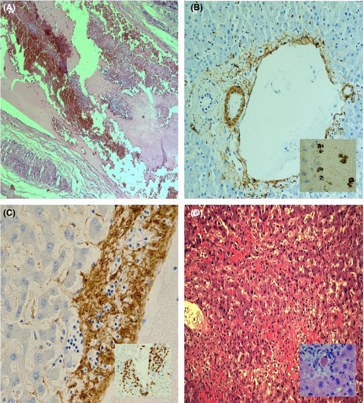
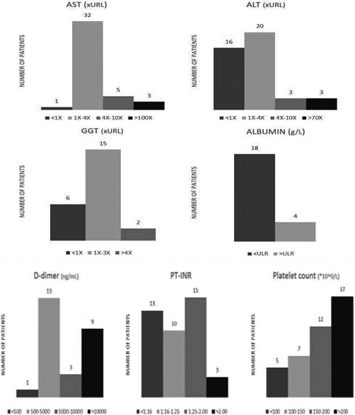
SARS2-CoV-2 breakout in Italy caused a huge number of severely ill patients with a serious increase in mortality. Although lungs seem to be the main target of the infection, very few information are available about liver involvement, possibly evocating a systemic disease. Post-mortem wedge liver biopsies from 48 patients died from severe pulmonary COVID-19 disease with respiratory failure were collected from two main hospitals in northern Italy. No patient had clinical symptoms of liver disease or signs of liver failure before and during hospitalization; for each of them liver function tests were available. All liver samples showed minimal inflammation features. Histological pictures compatible with vascular alterations were observed, characterized by increase in number of portal vein branches associated with lumen massive dilatation, partial or complete luminal thrombosis of portal and sinusoidal vessels, fibrosis of portal tract, focally markedly enlarged and fibrotic. SARS-CoV-2 was found in 15 of 22 samples tested by in situ hybridization method. Our preliminary results confirm the clinical impression that liver failure is not a main concern and this organ is not the target of significant inflammatory damage. Histopathological findings are highly suggestive for marked derangement of intrahepatic blood vessel network secondary to systemic changes induced by virus that could target not only lung parenchyma but also cardiovascular system, coagulation cascade and endothelial layer of blood vessels. It still remains unclear if the mentioned changes are directly related to virus infection or if SARS-CoV-2 triggers a series of reactions leading to striking vascular alterations.

Keywords: SARS-Cov-2 infection and liver biopsy; liver histopathology; liver morphology in COVID 19 disease.


Conflict of interest statement

None.

Figures

FIGURE 1
FIGURE 1
A: Regular liver architecture with scattered portal and lobular lymphocytes, moderate portal fibrosis; widened portal vein with fibrotic walls; bile duct without significant histological alteration. Compare normal portal tract in cadaveric liver donor (inset) (H&E, 100X); B: diffuse alterations of intrahepatic vascular structures characterized by severe dilatation and complete luminal thrombosis (H&E, 100X); C: portal vein and periportal sinusoids occlusive thrombosis (H&E, 100X); D: CD34 expression in abnormal portal vein branches endothelium and diffuse network of sinusoids in all parts of the lobule (100X)
FIGURE 2
FIGURE 2
A: Roughly enlarged portal field with proliferations of portal veins and luminal severe dilatation (H&E, 100X); B: severe portal vein wall fibrosclerosis (H&E, 100X), highlighted by trichrome stain (inset) (100X); C: portal veins showing lumen focally herniated in periportal liver parenchyma and completely coated by hepatocytes (CD34, 100X); D: activated Kupffer cells with large cytoplasm containing necrotic debris ( PAS diastase, 100X)
FIGURE 3
FIGURE 3
A: Example of genuine thrombosis: portal branch with clearly enlarged lumen obliterated by red cells mixed and stratified with lymphocytes and granulocytes (H&E, 100X); B: smooth muscle layer of portal vein lamina media extremely irregular fragmented (SMA, 100X); SAR‐CoV‐2 virions are demonstrated within vessel lumen and on endothelial cells (ISH) (inset); C: medium layer of a portal vein, partially lost and infiltrated by inflammatory cells lymphocytes, also attaching endothelial layer (SMA, 400X); CD3‐positive lymphocytes attack endothelium and medium vessel layer (inset); D: severe confluent haemorrhagic necrosis in a patient with elevation of ALT > 10 N (H&E, 100X); inset showing liver necrosis by apoptosis (H&E, 400X)
FIGURE 4
FIGURE 4
Biochemical changes in liver function tests (AST, ALT, GGT [fold‐x‐URL], albumin g/l) (top), PT‐INR, D‐Dimer and platelets (bottom). Data are not available for all patients as a result of a very short time of hospital stay. (URL: Upper Reference Limit)

References

    1. Yao XH, Li TY, He ZC, Ping YF, Liu HW, Yu SC, Mou HM, Wang LH, Zhang HR, Fu WJ, Luo T. A pathological report of three COVID‐19 cases by minimally invasive autopsies. Zhonghua Bing Li Xue Za Zhi. 2020;49:E009. 10.3760/cma.j.cn112151-20200312-00193. [Epub ahead of print] - DOI - PubMed
    1. Sun J, Aghemo A, Forner A, Valenti L. COVID‐19 and liver disease. Liver Int. 2020;40(6):1278‐1281. 10.1111/liv.14470 - DOI - PubMed
    1. Tang N, Li D, Wang X, et al. Abnormal coagulation parameters are associated with poor prognosis in patients with novel coronavirus pneumonia. J Thromb Haemost. 2020;18:844‐847. - PMC - PubMed
    1. Carsana L, Sonzogni A, Nasr A, et al. Pulmonary post‐mortem findings in a large series of COVID‐19 cases from Northern Italy. Lancet Infect Dis, accepted for publication. - PMC - PubMed
    1. Bangash MN, Patel J, Parekh D. COVID‐19 and the liver: little cause for concern. Lancet Gastroenterol Hepatol. 2020;5(6):529‐530. 10.1016/S2468-1253(20)30084-4 - DOI - PMC - PubMed

MeSH terms