. 2020 Sep;76(3):795-807.
doi: 10.1161/HYPERTENSIONAHA.120.15581. Epub 2020 Jul 13.

Endothelium-Macrophage Crosstalk Mediates Blood-Brain Barrier Dysfunction in Hypertension

Affiliations

Endothelium-Macrophage Crosstalk Mediates Blood-Brain Barrier Dysfunction in Hypertension

Monica M Santisteban et al. Hypertension. 2020 Sep.

Abstract

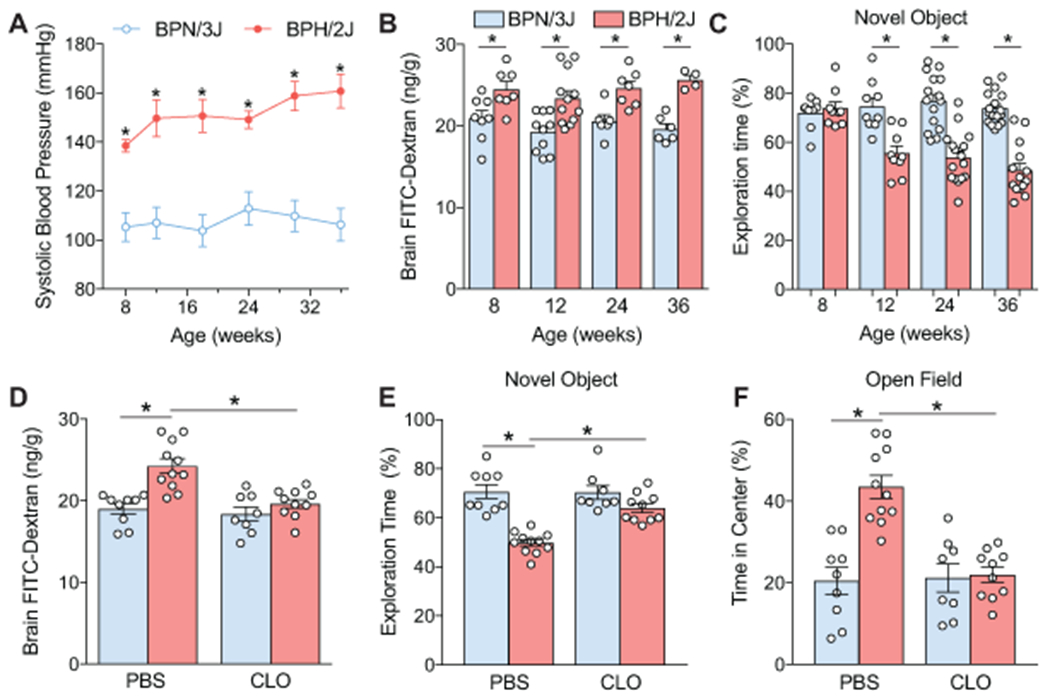
Hypertension is a leading cause of stroke and dementia, effects attributed to disrupting delivery of blood flow to the brain. Hypertension also alters the blood-brain barrier (BBB), a critical component of brain health. Although endothelial cells are ultimately responsible for the BBB, the development and maintenance of the barrier properties depend on the interaction with other vascular-associated cells. However, it remains unclear if BBB disruption in hypertension requires cooperative interaction with other cells. Perivascular macrophages (PVM), innate immune cells closely associated with cerebral microvessels, have emerged as major contributors to neurovascular dysfunction. Using 2-photon microscopy in vivo and electron microscopy in a mouse model of Ang II (angiotensin II) hypertension, we found that the vascular segments most susceptible to increased BBB permeability are arterioles and venules >10 µm and not capillaries. Brain macrophage depletion with clodronate attenuates, but does not abolish, the increased BBB permeability in these arterioles where PVM are located. Deletion of AT1R (Ang II type-1 receptors) in PVM using bone marrow chimeras partially attenuated the BBB dysfunction through the free radical-producing enzyme Nox2. In contrast, downregulation of AT1R in cerebral endothelial cells using a viral gene transfer-based approach prevented the BBB disruption completely. The results indicate that while endothelial AT1R, mainly in arterioles and venules, initiate the BBB disruption in hypertension, PVM are required for the full expression of the dysfunction. The findings unveil a previously unappreciated contribution of resident brain macrophages to increased BBB permeability of hypertension and identify PVM as a putative therapeutic target in diseases associated with BBB dysfunction.

Keywords: angiotensin II; arterioles; cerebrovascular circulation; cognitive dysfunction.


Conflict of interest statement

DISCLOSURES

CI serves on the strategic advisory board of Broadview Ventures. The other authors have no conflicts to disclose.

Figures

Figure 1.
Figure 1.. Ang II hypertension enhances BBB permeability.
A. Slow-pressor Ang II increases systolic BP gradually (n=10; 2-way ANOVA, Bonferroni’s multiple comparisons test; *p<0.001). B. Ang II hypertension enhances BBB permeability to 3kDa FITC-dextran (n=13-14; two-tailed unpaired t-test, *p<0.0001 vs saline). C. Ten minutes following 3kDa FITC-dextran administration, BBB leakage is visualized by 2-photon microscopy. Scale bar: 100μm. D. In cerebral microvascular preparation Ang II hypertension reduces the mRNA expression of the TJ proteins claudin-5 and occludin, but not ZO-1 (n=7-8). Expression of the vesicular transport regulator Mfsd2a is also suppressed (two-tailed unpaired t-test, claudin-5 n=10-12 *p=0.0275, occludin n=11-16 *p<0.0001, Mfsd2a n=11-12 *p=0.0002). E. In cerebral endothelial cell cultures, Ang II treatment (300nM; 24 hours) disrupts TJ as reflected by claudin-5 (green) internalization and ZO-1 (magenta) disorganization. Scale bar: 50μm. Abbreviations: A: arteriole; V: venule; SBP: systolic blood pressure.
Figure 2.
Figure 2.. Ang II hypertension induces tight junction remodeling and enhances endothelial vesicle density.
A. Electron micrographs of neocortical capillaries showing cerebral endothelial cells forming tight junctions (magenta outline) and containing endothelial vesicles (highlighted in orange and black arrows). Tight junction remodeling and increased vesicle density is observed in Ang II (right) vs saline (left) treated mice. Scale bar: 500nm. B. Tight junction length is reduced in both capillaries and arterioles in Ang II hypertension (Saline/Ang II: capillaries n=102, arterioles n=69/79; 2-way ANOVA, Interaction p=0.0509, Vessel Size Factor p=0.0039, Ang II Factor p<0.0001; Bonferroni’s multiple comparison test: Ang II vs Saline capillaries p=0.0422, arterioles p<0.0001, Saline capillaries vs Saline arterioles p=0.0050). C. Tortuosity ratio (see Methods) is reduced capillaries and arterioles in Ang II hypertension (Saline/Ang II: capillaries n=104/102, arterioles n=71/79; 2-way ANOVA, Interaction p=0.4364, Vessel Size Factor p=0.0577, Ang II Factor p<0.0001; Bonferroni’s multiple comparison test: Ang II vs Saline capillaries p<0.0001, arterioles p<0.0001). D. Cerebral endothelial vesicle density increases in Ang II hypertension (Saline/Ang II: capillaries n=104/60, arterioles n=41/44; 2-way ANOVA, Interaction p=0.0001, Vessel Size Factor p=0.0100, Ang II Factor p<0.0001; Bonferroni’s multiple comparison test: Ang II vs Saline capillaries p<0.0001, arterioles p<0.0001, Ang II capillaries vs Ang II arterioles p<0.0001). E. Vesicle density increases with vessel diameter in Ang II hypertension. Lighter symbols indicate capillaries, and darker symbols indicate arterioles. Line and shading indicate linear regression fit with 95% confidence interval. Multiple comparison test (95% confidence ANCOVA) was conducted between linear fit models (Saline vs Ang II p=0.0025).
Figure 3.
Figure 3.. PVM depletion attenuates BBB permeability in cerebral microvessels larger than 30μm.
A. PVM depletion with liposomal clodronate (CLO) attenuates the breakdown of the BBB in Ang II hypertension (n=8-9; 2-way ANOVA; Liposome Factor p=0.0029, Ang II Factor p<0.0001, Interaction p=0.0054; Bonferroni’s multiple comparison test: Saline-PBS vs Ang II-PBS p<0.0001, Saline-CLO vs Ang II-CLO p=0.0219, Ang II-PBS vs Ang II-CLO p=0.0007). B. Three-dimensional reconstruction of the vasculature labeled with 70kDa molecular weight Texas Red-dextran (magenta) and extravasated 3kDa molecular weight FITC-dextran (green). Extravascular and intravascular fluorescent signal is monitored over 10 minutes at the yellow line drawn on the vascular reconstruction above. C. Representative 2-photon images taken 5 minutes after injection of FITC dextran showing that BBB leakage in Ang II hypertension is prevented by PVM depletion. Scale bar is 100μm. D. Scatter of individual measurements plotting mean of extravascular signal intensity over 10 minutes versus depth from the surface of the brain. Top panels represent arterioles, and bottom panels venules; the diameter of the symbols (circles) is proportional to vessel diameter. Lines and shading indicate regression fit (logarithmic conversion on intensity axis) with 95% confidence interval. Multiple comparison test (95% confidence ANCOVA) was conducted between linear fit models (n=91-136; Artery: *p<0.01 vs Saline-PBS, #p<0.01 vs Ang II-PBS; Vein: *p<0.01 vs Saline-PBS, Ang II-PBS vs Ang II-CLO p=0.0687). E. Mean extravascular signal was compared over microvascular diameters in the first 60μm below the surface of the brain. Ang II hypertension does not enhance leakage of FITC-dextran in capillaries (2-way ANOVA, Interaction p=0.1562, Liposome Factor p=0.5159, Ang II Factor p=0.7333). Ang II hypertension enhances leakage of FITC-dextran in arterioles, which is attenuated by PVM depletion (2-way ANOVA, Interaction p=0.0786, Liposome Factor p=0.0014, Ang II Factor p<0.0001; Bonferroni’s multiple comparison test: Saline-PBS vs Ang II-PBS p=0.0001, Ang II-PBS vs Ang II-CLO p<0.0001).
Figure 4.
Figure 4.. PVM depletion restores BBB integrity and improves cognitive function in chronically hypertensive BPH/2J mice.
A. BPH/2J mice have elevated systolic BP as early as 8 weeks of age compared to normotensive BPN/3J controls (n=7-10; 2-way ANOVA, Bonferroni’s multiple comparisons test; *p<0.001). B. Enhanced leakage of FITC-Dextran is found in BPH/2J mice as early as 8 weeks of age (2-way ANOVA, Interaction p=0.6749, Age Factor p=0.2640, Genotype Factor p<0.0001; Bonferroni’s multiple comparison test: 8 weeks n=7-8 *p=0.0299; 12 weeks n=10-12 *p=0.0010; 24 weeks n=6-7 *p=0.0189; 36 weeks n=6-4 *p=0.0017). C. BPH/2J mice spend less time exploring a novel object starting at 12 weeks, and progressing through 36 weeks of age (2-way ANOVA, Interaction p<0.0001, Age Factor p=0.0009, Genotype Factor p<0.0001; Bonferroni’s multiple comparison test: 8 weeks n=9 p>0.9999; 12 weeks n=9 *p=0.0001; 24 weeks n=15-16 *p<0.0001; 36 weeks n=14-17 *p<0.0001). D. PVM depletion with liposomal clodronate (CLO) attenuates the BBB breakdown in BPH/2J mice (n n=8-11, 2-way ANOVA, Interaction p=0.0118, Liposome Factor p=0.0013, Genotype Factor p=0.0001; Bonferroni’s multiple comparison test: BPN-PBS vs BPH-PBS *p<0.0001, BPH-CLO vs BPH-PBS *p=0.0003). E. PVM depletion restores cognitive deficits assessed by novel object recognition test in BPH/2J mice (n=8-11, 2-way ANOVA, Interaction p=0.0022, Liposome Factor p=0.0030, Genotype Factor p<0.0001; Bonferroni’s multiple comparison test: BPN-PBS vs BPH-PBS *p<0.0001, BPH-CLO vs BPH-PBS *p=0.0002). F. PVM depletion restores the aversion toward the center area at the open-field test without affecting total ambulatory time in BPH/2J mice (n=8-11, 2-way ANOVA, Interaction p=0.0005, Liposome Factor p=0.0010, Genotype Factor p=0.0002; Bonferroni’s multiple comparison test: BPN-PBS vs BPH-PBS *p<0.0001, BPH-CLO vs BPH-PBS *p<0.0001).
Figure 5.
Figure 5.. AT1R and Nox2 on PVM contribute to the BBB breakdown in Ang II hypertension.
A. Depletion of AT1R or Nox2 on PVM in AT1R−/−→WT or Nox2−/−→WT chimeras attenuates the BBB dysfunction in Ang II hypertension (n=8-12, 2-way ANOVA, Interaction p=0.0275, Donor Genotype Factor p=0.0354, Ang II Factor p<0.0001; Bonferroni’s multiple comparison test: Ang II vs Saline [WT p<0.0001, AT1R−/− p=0.0384, Nox2−/− p=0.0047], chimera-Ang II vs WT-Ang II [AT1R−/− p=.0037, Nox2−/− p=0.0047]). B. Depletion of AT1R or Nox2 in PVM on does not affect the hypertensive response to Ang II infusion over 14 days (n=8-10, 2-way ANOVA, Bonferroni’s multiple comparison test: *p<0.01 Ang II vs saline). C. Introducing AT1R+/+ or Nox2+/+ PVM via bone marrow transplantation in AT1R−/− or Nox2−/− does not induce opening of the BBB during Ang II hypertension (n=6-12, 2-way ANOVA, Interaction p<0.0001, Recipient Genotype p<0.0001, Ang II p<0.0001; Bonferroni’s multiple comparison test: Ang II vs Saline WT p<0.0001, chimera-Ang II vs WT-Ang II [AT1R−/− p<0.0001, Nox2−/− p<0.0001]). D. WT→AT1R−/− mice do not develop hypertension during Ang II infusion, while WT→Nox2−/− have increased SBP during Ang II infusion except for a small attenuation at day 14 (n=6-12, 2-way ANOVA, Bonferroni’s multiple comparison test: *p<0.01 Ang II vs saline, #p=0.0392 WT→Nox2−/− Ang II vs WT→WT Ang II).
Figure 6.
Figure 6.. Cerebral endothelial AT1R are necessary for the BBB breakdown in Ang II hypertension.
A. AAV-BR1-iCre (1.8x10¹¹ VG) administered i.v. in Ai14 ROSA26TdTomato reporter mice induces Td-Tomato expression (magenta) overlapping with the endothelial-specific marker CD31 (green). B. Flow cytometry data showing that AAV-BR1-iCre induces recombination in endothelial cells (EC), but not microglia (MG) or brain macrophages (MΦ), attesting to the cell-type specificity of the viral transduction (n=3). C. Flow cytometry data demonstrating the specificity of AAV-BR1-iCre recombination in cortical (CTX) and hippocampal (Hipp) endothelial cells, but not in endothelial cells of the dura mater, which contains extracerebral vessels (n=3). D. AAV-BR1-iCre viral transduction maintains >75% efficiency in vessels larger than 50μm in diameter, but decreases in vessels >50μm. Percent of CD31+ endothelial area that is Td-Tomato + is plotted against vessel diameter. Line and shading indicate linear regression fit with 95% confidence interval. E. AAV-BR1-iCre (1.8x10¹¹ viral genome; i.v.) induces a 40% reduction in agtr1a genomic DNA when administered to AT1Aflox mice (n=7, two-tailed unpaired t-test, *p<0.0001 vs WT + iCre EC). F. AT1R knockdown in cerebral endothelial cells prevents the breakdown of BBB in Ang II hypertension (n=7- 9, 2-way ANOVA, Interaction p=0.0009, Genotype p=0.0007, Ang II p<0.0001; Bonferroni’s multiple comparison test: WT + iCre Saline vs Ang II *p<0.0001; WT + iCre Ang II vs AT1Aflox + iCre Ang II p<0.0001).

References

    1. WHO. A global brief on hypertension : Silent killer, global public health crisis. World health organization. 2013
    1. Forouzanfar MH, Liu P, Roth GA, Ng M, Biryukov S, Marczak L, et al. Global burden of hypertension and systolic blood pressure of at least 110 to 115 mmHg, 1990-2015. Jama. 2017;317:165–182 - PubMed
    1. Dorans KS, Mills KT, Liu Y, He J. Trends in prevalence and control of hypertension according to the 2017 american college of cardiology/american heart association (acc/aha) guideline. J Am Heart Assoc. 2018;7:e008888. - PMC - PubMed
    1. Iadecola C, Gottesman RF. Neurovascular and cognitive dysfunction in hypertension. Circ Res. 2019;124:1025–1044 - PMC - PubMed
    1. Pires PW, Dams Ramos CM, Matin N, Dorrance AM. The effects of hypertension on the cerebral circulation. Am J Physiol Heart Circ Physiol. 2013;304:H1598–1614 - PMC - PubMed

Publication types

MeSH terms

Substances