Case Reports
. 2020 Sep 24;82(9):1271-1276.
doi: 10.1292/jvms.19-0605. Epub 2020 Jul 13.

Treatment with rivaroxaban and monitoring of coagulation profiles in two dogs with venous thromboembolism

Affiliations
Case Reports

Treatment with rivaroxaban and monitoring of coagulation profiles in two dogs with venous thromboembolism

Mona Uchida et al. J Vet Med Sci. .

Abstract

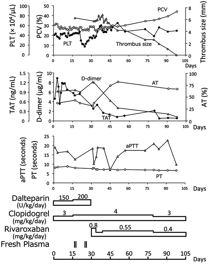
Two dogs with immune-mediated hemolytic anemia complicated with thromboembolism were presented. Both of the dogs were initially treated with immunosuppressive therapy in conjunction with dalteparin and clopidogrel. Although the immunosuppressive therapy was effective, peritoneal effusion due to thromboembolism was observed during the course of the disease in these dogs. After initiation of rivaroxaban treatment, peritoneal effusion decreased immediately in parallel with the normalization of D-dimer, antithrombin (AT), and thrombin-antithrombin complex (TAT). Hematochezia, cutaneous hemorrhage, and hematuria were observed as adverse events after administration of rivaroxaban in one case. Rivaroxaban was effective for the control of thromboembolism secondary to immune-mediated hemolytic anemia, and D-dimer, AT, and TAT were useful to monitor the status of thromboembolic disease in dogs.

Keywords: direct oral anticoagulant; heparin resistance; immune-mediated hemolytic anemia; thrombotic marker; venous thromboembolism.


Figures

Fig. 1.
Fig. 1.
Diagnostic imaging of thrombi in Dog 1. Ultrasonography of the splenic vein (A, E) and transverse image of the abdominal computed tomography (CT) (B, C, D). The thrombus (arrow) in the splenic vein was visualized by ultrasonography (A), but disappeared after treatment (E). In the abdominal CT scan in the post-contrast phase, thrombi (arrows) were found in the portal vein (B), the splenic vein (C), and the caudal mesenteric vein (D).
Fig. 2.
Fig. 2.
Treatment and course of disease in Dog 1. Packed cell volume (PCV), the number of platelets (PLT), activated partial thromboplastin time (aPTT), prothrombin time (PT), antithrombin (AT), D-dimer, and thrombin-antithrombin complex (TAT) were measured until 98 days after the first presentation. The maximum short diameter of the thrombus in the splenic vein was measured to indicate the thrombus size by ultrasonography. Names and doses of antithrombotic agents are shown under the graphs of coagulation/fibrinolysis parameters.
Fig. 3.
Fig. 3.
Treatment and course of disease in Dog 2. Packed cell volume (PCV), the number of platelets (PLT), activated partial thromboplastin time (aPTT), prothrombin time (PT), antithrombin (AT), D-dimer, and thrombin-antithrombin complex (TAT) were measured until 34 days after the first presentation. Names and doses of antithrombotic agents are shown under the graphs of coagulation/fibrinolysis parameters.

References

    1. Bae J., Kim H., Kim W., Kim S., Park J., Jung D. I., Yu D.2019. Therapeutic monitoring of rivaroxaban in dogs using thromboelastography and prothrombin time. J. Vet. Intern. Med. 33: 1322–1330. doi: 10.1111/jvim.15478 - DOI - PMC - PubMed
    1. Barrett Y. C., Wang Z., Frost C., Shenker A.2010. Clinical laboratory measurement of direct factor Xa inhibitors: anti-Xa assay is preferable to prothrombin time assay. Thromb. Haemost. 104: 1263–1271. doi: 10.1160/TH10-05-0328 - DOI - PubMed
    1. Beyer-Westendorf J., Förster K., Pannach S., Ebertz F., Gelbricht V., Thieme C., Michalski F., Köhler C., Werth S., Sahin K., Tittl L., Hänsel U., Weiss N.2014. Rates, management, and outcome of rivaroxaban bleeding in daily care: results from the Dresden NOAC registry. Blood 124: 955–962. doi: 10.1182/blood-2014-03-563577 - DOI - PMC - PubMed
    1. Boisclair M. D., Lane D. A., Wilde J. T., Ireland H., Preston F. E., Ofosu F. A.1990. A comparative evaluation of assays for markers of activated coagulation and/or fibrinolysis: thrombin-antithrombin complex, D-dimer and fibrinogen/fibrin fragment E antigen. Br. J. Haematol. 74: 471–479. doi: 10.1111/j.1365-2141.1990.tb06337.x - DOI - PubMed
    1. Brotman D. J., Girod J. P., Posch A., Jani J. T., Patel J. V., Gupta M., Lip G. Y., Reddy S., Kickler T. S.2006. Effects of short-term glucocorticoids on hemostatic factors in healthy volunteers. Thromb. Res. 118: 247–252. doi: 10.1016/j.thromres.2005.06.006 - DOI - PubMed

Publication types

MeSH terms