Novel Clinical and Radiological Findings in a Family with Autosomal Recessive Omodysplasia
- PMID: 32655339
- PMCID: PMC7325126
- DOI: 10.1159/000506384
Novel Clinical and Radiological Findings in a Family with Autosomal Recessive Omodysplasia
Abstract
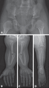
Autosomal recessive omodysplasia (GPC6-related) is a rare short-limb skeletal dysplasia caused by biallelic mutations in the GPC6 gene. Affected individuals manifest with rhizomelic short stature, decreased mobility of elbow and knee joints as well as craniofacial anomalies. Both upper and lower limbs are severely affected. These manifestations contrast with normal height and limb shortening restricted to the arms in autosomal dominant omodysplasia (FZD2-related). Here, we report 2 affected brothers of Pakistani descent from Denmark with GPC6-related omodysplasia, aiming to highlight the clinical and radiological findings. A homozygous deletion of exon 6 in the GPC6 gene was detected. The pathognomonic radiological findings were distally tapered humeri and femora as well as severe proximal radioulnar diastasis. On close observations, we identified a recurrent and not previously described type of abnormal patterning in all long bones.
Keywords: GPC6; Omodysplasia; Radiological feature; Recessive omodysplasia; Skeletal dysplasia.
Copyright © 2020 by S. Karger AG, Basel.
Figures


References
-
- Albano LM, Oliveira LA, Bertola DR, Mazzu JF, Kim CA. Omodysplasia: the first reported Brazilian case. Clinics (Sao Paulo) 2007;62:531–534. - PubMed
-
- al Gazali LI, Aboual-Asaad F. Autosomal recessive omodysplasia. Clin Dysmorphol. 1995;4:52–56. - PubMed
-
- Barrow M, Fitzsimmons JS. A new syndrome. Short limbs, abnormal facial appearance, and congenital heart defect. Am J Med Genet. 1984;18:431–433. - PubMed
-
- Baxova A, Maroteaux P, Barosova J, Netriova I. Parental consanguinity in two sibs with omodysplasia. Am J Med Genet. 1994;49:263–265. - PubMed
LinkOut - more resources
Full Text Sources

