. 2020 Jun 23:14:176.
doi: 10.3389/fncel.2020.00176. eCollection 2020.

CB1 Cannabinoid Receptors Stimulate Gβγ-GRK2-Mediated FAK Phosphorylation at Tyrosine 925 to Regulate ERK Activation Involving Neuronal Focal Adhesions

Affiliations

CB1 Cannabinoid Receptors Stimulate Gβγ-GRK2-Mediated FAK Phosphorylation at Tyrosine 925 to Regulate ERK Activation Involving Neuronal Focal Adhesions

George D Dalton et al. Front Cell Neurosci. .

Abstract

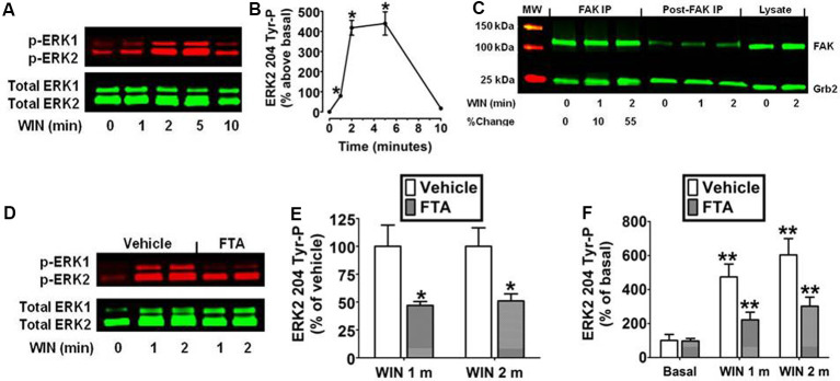
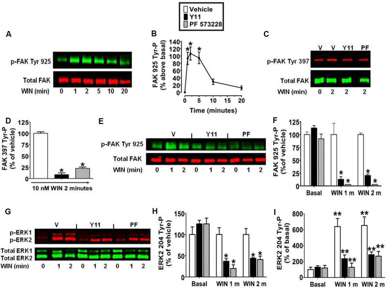
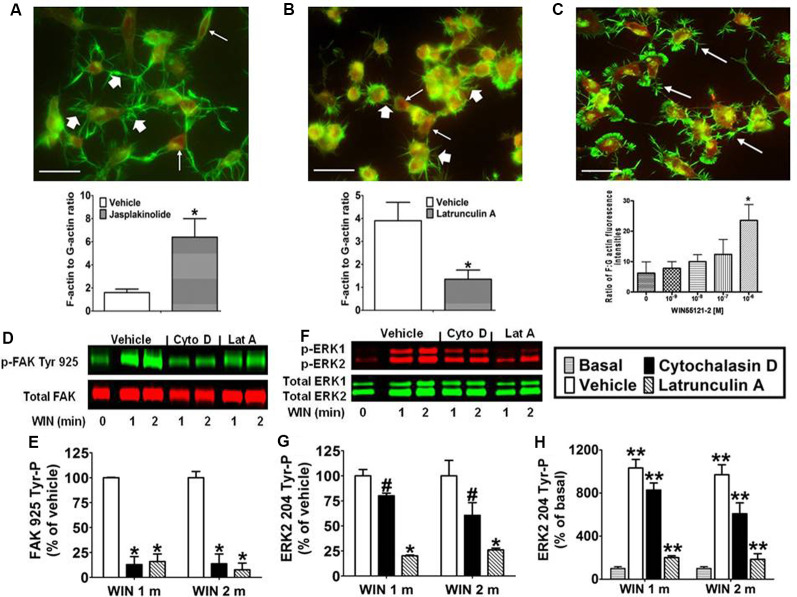
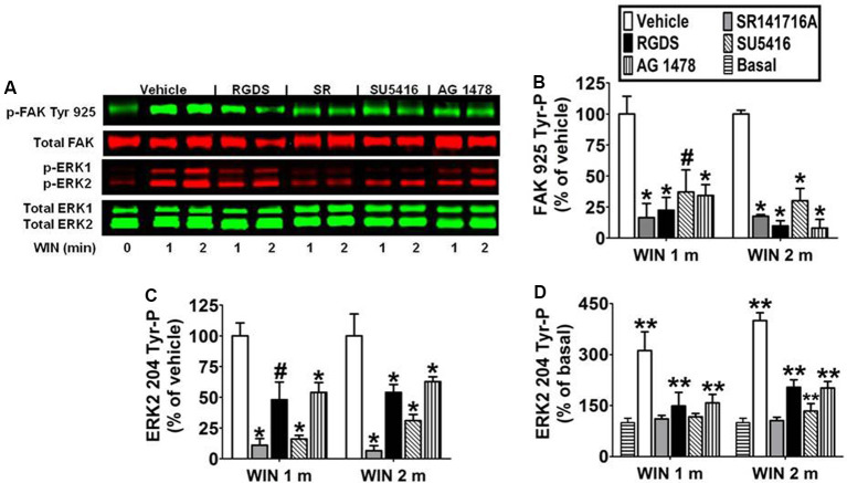
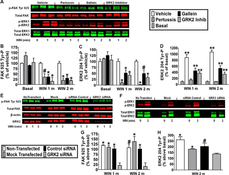
CB1 cannabinoid receptors (CB1) are abundantly expressed in the nervous system where they regulate focal adhesion kinase (FAK) and the mitogen-activated protein kinases (MAPK) extracellular signal-regulated kinase 1 and 2 (ERK1/2). However, the role of CB1-stimulated FAK 925 tyrosine phosphorylation (Tyr-P) in regulating ERK1/2 activation remains undefined. Here, immunoblotting analyses using antibodies against FAK phospho-Tyr 925 and ERK2 phospho-Tyr 204 demonstrated CB1-stimulated FAK 925 Tyr-P and ERK2 204 Tyr-P (0-5 min) which was followed by a decline in Tyr-P (5-20 min). CB1 stimulated FAK-Grb2 association and Ras-mediated ERK2 activation. The FAK inhibitors Y11 and PF 573228 abolished FAK 925 Tyr-P and partially inhibited ERK2 204 Tyr-P. FAK 925 Tyr-P and ERK2 204 Tyr-P were adhesion-dependent, required an intact actin cytoskeleton, and were mediated by integrins, Flk-1 vascular endothelial growth factor receptors, and epidermal growth factor receptors. FAK 925 Tyr-P and ERK2 204 Tyr-P were blocked by the Gβγ inhibitor gallein, a GRK2 inhibitor, and GRK2 siRNA silencing, suggesting Gβγ and GRK2 participate in FAK-mediated ERK2 activation. Together, these studies indicate FAK 925 Tyr-P occurs concurrently with CB1-stimulated ERK2 activation and requires the actin cytoskeleton and Gi/oβγ-GRK2-mediated cross-talk between CB1, integrins, and receptor tyrosine kinases (RTKs).

Keywords: CB1; ERK; FAK; GRK2; Grb2; Gβγ.


Figures

Figure 1
Figure 1
Cannabinoid receptors (CB1) stimulate Grb2/Sos1/Ras-dependent activation of extracellular signal-regulated kinase 2 (ERK2) phosphorylation at tyrosine 204 in N18TG2 cells. (A,B) Cells were treated with 0.01 μM WIN55212-2 (WIN) at 37°C for 1, 2, 5, and 10 min (m). Cell lysates were analyzed using western blots and representative blot images and analysis of ERK2 204 Tyr-P (normalized to total ERK2 levels) are shown. Data are reported as mean ± SEM of the % change over basal from three separate experiments. (C) Cells were treated with 0.01 μM WIN for 1 or 2 m at 37°C. Proteins from total cell lysate (500 μg) were immunoprecipitated with FAK antibodies and were immunoblotted with FAK and Grb2 antibodies. Data are the % change from basal Grb2 levels associated with FAK (normalized to total FAK at each time point). MW, molecular weight size marker. (D–F) Cells were pretreated for 15 m with a vehicle or the Ras inhibitor farnesylthiosalicylic acid (FTA, 10 μM) before treatment with 0.01 μM WIN for 1 or 2 m at 37°C. Cell lysates were analyzed using western blots and representative blot images and analysis of ERK2 204 Tyr-P (normalized to total ERK2 levels) are shown. Data are reported as mean ± SEM of panel (E) % of vehicle-treated ERK2 204 Tyr-P at the same time point or (F) the % of basal/time 0 ERK2 204 Tyr-P from three separate experiments. Significance was assessed using Student’s t-test [*p < 0.01 indicates significantly different from vehicle-treated (at the same time point); **p < 0.05 indicates significantly different from basal/time 0]. For each dataset, cells were cultured and experiments were completed on at least three separate occasions.
Figure 2
Figure 2
Inhibition of focal adhesion kinase (FAK) 925 Tyr-P abrogates CB1-stimulated ERK2 204 Tyr-P in N18TG2 cells. Cells were treated with 0.01 μM WIN55212-2 (WIN) at 37°C for 1, 2, 5, 10, or 20 min (m). (A,B) Cell lysates were analyzed using western blots and representative blot images and analysis of FAK 925 Tyr-P (normalized to total FAK levels) are shown. *p < 0.01 indicates significantly different from basal/time 0 using Student’s t-test. (C–I) Cells were pretreated for 15 m with 10 nM FAK inhibitor [Y11 or PF 573228 (PF)] before treatment with 0.01 μM WIN. Cell lysates were analyzed using western blots and representative blot images and analysis of (C,D) FAK 397 Tyr-P, (E,F) FAK 925 Tyr-P, and (G–I) ERK2 204 Tyr-P (normalized to total FAK or ERK2 levels) are shown. Data are reported as mean ± SEM of (B) the % change over basal FAK 925 Tyr-P, (D,F,H) the % of vehicle-treated FAK 397, FAK 925 and ERK2 204 Tyr-P at the same time point, and (I) the % of basal/time 0 from three separate experiments. For 2C-I, significance was assessed using One Way ANOVA followed by Dunnett’s multiple comparisons posthoc test [*p < 0.01 indicates significantly different from vehicle-treated (at the same time point); **p < 0.05 indicates significantly different from basal/time 0]. For each dataset, cells were cultured and experiments were completed on at least three separate occasions.
Figure 3
Figure 3
CB1-stimulated FAK phosphorylation at tyrosine 925 and ERK2 phosphorylation at tyrosine 204 requires an intact actin cytoskeleton in N18TG2 cells. (A–C) Cells grown on glass coverslips were treated with jasplakinolide (4 nM, 10 min), latrunculin A (0.01 μg/ml, 30 min), or WIN55212-2 (1–1,000 nM, 20 min). Actin remodeling was detected by fluorescent double labeling of F-actin (Alexa Fluor 488 phalloidin, short white arrow) and G-actin [Texas Red DNase I, long white arrow; neurite outgrowths, white arrow in panel (C)]. F-actin to G-actin ratios were quantified from green and red staining intensities using Image Pro 4.5 software. *p < 0.01 indicates significantly different from vehicle-treated using Student’s t-test. (D–H) Cells were pretreated with cytochalasin D (2 μM, Cyto D) or latrunculin A (1 μM, Lat A) before treatment with 0.01 μM WIN55212-2 (WIN) for 1 or 2 min (m) at 37°C. Cell lysates were analyzed using western blots and representative blot images and analysis of FAK 925 Tyr-P (normalized to total FAK levels) and ERK2 204 Tyr-P (normalized to total ERK2 levels) are shown. Data are reported as mean ± SEM of (E) % of vehicle-treated FAK 925 Tyr-P at the same time point, (G) % of vehicle-treated ERK2 204 Tyr-P at the same time point, and (H) % of basal/time 0 ERK2 204 Tyr-P from three separate experiments. Significance was assessed using One Way ANOVA followed by Dunnett’s multiple comparisons posthoc test [#p < 0.05, *p < 0.01 indicates significantly different from vehicle-treated (at the same time point); **p < 0.05 indicates significantly different from basal/time 0]. For each dataset in panels (D–H), cells were cultured and experiments were completed on at least three separate occasions.
Figure 4
Figure 4
CB1-stimulated FAK phosphorylation at tyrosine 925 and ERK2 phosphorylation at tyrosine 204 are mediated by integrins and receptor tyrosine kinases in N18TG2 cells. (A–D) Cells were pretreated for 15 min (m) with the integrin antagonist RGDS peptide (100 μM), CB1 antagonist SR141716A (1 μM, SR), Flk-1 VEGFR antagonist SU 5416 (1 μM), or EGFR antagonist AG 1478 (2 μM) before treatment with 0.01 μM WIN55212-2 (WIN) for 1 or 2 m at 37°C. Cell lysates were analyzed using western blots and representative blot images are shown. Data are reported as mean ± SEM of (B) % of vehicle-treated FAK 925 Tyr-P (normalized to total FAK levels) at the same time point, (C) % of vehicle-treated ERK2 204 Tyr-P (normalized to total ERK2 levels) at the same time point, and (D) % of basal/time 0 ERK2 204 Tyr-P (normalized to total ERK2 levels). Significance was assessed using One Way ANOVA followed by Dunnett’s multiple comparisons posthoc test [#p < 0.05, *p < 0.01 indicates significantly different from vehicle-treated (at the same time point); **p < 0.05 indicates significantly different from basal/time 0]. For each dataset, cells were cultured and experiments were completed on at least three separate occasions.
Figure 5
Figure 5
CB1-stimulated FAK phosphorylation at tyrosine 925 and ERK2 phosphorylation at tyrosine 204 are mediated by Gβγ and GRK2 in N18TG2 cells. (A–D) Cells were pretreated with the Gi/o inhibitor pertussis toxin (100 ng/mL, 20 h), Gβγ inhibitor gallein (10 μM, 15 min), or GRK2 inhibitor (1 μM, 15 min) before treatment with 0.01 μM WIN55212-2 (WIN) for 1 or 2 min (m) at 37°C. Cell lysates were analyzed using western blots and representative blot images are shown. Data are reported as mean ± SEM of (B) % of vehicle-treated FAK 925 Tyr-P (normalized to total FAK levels) at the same time point, (C) % of vehicle-treated ERK2 204 Tyr-P (normalized to total ERK2 levels) at the same time point, and (D) % of basal/time 0 ERK2 204 Tyr-P (normalized to total ERK2 levels). Significance was assessed using One Way ANOVA followed by Dunnett’s multiple comparisons posthoc test [*p < 0.01, #p < 0.05 indicates significantly different from vehicle-treated (at the same time point); **p < 0.05 indicates significantly different from basal/time 0]. (E–H) Cells (2 × 10⁵) were transfected with no siRNA (mock transfection), GRK2-specific siRNA (100 nM), or negative control siRNA (100 nM) before treatment with 0.01 μM WIN for 2 min at 37°C. Immunoblot analysis was performed and data are reported as mean ± SEM of the % change over basal (G) FAK 925 Tyr-P (normalized to total FAK levels) and (H) ERK2 204 Tyr-P (normalized to total ERK2 levels). Significance was assessed using One Way ANOVA followed by Dunnett’s multiple comparisons posthoc test (*p < 0.01, #p < 0.05 indicates significantly different from GRK2 siRNA at the same time point). For each dataset, cells were cultured and experiments were completed on at least three separate occasions.

References

    1. Abedi H., Zachary I. (1997). Vascular endothelial growth factor stimulates tyrosine phosphorylation and recruitment to new focal adhesions of focal adhesion kinase and paxillin in endothelial cells. J. Biol. Chem. 272, 15442–15451. 10.1074/jbc.272.24.15442 - DOI - PubMed
    1. Allingham J. S., Klenchin V. A., Rayment I. (2006). Actin-targeting natural products: structures, properties and mechanisms of action. Cell. Mol. Life Sci. 63, 2119–2134. 10.1007/s00018-006-6157-9 - DOI - PMC - PubMed
    1. Bisogno T., Sepe N., Melck D., Maurelli S., De Petrocellis L., Di Marzo V. (1997). Biosynthesis, release and degradation of the novel endogenous cannabimimetic metabolite 2-arachidonoylglycerol in mouse neuroblastoma cells. Biochem. J. 322, 671–677. 10.1042/bj3220671 - DOI - PMC - PubMed
    1. Bradford M. M. (1976). A rapid and sensitive method for the quantitation of microgram quantities of protein utilizing the principle of protein-dye binding. Anal. Biochem. 72, 248–254. 10.1006/abio.1976.9999 - DOI - PubMed
    1. Breivogel C. S., Lambert J. M., Gerfin S., Huffman J. W., Razdan R. K. (2008). Sensitivity to Δ9-tetrahydrocannabinol is selectively enhanced in β-arrestin2 −/− mice. Behav. Pharmacol. 19, 298–307. 10.1097/FBP.0b013e328308f1e6 - DOI - PMC - PubMed