. 2020 Jun 23:11:685.
doi: 10.3389/fphys.2020.00685. eCollection 2020.

Repetitive Bouts of Exhaustive Exercise Induces a Systemic Inflammatory Response and Multi-Organ Damage in Rats

Affiliations

Repetitive Bouts of Exhaustive Exercise Induces a Systemic Inflammatory Response and Multi-Organ Damage in Rats

Peng Liao et al. Front Physiol. .

Abstract

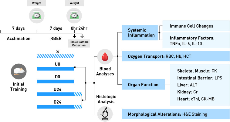
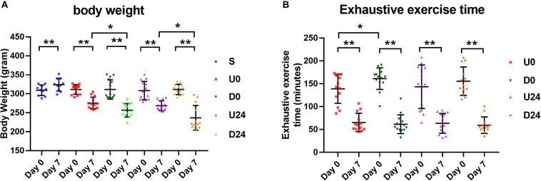
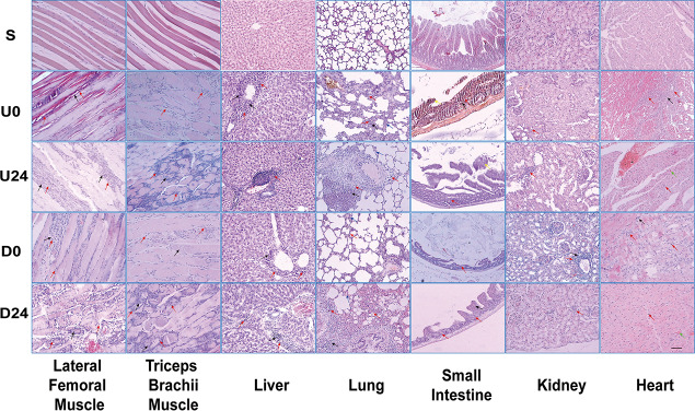
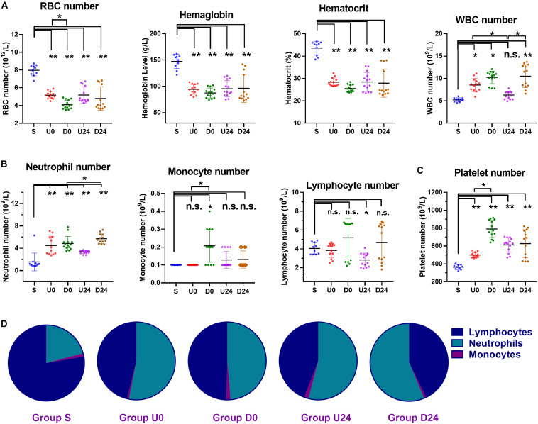
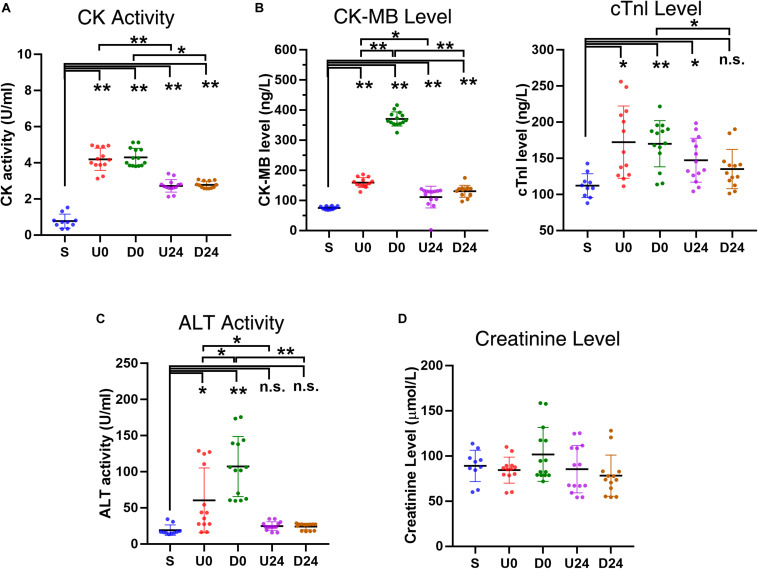
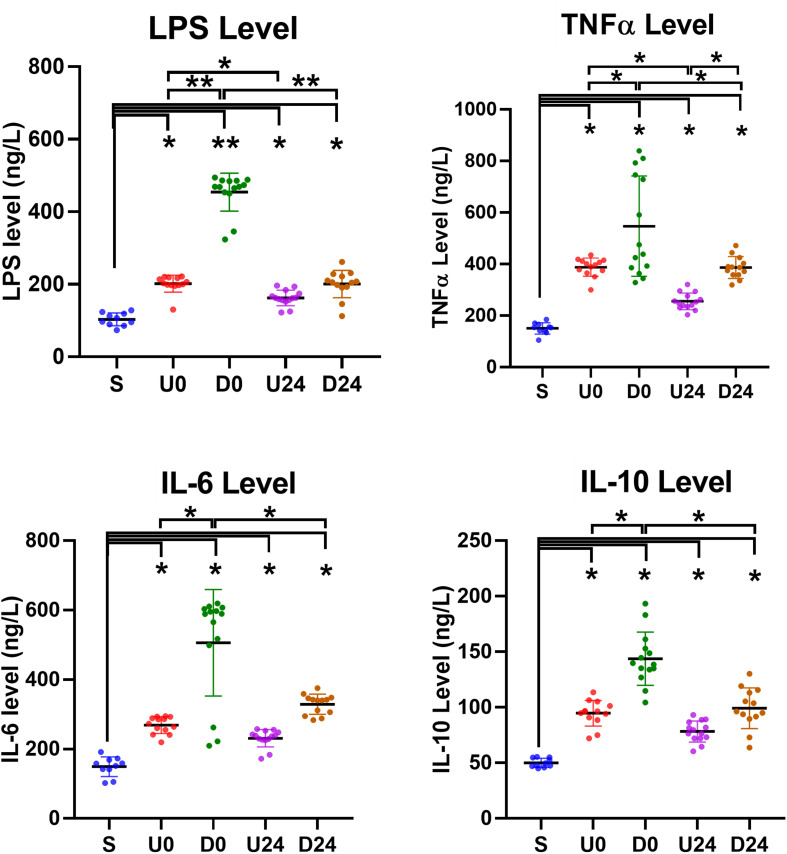
Multiple organ dysfunction syndrome can follow severe infection or injury, but its relationship to exercise is not well understood. Previous studies have observed that prolonged strenuous exercise can lead to transiently increased level and/or activity of markers for systemic inflammatory response and multiple organ damage. However, few studies have analyzed the pathogenesis of the inflammatory response and subsequent multi-organ injury in exhaustive exercise conditions. In this study, we established a rat model of repetitive bouts of exhaustive running (RBER) and investigated its effects on multiple organ damage. Rats were subjected to RBER in either uphill or downhill running modes daily for a period of 7 days. Morphologically, RBER causes tissue structural destruction and infiltration of inflammatory cells in the skeletal muscles and many visceral organs. RBER also causes sustained quantitative changes in leukocytes, erythrocytes, and platelets, and changes in the concentration of blood inflammatory factors. These inflammatory alterations are accompanied by increases in serum enzyme levels/activities which serve as functional markers of organ damage. In general, RBER in the downhill mode seemed to cause more damage evaluated by the above-mentioned measures than that produced in the uphill mode. A period of rest could recover some degree of damage, especially for organs such as the heart and kidneys with strong compensatory capacities. Together, our data suggest that, as a result of multi-organ interactions, RBER could cause a sustained inflammatory response for at least 24 h, resulting in tissue lesion and ultimately multiple organ dysfunction.

Keywords: exhaustive exercise; microhemorrhage; multi-organ dysfunction; necrosis; systemic inflammation.


Figures

FIGURE 1
FIGURE 1
The experimental design and conduct of this study is exhibited here. All animals were trained for 7 days during the acclimation period. Animals were then randomly assigned to the non-exercise sedentary group, or to 1 of 4 exercise groups. Animals in U0 and D0 were trained using the repetitive bouts of exhaustive running (RBER) model for 7 days, via uphill and downhill running respectively, and sacrificed immediately after. Animals in the U24 and D24 were similarly trained, but were sacrificed after 24 h of rest. Tissues were collected following sacrifice and processed for various blood and histologic analyses. S, sedentary; U0, uphill training, samples collected immediately; U24, uphill, 24 h later; D0, downhill, immediately; D24, downhill, 24 h later.
FIGURE 2
FIGURE 2
Changes in body weight and exhaustive time in different groups. (A) Except the sedentary group (S), the average body weight of rats in each exercise group was decreased significantly after RBER. (B) Exhaustive exercise time in each exercise group was significantly reduced at day 7 after RBER. Data is represented as mean ± SD in the figures. U0, uphill training, analyzed immediately; U24, uphill, 24 h later; D0, downhill, immediately; D24, downhill, 24 h later. *P < 0.05; **P < 0.01; n ≥ 10.
FIGURE 3
FIGURE 3
Light microscopy demonstrating morphologic changes in various tissues/organs after RBER. Arrows: Black = necrosis; Red = inflammatory infiltration; Green = microhemorrhage; Blue = alveolar septum thickening; Yellow = detached intestinal mucosa. S, sedentary; U0, uphill training, tissues collected immediately; U24, uphill, 24 h later; D0, downhill, immediately; D24, downhill, 24 h later. n ≥ 10. Scale bar = 100 μm.
FIGURE 4
FIGURE 4
Changes in the numbers and percentages of blood cells in different groups. (A) Compared with sedentary group S, the hemoglobin level, hematocrit, and number of red blood cells (RBC) were all decreased in each exercise group, while white blood cells (WBCs) were increased. (B) The WBC differential demonstrated a significant increase in number of neutrophils in each exercise group. The number of monocytes was roughly unaffected, except in group D0 which demonstrated a significant increase. The number of lymphocytes was roughly unaffected, except in group U24 which demonstrated a significant decrease. (C) The platelet numbers were increased in all exercise groups. (D) The percentage of neutrophils was increased each exercise group (all P < 0.01). As a result, the percentage of lymphocytes was decreased in each exercise group (all P < 0.01). S, sedentary; U0, uphill training, samples collected immediately; U24, uphill, 24 h later; D0, downhill, immediately; D24, downhill, 24 h later. *P < 0.05; **P < 0.01, n.s., not significant. n ≥ 10.
FIGURE 5
FIGURE 5
Changes in serum enzyme activities and levels after RBER. (A) Serum creatine kinase (CK) activity was greatly increased in each exercise group, reflecting skeletal muscle damage in the rats following exercise (B) Upregulation of specific myocardial marker CK-myocardial band (CK-MB) and cardiac troponin I (cTnI) in each exercise group, indicative of myocardial injury after exercise. The upregulation noted in U0 and D0 was largely recovered after 24 h of rest as shown in U24 and D24. (C) The serum alanine aminotransferase (ALT) activity was increased in U0 and D0, but not in U24 and D24, implying that damage of liver cells occurred acutely after exercise but was recovered after 24 h of rest. (D) The creatinine level was largely unaffected, indicating that the injury to kidney was minor in all exercise groups. S, sedentary; U0, uphill training, samples collected immediately; U24, uphill, 24 h later; D0, downhill, immediately; D24, downhill, 24 h later. *P < 0.05; **P < 0.01, n.s., not significant. n ≥ 10.
FIGURE 6
FIGURE 6
Serum changes in inflammation-related factors. The serum levels of lipopolysaccharide (LPS), tumor necrosis factor-alpha (TNF-α), interleukin (IL)-6 and IL-10 were all upregulated in each exercise group. They were relatively higher in downhill running groups than those in corresponding uphill groups. The degrees of increases were also significantly or insignificantly alleviated after 24 h of rest in U24 and D24 groups. S, sedentary; U0, uphill training, samples collected immediately; U24, uphill, 24 h later; D0, downhill, immediately; D24, downhill, 24 h later. *P < 0.05; **P < 0.01. n ≥ 10.

Similar articles

Cited by

References

    1. Alves A. J., Viana J. L., Cavalcante S. L., Oliveira N. L., Duarte J. A., Mota J., et al. (2016). Physical activity in primary and secondary prevention of cardiovascular disease: overview updated. World J. Cardiol. 8 575–583. - PMC - PubMed
    1. American College of Sports Medicine Armstrong L. E., Casa D. J., Millard-Stafford M., Moran D. S., Pyne S. W., et al. (2007). American college of sports medicine position stand. exertional heat illness during training and competition. Med. Sci. Sports Exerc. 39 556–572. 10.1249/mss.0b013e31802fa199 - DOI - PubMed
    1. Behringer M., Kilian Y., Montag J., Geesmann B., Mester J. (2016). Plasma concentration of high-mobility group box 1 (HMGB1) after 100 drop to vertical jumps and after a 1200-km bicycle race. Res. Sports Med. 24 119–129. 10.1080/15438627.2015.1126275 - DOI - PubMed
    1. Behringer M., Montag J., Franz A., Mccourt M. L., Mester J., Nosaka K. K. (2014). Exhaustive exercise–a near death experience for skeletal muscle cells? Med. Hypotheses 83 758–765. 10.1016/j.mehy.2014.10.005 - DOI - PubMed
    1. Booth F. W., Roberts C. K., Laye M. J. (2012). Lack of exercise is a major cause of chronic diseases. Compr. Physiol. 2 1143–1211. - PMC - PubMed

LinkOut - more resources