. 2020 Jun 16:11:1262.
doi: 10.3389/fimmu.2020.01262. eCollection 2020.

Internalization and Intoxication of Human Macrophages by the Active Subunit of the Aggregatibacter actinomycetemcomitans Cytolethal Distending Toxin Is Dependent Upon Cellugyrin (Synaptogyrin-2)

Affiliations

Internalization and Intoxication of Human Macrophages by the Active Subunit of the Aggregatibacter actinomycetemcomitans Cytolethal Distending Toxin Is Dependent Upon Cellugyrin (Synaptogyrin-2)

Kathleen Boesze-Battaglia et al. Front Immunol. .

Abstract

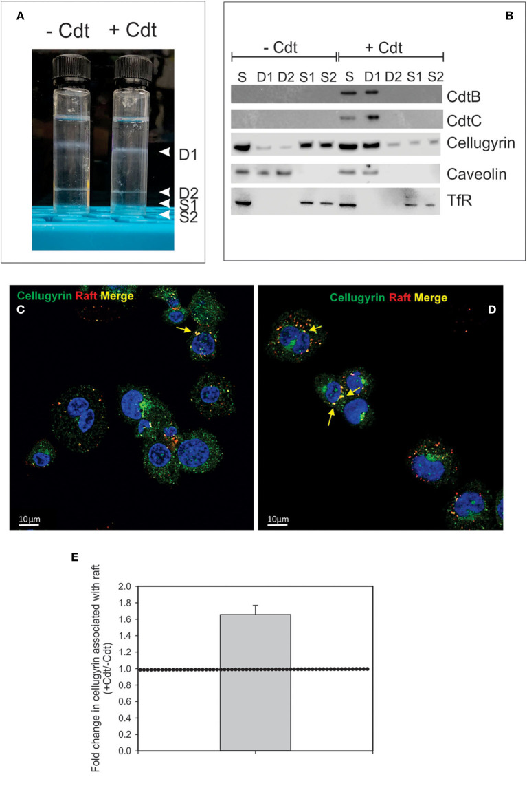
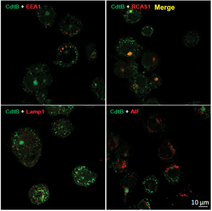
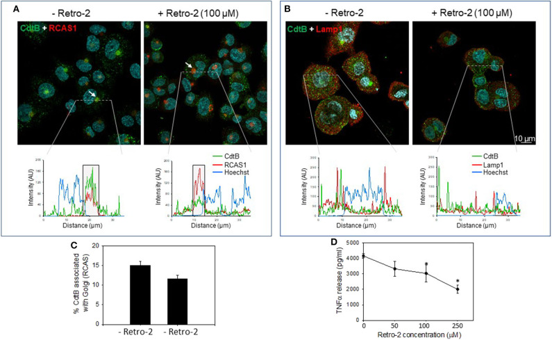
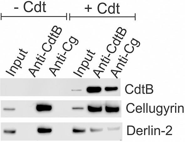
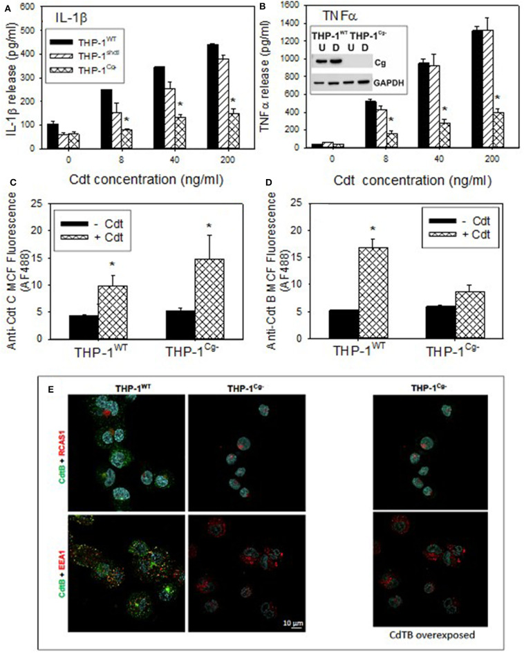
The Aggregatibacter actinomycetemcomitans cytolethal distending toxin (Cdt) is a heterotrimeric AB2 toxin capable of inducing cell cycle arrest and apoptosis in lymphocytes and other cell types. Recently, we have demonstrated that human macrophages are resistant to Cdt-induced apoptosis but are susceptible to toxin-induced pro-inflammatory cytokine response involving activation of the NLRP3 inflammasome. Exposure to Cdt results in binding to the cell surface followed by internalization and translocation of the active subunit, CdtB, to intracellular compartments. Internalization involves hijacking of retrograde pathways; treatment of cells with Retro-2 leads to a decrease in CdtB-Golgi association. These events are dependent upon toxin binding to cholesterol in the context of lipid rich membrane microdomains often referred to as lipid rafts. We now demonstrate that within 1 h of exposure of macrophages to Cdt, CdtB is internalized and found primarily within lipid rafts; concurrently, cellugyrin (synaptogyrin-2) also translocates into lipid rafts. Further analysis by immunoprecipitation indicates that CdtB associates with complexes containing both cellugyrin and Derlin-2. Moreover, a human macrophage cell line deficient in cellugyrin expression (THP-1Cg-) challenged with Cdt failed to internalize CdtB and was resistant to the Cdt-induced pro-inflammatory response. We propose that lipid rafts along with cellugyrin play a critical role in the internalization and translocation of CdtB to critical intracellular target sites in human macrophages. These studies provide the first evidence that cellugyrin is expressed in human macrophages and plays a critical role in Cdt toxicity of these cells.

Keywords: Aggregatibacter actinomycetemcomitans; bacterial toxins; cellugyrin; cytolethal distending toxin; lipid rafts; macrophages.


Figures

Figure 1
Figure 1
Cdt-induces translocation of cellugyrin to cholesterol rich microdomains. THP-1WT-derived macrophage were treated with medium or Cdt (1 μg/ml) for 1 h. Cells were harvested, washed, and cholesterol rich microdomains isolated as detergent resistant membranes (DRM) as described in Materials and Methods. (A) Shows the two DRM zones, DRM1 (D1) and DRM2 (D2), on the sucrose gradient; additionally two soluble fractions were collected, designated S1 and S2, which contain the bottom 0.5 ml (each) of the gradient. (B) Each fraction was analyzed by Western blot for the presence of CdtB, CdtC, cellugyrin, caveolin (lipid raft marker) and the transferrin receptor (TfR) (non-raft membrane marker). S refers to unfractionated start material. (C,D) Representative confocal images of differentiated untreated THP-1WT cells (Ctl; C) or treated with 1 μg/ml Cdt (+Cdt; D) and stained for rafts (red) and cellugyrin (green). (E) Shows the fold change in cellugyrin associated with lipid rafts in Cdt treated cells relative to untreated cells (normalized to total cellugyrin). Dashed horizontal line represents unity. Results are from three experiments (4–7 fields per experiment); p <0.05 (two-tailed one sample t-test). The degree of overlap between cellugyrin and lipid raft staining was estimated by Pearson's correlation coefficient of 0.72 ± 0.04 and 0.73 ± 0.03 (mean ± SEM), n = 10 ROI for control and Cdt-treated cells, respectively.
Figure 2
Figure 2
CdtB localizes to early endosomes and Golgi. Confocal micrographs of differentiated THP-1WT cells treated with Cdt holotoxin and immunostained for the indicated organelle markers (red), CdtB (green), and merge (yellow). EEA1, early endosome; RCAS1, Golgi marker; Lamp1, lysosome marker; AIF, mitochondrial marker.
Figure 3
Figure 3
Retro-2 blocks retrograde transport of CdtB to Golgi. Confocal images showing maximum intensity projections (from 6 μn Z stack) of differentiated THP-1WT cells pre-treated for 1 h with medium alone (left) or with Retro-2 (100 μM, right); both samples then received Cdt (1 μg/ml). After 1 h, cells were stained for CdtB and RCAS1 or Lamp1. Nuclei stained with Hoechst are pseudo-colored in cyan. (A) Maximum intensity projections for CdtB (green) and RCAS1 (red). Bottom graphs represent intensity profiles for CdtB (green), RCAS1 (red), and Hoechst nuclear stain (blue) across dotted lines depicted. Boxed regions correspond to RCAS1 and CdtB positive structures (marked by white arrows in the top panel). (B) Maximum intensity projections for CdtB (green) and Lamp1 (red). Bottom graphs represent intensity profiles for CdtB (green), Lamp1 (red), and nuclear stain (blue) across dotted lines depicted. AU, arbitrary units. All images are representative of four independent experiments (3–4 fields per experiment). (C) Shows the results of the analysis for the percent of CdtB associated with Golgi with and without Retro-2. Data are plotted as mean ± SEM. (D) THP-1WT macrophages were treated with Retro-2 and Cdt as described above; 5 h later supernatants were analyzed by ELISA for TNFα. Results are the average (pg/ml) TNFα ±SEM of three experiments; *indicates statistical significance (p <0.05).
Figure 4
Figure 4
Immunoprecipitation of cellugyrin, Derlin-2, and CdtB. THP-1WT cells were treated with medium or Cdt (2 μg/ml) for 2 h and then washed and homogenized as described in Materials and Methods. Supernatants were immunoprecipitated with either immobilized anti-CdtB or anti-cellugyrin. The bound protein was eluted and analyzed by Western blot for the presence of CdtB, celluygrin, and Derlin-2. Results are representative of three experiments.
Figure 5
Figure 5
Comparison of THP-1WT- and THP-1Cg− -derived macrophages on susceptibility to Cdt-induced pro-inflammatory response, Cdt holotoxin binding and CdtB internalization. (A,B) Macrophages derived from THP-1WT (solid bars) THP-1schctl (hatched bars) and THP-1Cg− (cross-hatched bars) cells were treated with varying amounts of Cdt for 5 h; supernatants were harvested and analyzed by ELISA for IL-1β (A) and TNFα (B). Data are plotted as the mean ± SEM (pg/ml) and represent the results from three experiments. Inset in (B) shows Western blot analysis of cellugyrin in undifferentiated (U) and differentiated (D) cells derived from THP-1WT- and THP-1Cg− cells. (C) Shows the results of flow cytometric analysis of Cdt binding to THP-1WT- and THP-1Cg− -derived macrophages. Cells were incubated for 60 min at 5°C with Cdt (1 μg/ml), washed and stained for the presence of cell surface associated Cdt using anti-CdtC mAb conjugated to AlexaFlour488. The mean channel fluorescence [mcf; (mean±SEM)] for three experiments is shown. (D) Shows the flow cytometric analysis of CdtB internalization. THP-1WT- and THP-1Cg− -derived macrophages were incubated with Cdt as above except at 37°C. Cells were washed, fixed, permeabilized and stained with anti-CdtB mAb conjugated to AlexaFluor488. The mcf (mean ± SEM) for three experiments is shown. (E) Shows the confocal microscopic analysis of CdtB localization to early endosomes or Golgi in THP-1WT- and THP-1Cg− cells. Confocal micrographs of Cdt-treated cells immunostained for CdtB (green) and RCAS1 (red, top) or EEA1 (red, bottom). CdtB signal (green) was enhanced in the panel on the right. Nuclei stained with Hoechst are pseudo-colored in cyan.
Figure 6
Figure 6
Schematic model showing proposed CdtB-cellugyrin interaction. Cdt holotoxin binds to cells via cholesterol in the context of membrane lipid rafts. CdtB internalization is further dependent upon its ability to interact with cholesterol. As a result of exposure to Cdt, cellugyrin (shown in red) containing SLMVs translocate from cytosol to membrane lipid rafts. We propose that this translocation leads to association of CdtB with the cellugyrin-containing SLMVs. This interaction may involve direct binding to cellugyrin either on extra- or intra-vesicular loops or indirect association via an unidentified binding partner (shown in black). We further propose that CdtB is transported via SLMVs to intracellular target sites; for example sites containing PIP3 pools where the enzymatically active CdtB subunit is released from SLMVs and is then able to degrade the signaling lipid resulting in PI-3K blockade and toxicity.

References

    1. Scuron MD, Boesze-Battaglia K, Dlakic M, Shenker BJ. The cytolethal distending toxin contributes to microbial virulence and disease pathogenesis by acting as a tri-perditious toxin. Front Cell Infect Microbiol. (2016) 6:168. 10.3389/fcimb.2016.00168 - DOI - PMC - PubMed
    1. Smith JL, Bayles DO. The contribution of cytolethal distending toxin to bacterial pathogenesis. Crit Rev Microbiol. (2006) 32:227–48. 10.1080/10408410601023557 - DOI - PubMed
    1. Ahmed HJ, Svensson LA, Cope LD, Latimer JL, Hansen EJ, Ahlman K, et al. Prevalence of cdtABC genes encoding cytolethal distending toxin among Haemophilus ducreyi and Actinobacillus actinomycetemcomitans strains. J Med Microbiol. (2001) 50:860–4. 10.1099/0022-1317-50-10-860 - DOI - PubMed
    1. Fox JG, Rogers AB, Whary MT, Ge Z, Taylor NS, Xu S, et al. Gastroenteritis in NF-kappaB-deficient mice is produced with wild-type Camplyobacter jejuni but not with C. jejuni lacking cytolethal distending toxin despite persistent colonization with both strains. Infect Immun. (2004) 72:1116–25. 10.1128/iai.72.2.1116-1125.2004 - DOI - PMC - PubMed
    1. Ge Z, Feng Y, Whary MT, Nambiar PR, Xu S, Ng V, et al. Cytolethal distending toxin is essential for Helicobacter hepaticus colonization in outbred Swiss Webster mice. Infect Immun. (2005) 73:3559–67. 10.1128/IAI.73.6.3559-3567.2005 - DOI - PMC - PubMed

Publication types

LinkOut - more resources