

Pancreatic neuroendocrine neoplasms: current state and ongoing controversies on terminology, classification and prognostication
- PMID: 32655934
- PMCID: PMC7340800
- DOI: 10.21037/jgo.2020.03.07
Pancreatic neuroendocrine neoplasms: current state and ongoing controversies on terminology, classification and prognostication
Abstract
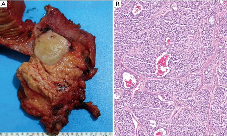
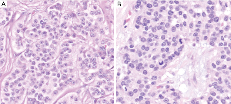
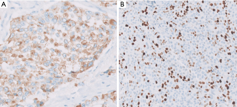
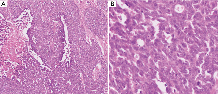
Significant improvements have taken place in our understanding of classification neuroendocrine neoplasms of the pancreas in the past decade. These are now regarded in three entirely separate categories: (I) neuroendocrine tumors (PanNETs) are by definition well differentiated, the pancreatic counterpart of carcinoids; (II) neuroendocrine carcinomas, which are poorly differentiated (PDNEC), the pancreatic examples of small cell carcinomas or large cell neuroendocrine carcinomas; (III) other neoplasms that have neuroendocrine differentiation or a distinct neuroendocrine component. PanNETs are by far the most common. They are now regarded as malignancies (albeit often curable when low grade and low stage) with the exception of minute incidental proliferations (tumorlets, or dysplastic-like changes) seen in the setting of some syndromes like MEN. PanNETs are staged based on their size, and for small T1 tumors, watchful waiting is now being considered, although these tumors are also known to show about 10% metastatic rate and/or progression, creating concerns about this approach. PanNETs are graded into 3, based on the proliferative activity, mostly based on the Ki-67 index, and also partly mitotic activity, although the latter seldom if ever is the determinant of the final grade. Neuroendocrine neoplasms with well differentiated morphology but Ki-67 >20% are now regarded as PanNET Grade 3 (G3); they have been shown to have a prognosis significantly worse than lesser grade PanNETs but still incomparably better than frank PDNECs, the latter typically has Ki-67 >50% (often much higher) and require platinum-based chemotherapy. There are also cases that are ambiguous between PanNET-G3 and PDNEC, and very rarely transformation of the former to the latter appears to occur. For low grade (G1/G2) PanNETs, more refined criteria to further prognosticate this group are needed. Morphologic variants being recognized may bring new perspectives to this group.
Keywords: Pancreas; WHO 2019; carcinoma; classification; neuroendocrine neoplasm; pathology; prognosis; terminology; tumor.
2020 Journal of Gastrointestinal Oncology. All rights reserved.
Conflict of interest statement
Conflicts of Interest: All authors have completed the ICMJE uniform disclosure form (available at http://dx.doi.org/10.21037/jgo.2020.03.07). The series “Pancreatic Neuroendocrine Tumors” was commissioned by the editorial office without any funding or sponsorship. CNC and DBE served as the unpaid Guest Editors of the series. The other authors have no other conflicts of interest to declare.
Figures
References
-
- Gill A, Klimstra D, Lam A, et al. Tumors of the pancreas. In: WHO Classification of Tumors: Digestive System Tumours. 5th edition. Lyon (France): International Agency for Research on Cancer, 2019;295-376.
Publication types
LinkOut - more resources
Full Text Sources
Miscellaneous