Case Reports
. 2020 Jun 8;12(6):e8512.
doi: 10.7759/cureus.8512.

Cryptococcal Choroid Plexitis and Non-Communicating Hydrocephalus

Affiliations
Case Reports

Cryptococcal Choroid Plexitis and Non-Communicating Hydrocephalus

Kyle P O'Connor et al. Cureus. .

Abstract

Cryptococcus neoformans is a fungus that commonly invades the central nervous system. While the choroid plexus, the site of the blood-cerebrospinal fluid barrier, serves as one potential entry point for the pathogen, disease involvement of the choroid plexus itself remains a very rare manifestation of Cryptococcus infection. In cases in which choroid plexus involvement blocks cerebrospinal fluid flow, obstructive hydrocephalus may occur. Here we report the case of a 63-year-old woman who presented with choroid plexitis causing obstructive hydrocephalus at the foramen of Monro. Endoscopic biopsy confirmed Cryptococcus neoformans, and the patient was successfully treated with amphotericin, flucytosine, and fluconazole. With proper recognition and treatment of this pathology, patients can fully recover from this condition.

Keywords: biopsy; cryptococcus neoformans; endoscopic; hydrocephalus; infection.


Conflict of interest statement

The authors have declared that no competing interests exist.

Figures
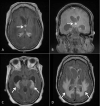
Figure 1
Figure 1. MRI Findings of Choroid Plexitis
(A) Axial T1-weighted post contrast MRI and (B) coronal T1-weighted post contrast MRI demonstrating a hyperintense nodularity at the foramen of Monro (arrows). (C) Axial T1-weighted post contrast MRI demonstrating involvement of the choroid plexus within the temporal horn of the lateral ventricle (arrows). (D) Axial section of T2-weighted FLAIR MRI demonstrating transependymal flow (arrows). FLAIR, Fluid-attenuated inversion recovery.
Figure 2
Figure 2. Hematoxylin and Eosin and Methenamine-Silver Staining of Biopsy Sample
Hematoxylin and eosin (A) and Gomori methenamine-silver (B) stains (both X20) demonstrate the presence of Cryptococcus. Encapsulated Cryptococcus yeasts are indicated by the black arrows and budding yeasts are indicated by the white arrows.
Figure 3
Figure 3. Mucicarmine Staining of Biopsy Sample
Mucicarmine staining (X40) demonstrates the presence of a mucicarmine-positive (pink) capsule. An encapsulated Cryptococcus yeast is indicated by the black arrow and budding yeasts are indicated by the white arrow.

References

    1. The choroid plexus-a multi-role player during infectious diseases of the CNS. Schwerk C, Tenenbaum T, Kim KS, Schroten H. Front Cell Neurosci. 2015;9:80. - PMC - PubMed
    1. Molecular mechanisms of cryptococcal meningitis. Liu T-B, Perlin DS, Xue C. Virulence. 2012;3:173–181. - PMC - PubMed
    1. Cryptococcosis. Maziarz EK, Perfect JR. Infect Dis Clin North Am. 2016;30:179–206. - PMC - PubMed
    1. Cryptococcal choroid plexitis: rare imaging findings of central nervous system cryptococcal infection in an immunocompetent individual. Kumari R, Raval M, Dhun A. Br J Radiol. 2010;83:14–17. - PMC - PubMed
    1. MRI features of choroid plexitis. Cho IC, Chang KH, Kim YH, Kim SH, Yu IK, Han MH. Neuroradiology. 1998;40:303–307. - PubMed

Publication types

LinkOut - more resources