Review
. 2020 Jun 16:8:452.
doi: 10.3389/fcell.2020.00452. eCollection 2020.

The Neurovascular Unit in Glaucomatous Neurodegeneration

Affiliations
Review

The Neurovascular Unit in Glaucomatous Neurodegeneration

Lauren K Wareham et al. Front Cell Dev Biol. .

Abstract

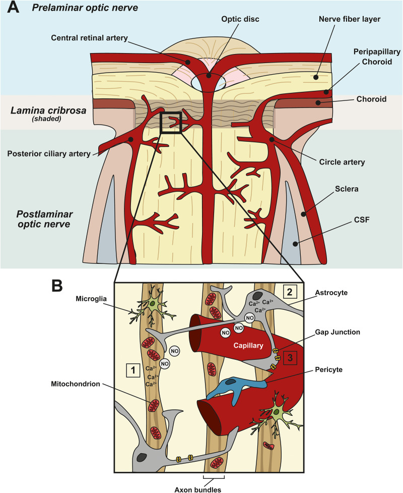
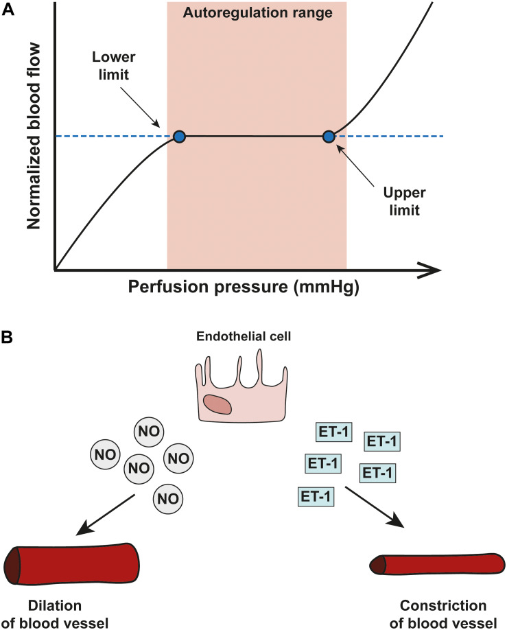
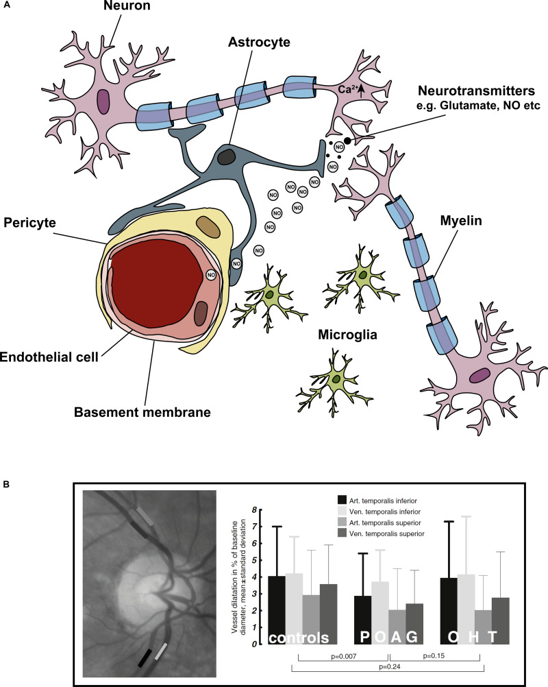
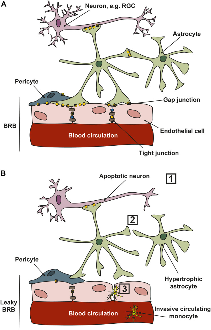
Glaucoma is a neurodegenerative disease of the visual system and leading cause of blindness worldwide. The disease is associated with sensitivity to intraocular pressure (IOP), which over a large range of magnitudes stresses retinal ganglion cell (RGC) axons as they pass through the optic nerve head in forming the optic projection to the brain. Despite clinical efforts to lower IOP, which is the only modifiable risk factor for glaucoma, RGC degeneration and ensuing loss of vision often persist. A major contributor to failure of hypotensive regimens is the multifactorial nature of how IOP-dependent stress influences RGC physiology and structure. This stress is conveyed to the RGC axon through interactions with structural, glial, and vascular components in the nerve head and retina. These interactions promote pro-degenerative pathways involving biomechanical, metabolic, oxidative, inflammatory, immunological and vascular challenges to the microenvironment of the ganglion cell and its axon. Here, we focus on the contribution of vascular dysfunction and breakdown of neurovascular coupling in glaucoma. The vascular networks of the retina and optic nerve head have evolved complex mechanisms that help to maintain a continuous blood flow and supply of metabolites despite fluctuations in ocular perfusion pressure. In healthy tissue, autoregulation and neurovascular coupling enable blood flow to stay tightly controlled. In glaucoma patients evidence suggests these pathways are dysfunctional, thus highlighting a potential role for pathways involved in vascular dysfunction in progression and as targets for novel therapeutic intervention.

Keywords: gap junctions; glaucoma; neurodegeneration; neurovascular coupling; neurovascular unit; vasculature.


Figures

FIGURE 1
FIGURE 1
Neurovascular coupling in the ONH and vascular dysfunction in glaucoma. (A) Schematic showing the blood supply at the ONH. Prelaminar, lamina cribrosa, and postlaminar sections are indicated. The primary blood supply to the ONH and retina comes from the choroid, the central retinal artery, the posterior ciliary artery and the circle artery. (B) Enlargement of the boxed area in (A) showing a capillary and its associated cells in the ONH. Astrocytes, pericytes, and endothelial cells of blood vessels constitute the neurovascular unit (NVU), to link local neuronal activity to vascular changes. In healthy tissue, when there is a spike in neuronal activity (1), or metabolic demand, it leads to an increased intracellular concentration of Ca2+ in neurons and (2) astrocytes. This, in turn, leads to the generation of nitric oxide (NO), a vasoactive gaseous messenger, which can diffuse to nearby blood vessels, altering blood flow. In glaucoma, apoptotic neurons and reactive astrocytes lead to the breakdown of this coupling. (3) In addition, in glaucoma, ischemia, and perfusion instability damages astrocyte–astrocyte gap junctions, leading to miscommunication between astrocytes and neurons.
FIGURE 2
FIGURE 2
Blood flow autoregulation in the eye. (A) A schematic showing an autoregulation curve that describes the relationship between normalized blood flow (y-axis) and perfusion pressure (x-axis). Autoregulation can only operate within a critical range of OPP and once OPP surpasses the optimal range (shown in pink), autoregulatory systems start to break down. (B) The two important vasoactive substances released by endothelial cells are nitric oxide (NO) and endothelin-1 (ET-1) and autoregulation of the vascular system in the eye relies on a delicate balance between the two; NO is a potent vasodilator released by smooth muscle cells and endothelial cells which acts via pericytes to dilate capillaries. Opposite in function is ET-1, a potent vasoconstrictor.
FIGURE 3
FIGURE 3
Cells of the ‘neurovascular unit’ and the light flicker response. Neurovascular coupling describes the coupling of neuronal activity to vascular responses. (A) Shows the cells comprising the NVU, these include neurons (in the eye specifically – RGCs), astrocytes, microglia, pericytes, and endothelial cells. In general, a spike in neuronal activity leads to an increase in intracellular Ca2+, which generates NO. NO diffuses to local blood vessel endothelial cells, activating K+ channels, which leads to downstream vasodilation and increased blood flow. The light flicker response demonstrates the tight coupling of neuronal activity (in response to light) and change in vessel diameters in the retina. In glaucoma, this light flicker response is diminished. (B) To measure the light flicker response in the retina, a fundus video is used (image left) where the temporal inferior artery and vein, and temporal superior artery and vein are clearly visible. Areas of analysis are shown in grayscale boxes. Graphical representation the light flicker response in control, ocular hypertension (OHT) and glaucoma patients shows diminished vessel response with disease. Figure adapted from Gugleta et al. (2013b).
FIGURE 4
FIGURE 4
Cell communication in the NVU. (A) In healthy tissues, retinal ganglion cell (RGC) neurons, astrocytes and endothelial cells communicate through gap junctions which permit the movement of electrical conductance (Na+, K+, and Ca2+) and small molecules such as ATP/ADP, glutamate, and glucose, and second messengers. Gap junctions exist in the retina and ONH between neurons, glia and between vascular cells. (B) In glaucoma, several changes occur that affect communication between the cells of the NVU. (1) RGCs are vulnerable to stressors that lead to apoptosis; (2) cell–cell communication is lost as astrocytes and other glial cells become reactive and gap junction expression is reduced; (3) a reduction in gap junction expression, and loss of tight junctions between vascular cells leads to a leaky blood–retinal-barrier (BRB), allowing the infiltration of circulating monocytes into the retina and ONH.

References

    1. Ahmad S. S. (2016). Controversies in the vascular theory of glaucomatous optic nerve degeneration. Taiwan J. Ophthalmol. 6 182–186. 10.1016/j.tjo.2016.05.009 - DOI - PMC - PubMed
    1. Akil H., Huang A. S., Francis B. A., Sadda S. R., Chopra V. (2017). Retinal vessel density from optical coherence tomography angiography to differentiate early glaucoma, pre-perimetric glaucoma and normal eyes. PLoS One 12:e0170476. 10.1371/journal.pone.0170476 - DOI - PMC - PubMed
    1. Akopian A., Kumar S., Ramakrishnan H., Roy K., Viswanathan S., Bloomfield S. A. (2017). Targeting neuronal gap junctions in mouse retina offers neuroprotection in glaucoma. J. Clin. Invest. 127 2647–2661. 10.1172/JCI91948 - DOI - PMC - PubMed
    1. Alarcon-Martinez L., Yilmaz-Ozcan S., Yemisci M., Schallek J., Kilic K., Villafranca-Baughman D., et al. (2019). Retinal ischemia induces alpha-Sma-mediated capillary pericyte contraction coincident with perivascular glycogen depletion. Acta Neuropathol. Commun. 7:134. 10.1186/s40478-019-0761-z - DOI - PMC - PubMed
    1. Alm A., Bill A. (1973). Ocular and optic nerve blood flow at normal and increased intraocular pressures in monkeys (Macaca irus): a study with radioactively labelled microspheres including flow determinations in brain and some other tissues. Exp. Eye Res. 15 15–29.10.1016/0014-4835(73)90185-1 - DOI - PubMed