. 2020 Jul 1;6(27):eaba4526.
doi: 10.1126/sciadv.aba4526. eCollection 2020 Jul.

Esophageal extracellular matrix hydrogel mitigates metaplastic change in a dog model of Barrett's esophagus

Affiliations

Esophageal extracellular matrix hydrogel mitigates metaplastic change in a dog model of Barrett's esophagus

Juan Diego Naranjo et al. Sci Adv. .

Abstract

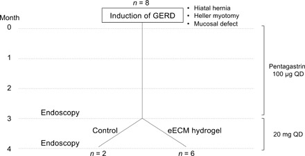
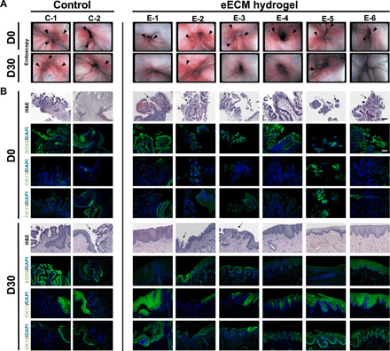
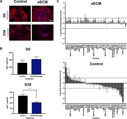
Chronic inflammatory gastric reflux alters the esophageal microenvironment and induces metaplastic transformation of the epithelium, a precancerous condition termed Barrett's esophagus (BE). The microenvironmental niche, which includes the extracellular matrix (ECM), substantially influences cell phenotype. ECM harvested from normal porcine esophageal mucosa (eECM) was formulated as a mucoadhesive hydrogel, and shown to largely retain basement membrane and matrix-cell adhesion proteins. Dogs with BE were treated orally with eECM hydrogel and omeprazole (n = 6) or omeprazole alone (n = 2) for 30 days. eECM treatment resolved esophagitis, reverted metaplasia to a normal, squamous epithelium in four of six animals, and downregulated the pro-inflammatory tumor necrosis factor-α+ cell infiltrate compared to control animals. The metaplastic tissue in control animals (n = 2) did not regress. The results suggest that in vivo alteration of the microenvironment with a site-appropriate, mucoadhesive ECM hydrogel can mitigate the inflammatory and metaplastic response in a dog model of BE.


Figures

Fig. 1
Fig. 1. Study overview.
A total of eight dogs underwent a reflux-inducing procedure in which a hiatal hernia, a Heller myotomy, and a mucosal defect were created. Animals received pentagastrin daily to increase the acidity of gastric secretions for 3 months. Following pentagastrin administration, animals were evaluated by endoscopy, and pentagastrin was replaced with omeprazole, a PPI commonly used to decrease the acidity of gastric secretions. Animals were randomly divided into two groups: (i) control (n = 2), to evaluate the effect of removing pentagastrin and starting omeprazole, and (ii) eECM treatment plus omeprazole (n = 6) for 30 days. The macroscopic, microscopic, and clinical outcomes of each eECM treatment animal were compared to pretreatment values for the same animal and to outcomes in the two animals in the control group. QD, one time a day.
Fig. 2
Fig. 2. eECM hydrogel viscoelastic and mucoadhesive properties are concentration dependent.
Rheological analysis was performed (n = 3, means ± SD). (A) eECM pre-gel viscosity at 10°C. (B) eECM hydrogel maximum storage modulus (G′), loss modulus (G″), and (C) time to 50% gelation after temperature was raised to 37°C. (D) Representative graphs of the storage modulus, loss modulus, and complex viscosity η*(ω) of the eECM hydrogels at 37°C, plotted over angular frequency. The ex vivo mucoadhesion of eECM hydrogel to esophageal mucosa was measured by (E) tensile testing (n = 3, means ± SD) and (F) after laminar flow of water for 6 and 24 hours. Gel thickness (H&E staining, black dotted lines) after laminar flow was quantified (n = 3, means ± SEM). Scale bar, 500 μm. eECM hydrogel (12 mg/ml) was dyed blue, brought to a temperature of 15°C, and delivered in vivo to the canine proximal and distal esophagus: (G) using an endoscopic catheter (50 ml, t = 0 min), (H) orally (50 ml, t = 50 min), and (I) orally at the volume used in the animal study (25 ml, t = 50 min). Photo credit: Juan Diego Naranjo, University of Pittsburgh. (J) Summary of the in vivo deliverability testing conditions at the different ECM hydrogel temperatures. Viscosity: *P ≤ 0.05, ****P ≤ 0.0001 by two-way ANOVA and post hoc Tukey’s test. Storage and loss modulus: *P ≤ 0.05, **P ≤ 0.01, ***P ≤ 0.001 by one-way ANOVA and post hoc Tukey’s test. Mucoadhesive strength: *P ≤ 0.05 by one-way ANOVA and post hoc Tukey’s test.
Fig. 3
Fig. 3. Proteomic signature of eECM.
(A) Absolute quantification of ECM proteins, binned by gene ontology class, for native esophageal mucosa tissue and eECM. Data are means ± SEM (n = 3). Top classes of proteins that showed retention of ECM proteins in eECM compared to native esophageal mucosa tissue included (B) basement membrane, (C) fibrillar collagens, and (D) microfibril-associated proteins (boxed). Proteins within the gene ontology classes are further shown (B to D). *P ≤ 0.05, **P ≤ 0.01, ***P ≤ 0.001, ****P ≤ 0.0001 by two-way ANOVA for treatment and protein functional class or protein type, with post hoc multiple comparisons test.
Fig. 4
Fig. 4. Effect of eECM hydrogel treatment on the macroscopic appearance of the mucosa and esophageal epithelial cell phenotype after 30 days.
(A) Endoscopies of the lower esophagus/GEJ taken at D0 (before treatment) and at D30 (after treatment) for control and eECM hydrogel animals. Dogs had varied degrees of esophagitis and ulceration (arrowheads) before treatment. No improvement was seen in the animals that only received omeprazole (control). Improvement was seen in all animals after 30 days of eECM hydrogel treatment, including some with complete macroscopic resolution (E-1, E-3, E-4, and E-6). For the remaining treatment animals (E-2 and E-5), there was partial or total resolution of esophagitis with partial healing of the biopsy site. Photo credit: Juan Diego Naranjo, University of Pittsburgh. (B) Biopsies were taken before treatment (D0), and location-matched samples were collected after 30 days of treatment (D30) for control animals and eECM hydrogel–treated animals. Samples at D0 and D30 were stained with hematoxylin and eosin (H&E), Barrett’s marker Sox9, or normal esophageal squamous epithelial markers CK13 and CK14. Arrows indicate goblet cells characteristic of intestinal metaplasia and BE. Scale bar, 100 μm.
Fig. 5
Fig. 5. Effect of eECM hydrogel treatment on TNFα+proinflammatory cell infiltrate and cytokine expression after 30 days.
(A) TNFα is a proinflammatory cytokine up-regulated in gastroesophageal reflux disease progression. TNFα immunolabeling with DAPI counterstain was performed on samples collected before and after 30 days of treatment for control and eECM hydrogel–treated animals. Representative images are shown. (B) Three pictures per animal were imaged and quantified using CellProfiler and compared to control. Scale bar, 100 μm. Data are means ± SEM for control (n = 2) and eECM hydrogel treatment (n = 6), with three technical replicates per sample. ***P ≤ 0.001 by Student’s unpaired t test. (C) Differential expression of cytokines in the eECM hydrogel and control animals’ esophageal tissue after 30 days of treatment, and normalized by the animals’ own proximal, normal esophageal tissue using a cytokine antibody microarray. Differentially expressed cytokines were defined as a >2- or <−2-fold change of eECM treatment or control compared to normal (dashed line).
Fig. 6
Fig. 6. Proteomic signature of eECM-treated tissue.
Principal components analysis (PCA) of “distal” tissue and “normal” tissue of eECM-treated dogs at D30 for (A) targeted proteomics and (B) global proteomics, showing the overlap between the remodeled distal tissue (E-Distal) and normal tissue (E-Normal) (n = 6). (C) Absolute quantification of ECM proteins, binned by gene ontology class, for distal and normal tissue of eECM-treated dogs at D30. Data are means ± SEM (n = 6). Expression of (D) basement membrane proteins (E), fibrillar collagen proteins, and (F) microfibril-associated proteins is expressed as a ratio of distal/normal for individual proteins within each class for the eECM-treated animals at D30. *P ≤ 0.05, **P ≤ 0.01, ****P ≤ 0.0001 by two-way ANOVA for tissue type and protein functional class or protein type, with post hoc multiple comparisons test.

References

    1. Bissell M. J., Hall H. G., Parry G., How does the extracellular matrix direct gene expression? J. Theor. Biol. 99, 31–68 (1982). - PubMed
    1. Schultz G. S., Davidson J. M., Kirsner R. S., Bornstein P., Herman I. M., Dynamic reciprocity in the wound microenvironment. Wound Repair Regen. 19, 134–148 (2011). - PMC - PubMed
    1. Sellaro T. L., Ranade A., Faulk D. M., McCabe G. P., Dorko K., Badylak S. F., Strom S. C., Maintenance of human hepatocyte function in vitro by liver-derived extracellular matrix gels. Tissue Eng. Part A 16, 1075–1082 (2010). - PMC - PubMed
    1. Zhang Y., He Y., Bharadwaj S., Hammam N., Carnagey K., Myers R., Atala A., Van Dyke M., Tissue-specific extracellular matrix coatings for the promotion of cell proliferation and maintenance of cell phenotype. Biomaterials 30, 4021–4028 (2009). - PMC - PubMed
    1. Keane T. J., DeWard A., Londono R., Saldin L. T., Castleton A. A., Carey L., Nieponice A., Lagasse E., Badylak S. F., Tissue-specific effects of esophageal extracellular matrix. Tissue Eng. Part A 21, 2293–2300 (2015). - PMC - PubMed

Publication types

LinkOut - more resources