. 2020 Jun;6(2):383-390.
doi: 10.21037/jss.2019.10.15.

Full endoscopic cervical spine surgery

Affiliations

Full endoscopic cervical spine surgery

Jian Shen et al. J Spine Surg. 2020 Jun.

Abstract

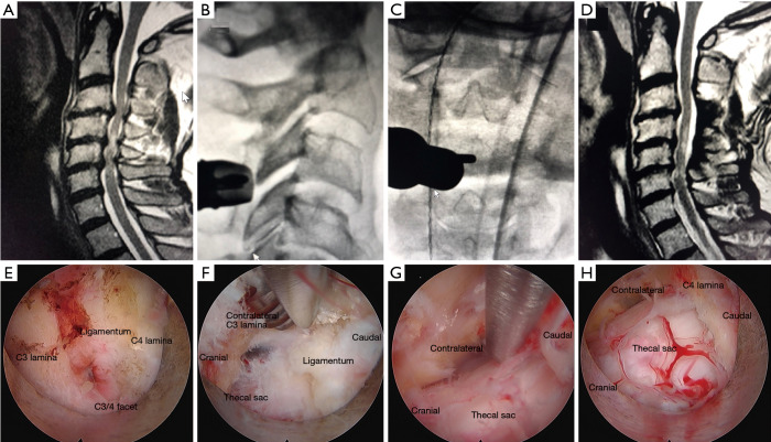
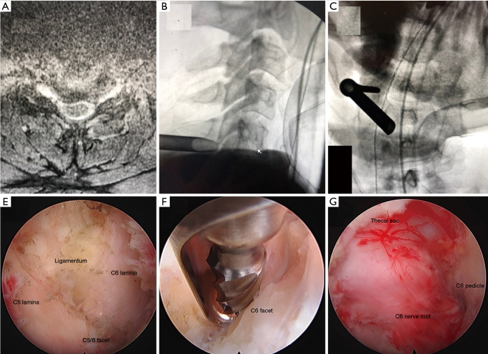
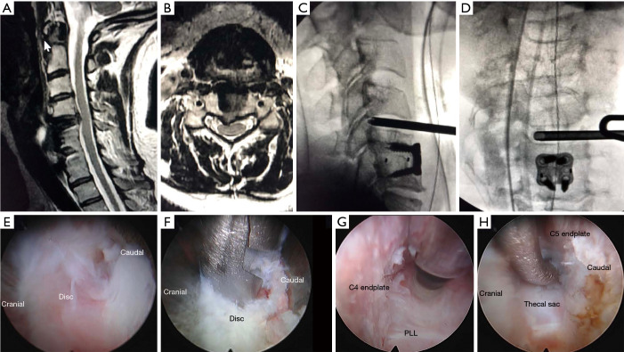
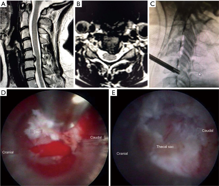
Background: The authors present 4 techniques for fully-endoscopic cervical spine surgery with accompanying case series: (I) posterior cervical unilateral laminectomy and bilateral decompression, (II) posterior cervical foraminotomy (PCF), (III) anterior cervical discectomy, and (IV) anterior transcorporeal discectomy.

Methods: We retrospectively reviewed fully endoscopic cervical spine surgery cases at one high-volume endoscopic center in the United States and present clinical data extracted from endoscopic spine surgery performed over a 6-year period with a minimum clinical follow up of 1 year.

Results: A series of 114 patients who underwent fully endoscopic cervical spine surgery between 2012 and 2018 is presented. Clinical results and technical data are presented.

Conclusions: Fully endoscopic cervical spine surgery is an emerging surgical technique for addressing cervical radiculopathy and myelopathy through a minimally invasive approach.

Keywords: Endoscopic spine surgery; cervical radiculopathy; minimally-invasive; myelopathy.


Conflict of interest statement

Conflicts of Interest: All authors have completed the ICMJE uniform disclosure form (available at http://dx.doi.org/10.21037/jss.2019.10.15). The series “Full-endoscopic Spine Surgery” was commissioned by the editorial office without any funding or sponsorship. The authors have no other conflicts of interest to declare.

Figures

Figure 1
Figure 1
Posterior cervical unilateral laminectomy and bilateral decompression. (A) Pre-operative T2-weighted sagittal MRI demonstrates severe spinal cord stenosis from C3–6 and cord myelomalacia; (B) lateral and (C) AP fluoroscopic images depicting the cannulated beveled tubular retractor with the Delta® working channel endoscope and Shrill® at the spinolaminar junction of C3–4; (D) post-operative T2 weighted sagittal MR image demonstrating the resolution of the cervical stenosis after the C3–6 endoscopic laminectomy; (E) endoscopic cameral view at the C3–4 spinolaminar junction. The C3 and C4 lamina, facet, and ligamentum flavum are demonstrated; (F) endoscopic camera view of the Shrill® drill performing the laminectomy; (G) endoscopic camera view of the Kerrison rongeur removing the contralateral ligamentum flavum; (H) endoscopic camera view of the bilaterally decompressed thecal sac. AP, anterior-posterior.
Figure 2
Figure 2
PCF. (A) Preoperative T2 axial MRI illustrating the right C5–6 foraminal stenosis; (B) lateral and (C) AP fluoroscopic views demonstrating the position of the beveled tubular retractor on the laminar-facet junction at C5–6; (D,E) endoscopic camera views of (D) the laminar-facet junction at C5–6, (E) the Shrill® drill removing the superior lateral edge of the C6 lamina, and (F) the decompressed C6 nerve root. PCF, posterior cervical foraminotomy; AP, anterior-posterior.
Figure 3
Figure 3
Anterior cervical discectomy. (A) Preoperative T2 sagittal and (B) T2 axial MR images demonstrating the C4–5 herniated disc superior to a previous C5-6 fusion; (C) lateral and (D) AP fluoroscopic images of the CESSYS® Cervical Hybrid endoscope and working channel in the C4–5-disc space; (E,F,G,H) endoscopic camera views of (E) the C4–5-disc herniation, (F) the endoscopic grasper removing the herniation, (G) the Shrill® drill drilling down the uncinate joint, and (H) the bendable grasper removing disc from the foramen. AP, anterior-posterior; PLL, posterior longitudinal ligament.
Figure 4
Figure 4
Anterior transcorporeal discectomy. (A) Preoperative T2 sagittal and (B) axial MR images of a C5–6 herniated disc; (C) lateral fluoroscopic image of the TESSYS® endoscope and tubular retractor placed through the body of C5; (D) endoscopic image of the disc herniation; (E) endoscopic image of the decompressed thecal sac after removal of the disc herniation. AP, anterior-posterior.

References

    1. Woods BI, Hilibrand AS. Cervical radiculopathy: epidemiology, etiology, diagnosis, and treatment. J Spinal Disord Tech 2015;28:E251-9. 10.1097/BSD.0000000000000284 - DOI - PubMed
    1. Rhee JM, Yoon T, Riew KD. Cervical radiculopathy. J Am Acad Orthop Surg 2007;15:486-94. 10.5435/00124635-200708000-00005 - DOI - PubMed
    1. Iyer A, Azad TD, Tharin S. Cervical spondylotic myelopathy. Clin Spine Surg 2016;29:408-14. 10.1097/BSD.0000000000000397 - DOI - PubMed
    1. Martins AN. Anterior cervical discectomy with and without interbody bone graft. J Neurosurg 1976;44:290-5. 10.3171/jns.1976.44.3.0290 - DOI - PubMed
    1. Rosenørn J, Hansen EB, Rosenørn MA. Anterior cervical discectomy with and without fusion. A prospective study. J Neurosurg 1983;59:252-5. 10.3171/jns.1983.59.2.0252 - DOI - PubMed